Download presentation
Presentation is loading. Please wait.

1
Radiologic Appropriateness Criteria
Sharal Mall, D.O. February 20, 2015

2
DISCLOSURES NONE

3
Pre-Test

4
1. Pt is a 62 y/o F c/o difficulty swallowing (dysphagia).
What to order next? 1. Pt is a 62 y/o F c/o difficulty swallowing (dysphagia). Upper GI study Barium Swallow/ Speech Evaluation Esophagram CT of the neck EGD
. Upper GI study. Barium Swallow/ Speech Evaluation. Esophagram. CT of the neck. EGD.")
5
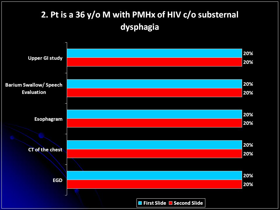
2. Pt is a 36 y/o M with PMHx of HIV c/o substernal dysphagia
What to order next? 2. Pt is a 36 y/o M with PMHx of HIV c/o substernal dysphagia Upper GI study Barium Swallow/ Speech Evaluation Esophagram CT of the chest EGD

6
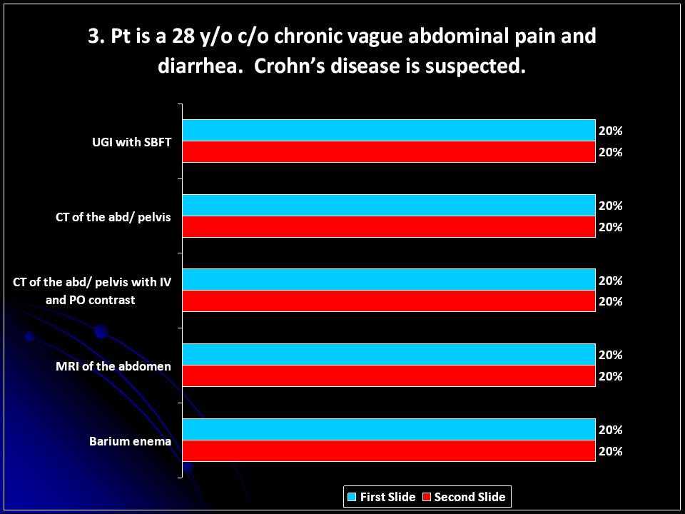
What to order next? 3. Pt is a 28 y/o c/o chronic vague abdominal pain and diarrhea. Crohn’s disease is suspected. UGI with SBFT CT of the abd/ pelvis CT of the abd/ pelvis with IV and PO contrast MRI of the abdomen Barium enema

7
What to order next? 4. Pt is a 72 y/o M with PMHx of multiple abdominal surgeries c/o distended stomach and pain. SBO is suspected. CT abd/ pelvis without contrast CT abd/ pelvis with PO contrast CT abd/ pelvis with IV contrast CT abd/ pelvis with PO and IV contrast Abdominal Xray series

8
What to order next? CXR Chest CT without contrast
5. Pt is a 54 y/o F c/o acute chest pain. No EKG changes and cardiac enzymes are normal. CXR Chest CT without contrast Chest CT with contrast Nuclear Stress Test study TEE

9
What to order next? CXR Invasive angiography CT abdomen EGD CT chest
6. Pt is a 67 y/o F with PMHx of EtOH abuse and liver cirrhosis. She is c/o hematemesis. CXR Invasive angiography CT abdomen EGD CT chest

10
What to order next? Repeat Xray CT MRI Bone scan US
7. Pt is 32 y/o runner c/o persistent L foot pain. Stress fx is suspected. Initial Xrays are normal. Repeat Xray CT MRI Bone scan US

11
What to order next? US IVP CT abd/ pelvis without contrast
8. Pt is a 53 y/o F with PMHx DM c/o back pain and fever. Acute pyelonephritis is suspected. US IVP CT abd/ pelvis without contrast CT abd/ pelvis with IV contrast CT abd/ pelvis with and without IV contrast

12
9. Pt is a 53 y/o F c/o painless hematuria. No PMHx of renal disease.
What to order next? 9. Pt is a 53 y/o F c/o painless hematuria. No PMHx of renal disease. IVP CT abd/ pelvis without contrast CT abd/ pelvis with IV contrast MRI US

13
10. Pt is 56 y/o M c/o vague Sx. Acute renal failure is suspected.
What to order next? 10. Pt is 56 y/o M c/o vague Sx. Acute renal failure is suspected. US CT abd/ pelvis without contrast CT abd/ pelvis with contrast MRI Nuclear Medicine Renogram

14
What to order next? CT abd/ pelvis without contrast
11. Pt is 53 y/o postmenopausal F c/o vague abdominal pain with suspected adnexal mass on physical exam. CT abd/ pelvis without contrast CT abd/ pelvis with contrast US MRI Xray

15
What to order next? Xray KUB CT abd/ pelvis without contrast
12. Pt is a 65 y/o M c/o acute onset of abdominal pain. PE yields a pulsatile abdominal mass. Xray KUB CT abd/ pelvis without contrast CT abd/ pelvis with contrast US colonoscopy

16
13. 78 y/o M presents with hypotension and rectal bleeding.
What to order next? y/o M presents with hypotension and rectal bleeding. CT abd/ pelvis CT abd/ pelvis with IV contrast RBC tagged nuclear medicine scan Colonoscopy Interventional Angiography

17
What to order next? Lt hand Xray Repeat Head CT MRI with Gd contrast
y/o M c/o sudden onset Lt hand numbness and weakness. Acute CVA is suspected. Initial head CT was ‘nonacute’. Lt hand Xray Repeat Head CT MRI with Gd contrast MRI without contrast US

18
What to order next? CT with IV contrast CT without IV contrast US Xray
15. An obese 57 y/o F with PMHx DM c/o acute edematous, painful and erythematous upper extremity. CT with IV contrast CT without IV contrast US Xray MRI

20
Plain Film X-Ray / Mammography
Cost: Both plain films and mammography are inexpensive Risk: Mortality and morbidity related to exposure to radiation Availability / Access Plain films are widely available Mammography is available during business hours Weight Limits The IVP table has a limit of 325 pounds. Alternatives to intravenous urography include CT, US, MRI, and cystoscopy/retrograde pyelography. Plain films have no fixed weight limit, but image quality declines with increasing patient weight and/or size. A departmental examination is always of better quality than a portable study due to better quality equipment and higher obtainable radiation parameters. Mammography has no specific weight limit.

21
Ultrasound Cost: Inexpensive Risk: Generally non-invasive
Essentially no mortality or morbidity Endovaginal, transrectal, transesophageal, and endocavitary US carry procedural risks related to introduction of the US probe Availability / Access: Relatively easily available

22
Ultrasound Prep: Patients should be NPO X 4 hours for GB, liver, pancreas, and biliary ductal examination Weight Limits: US has no definite fixed weight limit, but image quality and penetration decline as weight and/or size increases Vascular US has no weight limit per se, but very large limbs will have poor quality results or not be able to be meaningfully examined

23
CT (computed tomography)
Cost: Expensive Risk: Mortality and morbidity related to radiation exposure (especially children, breast tissue, lens, and ovaries/testes) Risk related to IV contrast use (death approximately 1 case per 40,000 – 100,000 uses) Oral contrast poses no real risk, except in pts who aspirate water soluable contrast
 Risk related to IV contrast use (death approximately 1 case per 40,000 – 100,000 uses) Oral contrast poses no real risk, except in pts who aspirate water soluable contrast.")
24
CT Availability / Access: Relatively easily available
Generally non-invasive All patients must be assessed for ability to receive IV contrast, even in “non-contrast” situations since changing circumstances may necessitate the use of IV contrast. All use of IV contrast requires IV access; of specific importance is that high-pressure contrast injections require at least an 18-gauge access with a catheter rated to accept high pressures! PICC lines and catheters of 20 gauge or less don’t meet these requirements unless specifically specified. This has particular importance for PE and other CT angiogram procedures (aorta, renal arteries etc.) and may render these CT procedures unable to be performed if suitable IV access isn’t available. New PICC lines are available that can accept high-pressure injections. “Power PICCs”
 and may render these CT procedures unable to be performed if suitable IV access isn’t available. New PICC lines are available that can accept high-pressure injections. Power PICCs")
25
CT Prep: Ideally patients should be NPO X 4 hours before administration of IV contrast, in case of contrast-induced vomiting, but this is optional if there is any urgency to the examination Weight Limits: Relative weight limit of 400 pounds Absolute weight limit of 450 pounds Patients with weight over 300 pounds or with protuberant abdomens tend to have degraded image quality PE studies in particular suffer quality image degradation as weight increases, thus, a V/Q scan is a better exam in large patients

26
MRI (Magnetic Resonance Imaging)
Cost: Very expensive Risk: Carries little mortality or morbidity due to electromagnetic exposure or IV contrast Availability / Access: Less easily available Small gauge IV access for IV contrast use is acceptable Prep: The patient doesn’t need to be NPO, but must be able to lay still In cases of claustrophobia or a non-cooperative patient, elective sedation may be required; this is arranged and ordered by the referring healthcare provider Weight Limits: The mobile MRI have weight limits of 300 pounds. Larger patients may require referral to an open MRI

27
Nuclear Medicine Cost: NM is moderately expensive Risk:
Carries minimal mortality and morbidity from the radiation and associated labeled radiopharmaceuticals Availability / Access: less available Small gauge IV access for IV radiopharmaceutical use is acceptable PET/CT is considered as a NM procedure and scheduled via the nuclear medicine department Please note that a cooperative patient who isn't artificially ventilated is required for the ventilation part of a lung V/Q scan; if these conditions aren't present, then an alternative PE examination should be selected

28
Nuclear Medicine Prep:
The patient should be NPO X 4 hours for GB, GB ejection fraction, and cardiac NM examinations Cardiac Stress Tests should also require the patient to be on a caffeine-free diet Patients on TPN or whom just ate may have false positive HIDA scans Weight Limits: Single head (camera) nuclear medicine table has a weight limit of 300 pounds Dual head NM table has a weight limit of 350 pounds PET/CT table has a weight limit of pounds There is progressive image degradation as weight increases due to soft tissue attenuation of the gamma rays or positrons being emitted by the radiopharmaceutical.
 nuclear medicine table has a weight limit of 300 pounds. Dual head NM table has a weight limit of 350 pounds. PET/CT table has a weight limit of pounds. There is progressive image degradation as weight increases due to soft tissue attenuation of the gamma rays or positrons being emitted by the radiopharmaceutical.")
29
Fluoroscopy (UGI-SBFT-BE)
Cost: moderately expensive Risk: A procedural risk of perforation (BE) Aspiration of oral contrast media (UGI & SBFT) Potential anaphylaxis due to allergic reaction to the latex balloon (BE) mortality and morbidity related to radiation exposure Availability / Access: Specific informed consent is not required less available after regular business hours Prep: Patients scheduled for UGI, SBFT, and BE examinations should be on restricted diet and/or bowel prep before their procedures. Details are available in the radiology department. Weight Limits The fluoroscopy table has a weight limit of 350 pounds Alternatives to fluoroscopy include EGD and Colonoscopy
 Aspiration of oral contrast media (UGI & SBFT) Potential anaphylaxis due to allergic reaction to the latex balloon (BE) mortality and morbidity related to radiation exposure. Availability / Access: Specific informed consent is not required. less available after regular business hours. Prep: Patients scheduled for UGI, SBFT, and BE examinations should be on restricted diet and/or bowel prep before their procedures. Details are available in the radiology department. Weight Limits. The fluoroscopy table has a weight limit of 350 pounds. Alternatives to fluoroscopy include EGD and Colonoscopy.")
30
Modified Swallow, Esophagram, UGI or SBFT?!?!
Modified speech swallowing exam- evaluates swallowing mechanism with liquid and solid barium preps as well as tablet form Esophagram- evaluates esophagus only and its motility UGI- evaluates esophagus, stomach and 1st and 2nd duodenum; may show reflux SBFT- only evaluates the small bowel (ie, esophagus and stomach are not evaluated)
")
31
Biopsy & Angiography / Interventional Procedures
Angio/interventional procedures are "mini surgeries“! Cost: Biopsy and angio/interventional procedures are expensive Risk: Significant procedural risks Associated mortality and morbidity from radiation exposure and contrast use (IV, intra-arterial, or intracavitary) Availability / Access: Availability is by consult to the angio/interventional service. Specific informed consent by the patient or guardian is required in all cases, unless the referring healthcare provider gives emergency consent Prep: There will be formal pre-op and post-op evaluation and monitoring of the patient to ensure maximum safety during and after the procedure All interventional procedures are usually offered as a “consult” service Weight Limits: Vascular US has no weight limit per se, but very large limbs will have poor quality results or not be able to be meaningfully examined
 Availability / Access: Availability is by consult to the angio/interventional service. Specific informed consent by the patient or guardian is required in all cases, unless the referring healthcare provider gives emergency consent. Prep: There will be formal pre-op and post-op evaluation and monitoring of the patient to ensure maximum safety during and after the procedure. All interventional procedures are usually offered as a consult service. Weight Limits: Vascular US has no weight limit per se, but very large limbs will have poor quality results or not be able to be meaningfully examined.")
33
IV Contrast (CT) CT examinations on patients with a history of severe allergy- e.g. anaphylaxis, throat swelling, difficulty breathing related to prior contrast exposure should never be performed with IV contrast. Informed consent will be obtained by the requesting physician or the radiology technician in the department. Prior to contrast administration, the patient or guardian is required to sign an informed consent explicitly stating that the risks, benefits, and alternative choices have been thoroughly explained to, and understood by the patient or appropriate guardian. All personnel administering contrast are required to confirm the presence of a consent prior to injection.

34
IV Contrast (CT) Immediate Adverse Reactions to CT Contrast Agents:
Mild (Incidence, 3%) Self limited without evidence of progression Hives, nasal stuffiness, itching, headache, shaking, dizziness Not necessarily due to contrast but reported as adverse event: Nausea and vomiting Moderate (Incidence, 0.04%) Clinical findings require treatment and careful observation for progression Tachycardia, bradycardia, hypertension, hypotension, dyspnea, bronchospasm, wheezing, laryngeal edema, pronounced cutaneous reaction Severe (Incidence 1-2 per 10,000 injections, 0.01%) Severe, life threatening symptoms, usually requires hospitalization Laryngeal edema, convulsions, profound hypertension, unresponsiveness
 Self limited without evidence of progression Hives, nasal stuffiness, itching, headache, shaking, dizziness Not necessarily due to contrast but reported as adverse event: Nausea and vomiting. Moderate (Incidence, 0.04%) Clinical findings require treatment and careful observation for progression Tachycardia, bradycardia, hypertension, hypotension, dyspnea, bronchospasm, wheezing, laryngeal edema, pronounced cutaneous reaction. Severe (Incidence 1-2 per 10,000 injections, 0.01%) Severe, life threatening symptoms, usually requires hospitalization Laryngeal edema, convulsions, profound hypertension, unresponsiveness.")
35
Factors that Increase the Risk of Adverse Reactions to Iodinated Contrast Agents
A. Systemic Reactions Previous adverse reaction History of asthma or bronchospasm History of allergy or atopy Anxiety Cardiac disease Medication (b-blockers) Hematologic and metabolic disease (sickle cell anemia, patients with thrombotic tendency, multiple myeloma, pheochromocytoma) B. Nephrotoxicity/ CIN Congestive heart failure (New York Heart Association class 3 & 4) Dehydration Renal disease, especially in diabetics treated with metformin (Glucophage) Medications (aspirin, NSAIDs)
 Hematologic and metabolic disease (sickle cell anemia, patients with thrombotic tendency, multiple myeloma, pheochromocytoma) B. Nephrotoxicity/ CIN. Congestive heart failure (New York Heart Association class 3 & 4) Dehydration. Renal disease, especially in diabetics treated with metformin (Glucophage) Medications (aspirin, NSAIDs)")
36
Contrast agents play an important and sometimes essential role in many kinds of imaging.
Clinicians are given the option of specifying whether contrast is to be used at the time of scheduling. However, if nothing is specified, the radiologist may make the decision at the time the examination is reviewed. Most pediatric radiology studies are protocoled by the radiologist.

37
Indications for IV Contrast in Computed Tomography
History suggests infection or abscess (head, abdomen, soft tissue) Evaluation of suspected inflammatory process Suspected bowel infarct and/ or ischemia History of malignancy or previous intracranial tumor Evaluation of metastatic disease Evaluation of solid organs (liver, kidney, spleen) esp. trauma! Evaluation of vascular pathology (embolus, dissection, thrombus, aneurysm, AVM) Proper evaluation of spinal canal in CT CT of the neck (for better distinction of adenopathy) CT of the mediastinum (better distinction of adenopathy) Proper evaluation of post-transplant patients Soft tissue mass or tumor
 Evaluation of suspected inflammatory process. Suspected bowel infarct and/ or ischemia. History of malignancy or previous intracranial tumor. Evaluation of metastatic disease. Evaluation of solid organs (liver, kidney, spleen) esp. trauma! Evaluation of vascular pathology (embolus, dissection, thrombus, aneurysm, AVM) Proper evaluation of spinal canal in CT. CT of the neck (for better distinction of adenopathy) CT of the mediastinum (better distinction of adenopathy) Proper evaluation of post-transplant patients. Soft tissue mass or tumor.")
38
Absolute Indications for IV contrast
infection/ abscess/ inflammation Metastatic disease/ history of cancer Vascular pathology Suspected solid organ injury

39
IV Contrast (CT) Screening
Risk of contrast related ARF/ CIN (contrast induced nephropathy) is similar for LOCM and HOCM in normal patients. Normal patients are classified as those patients with Cr < 1.4 mg/dl or well hydrated patients who are moderately impaired (Cr < 2 mg/dl) with or without diabetes, and may be injected. Patients undergoing hemodialysis should be scheduled for dialysis within 6 hours of the contrast injection. Diabetics who are on glucophage (Metformin) should be instructed to hold the medication for 48 hours following the CT, pending lab work. It is recommended that metformin be withheld for 24 hours before the test, however, recent data has shown that this is not a contraindication to IV contrast in emergency situations.
 is similar for LOCM and HOCM in normal patients. Normal patients are classified as those patients with Cr < 1.4 mg/dl or well hydrated patients who are moderately impaired (Cr < 2 mg/dl) with or without diabetes, and may be injected. Patients undergoing hemodialysis should be scheduled for dialysis within 6 hours of the contrast injection. Diabetics who are on glucophage (Metformin) should be instructed to hold the medication for 48 hours following the CT, pending lab work. It is recommended that metformin be withheld for 24 hours before the test, however, recent data has shown that this is not a contraindication to IV contrast in emergency situations.")
40
IV Contrast (CT) Screening
In patients with known renal disease, DM, multiple myeloma, one kidney, renal transplant, currently undergoing chemotx, gout, prior abnormal BUN or Cr, advanced heart failure or hypovolemia the GFR will be calculated. For patients who are on hemodialysis (GFR <30), dialysis is scheduled at the discretion of the nephrologist or ordering physician. The GFR is not calculated. In patients with GFR <30 mL/min IV contrast is contraindicated unless on hemodialysis. For patients with GFR mL/min, hydration protocol is performed. Patients with GFR are instructed to self hydrate before and after the scan.
, dialysis is scheduled at the discretion of the nephrologist or ordering physician. The GFR is not calculated. In patients with GFR <30 mL/min IV contrast is contraindicated unless on hemodialysis. For patients with GFR mL/min, hydration protocol is performed. Patients with GFR are instructed to self hydrate before and after the scan.")
41
IV Contrast (CT) Prophylaxis Protocol
For patients with GFR 30-60, hydration protocol of 500 cc normal saline is given intravenously. Mucomyst is not indicated and has never been proven to prevent CIN. For patients with a history of moderate or mild contrast reaction, premedication with Medrol is advised. (32 mg PO x 2, 12 hours and 2 hours before contrast administration) Alternatively, 200 mg of IV hydrocortisone can be used for NPO patients. If the patient is already on daily corticosteroids, prophylaxis is not needed. For patients with gout, multiple myeloma, one kidney, renal transplant or currently undergoing chemotx hydration protocol with 250 cc normal saline is given. The Iodine Myth: history of allergy to seafood/shellfish or topical iodine does not predispose to a contrast reaction and prophlaxis is not advised.
 Alternatively, 200 mg of IV hydrocortisone can be used for NPO patients. If the patient is already on daily corticosteroids, prophylaxis is not needed. For patients with gout, multiple myeloma, one kidney, renal transplant or currently undergoing chemotx hydration protocol with 250 cc normal saline is given. The Iodine Myth: history of allergy to seafood/shellfish or topical iodine does not predispose to a contrast reaction and prophlaxis is not advised.")
42
MRI contrast: Gadolinium
The only real indication for the use of Gd contrast is in the evaluation of metastasis, tumors or soft tissue mass lesions. Gd is also useful in evaluation of vascular pathology, although special MR techniques such as MRV or MRA do not require contrast. Gd arthrogram procedures are required to adequately evaluate the shoulder labrum, TFCC of the wrist and labrum of the hip. Overall incidence of acute reactions is 1 in 100,000 (0.01%) for normal patients and 1 in 10,000 for patients with GFR <30. No adverse reactions have ever been reported with intraarticular Gd contrast.
 for normal patients and 1 in 10,000 for patients with GFR <30. No adverse reactions have ever been reported with intraarticular Gd contrast.")
43
Nephrogenic Systemic Fibrosis
NSF is a debilitating acquired systemic disorder characterized by prominent skin manifestations (fibrosis), which typically spare the face, but also affects the lungs, esophagus, heart and skeletal muscles. The etiology of NSF remains unclear, but occurs exclusively in patients with chronic renal failure, and the incidence increases with repeated Gd exposure or higher doses. Skin lesions typically appear 1 week to 6 months after contrast exposure. There is no treatment for NSF, and immediate dialysis after Gd adminsitration shows no definite decrease in risk. Plasmaphoresis has been used with some success.
, which typically spare the face, but also affects the lungs, esophagus, heart and skeletal muscles. The etiology of NSF remains unclear, but occurs exclusively in patients with chronic renal failure, and the incidence increases with repeated Gd exposure or higher doses. Skin lesions typically appear 1 week to 6 months after contrast exposure. There is no treatment for NSF, and immediate dialysis after Gd adminsitration shows no definite decrease in risk. Plasmaphoresis has been used with some success.")
44
Nephrogenic Systemic Fibrosis
NSF shows diffuse increased muscle uptake on bone scan, and an edematous increased T2 signal within the skin, muscles and fascia on MR. Confirmation is typically made by skin biopsy. By 2010, approximately 600 cases have been reported up to that date. (out of 41 million doses) Most reported cases were associated with gadodiamide (Omniscan). Conemaugh Health System uses Optimark. (Multihance is used if the patient has a low GFR.) All patients are now screened before receiving Gd, and their GFR is calculated. Patients who have stage 3, 4 or 5 (end stage) renal disease should not receive Gd contrast.
 Most reported cases were associated with gadodiamide (Omniscan). Conemaugh Health System uses Optimark. (Multihance is used if the patient has a low GFR.) All patients are now screened before receiving Gd, and their GFR is calculated. Patients who have stage 3, 4 or 5 (end stage) renal disease should not receive Gd contrast.")
45
Nephrogenic Systemic Fibrosis
No consistently effective therapy exists to treat NSF. The CDC has so far failed to identify a single causative medication, but almost all cases have been reported in patients with GFR < 30 mL/min. For patients on hemodialysis, this should be performed ideally within 3-4 hours of receiving Gd contrast. In , virtually no new cases of NSF were reported after screening protocols were implemented, even after heightened awareness by the medical community. No other risk factors have been linked to the development of NSF.

47
Where’s the appendix?!?

48
There it is!

49
Barium vs. H2O soluble enteric contrast
Patients who are at risk for aspiration should not receive H2O soluble contrast. If aspirated, this can cause severe pulmonary edema or chemical pneumonitis. Patients with suspected bowel perforation should receive water or H2O soluble enteric contrast. Barium can cause chemical peritonitis and is not absorbed. No allergic reactions have ever been reported from barium contrast. H2O soluble contrast agents can be used for therapeutic enemas.

50
Breast Feeding or Pregnancy Contrast Issues
Pregnant women should not receive IV contrast of any kind, including nuclear isotopes. Iodinated contrast is harmful to the developing fetal thyroid and nuclear isotopes expose the fetus to systemic radiation. Enteric contrast is safe for any study, although the radiation risk of xray or CT exams is unknown. Breast feeding mothers can receive IV contrast, Gd contrast or nuclear medicine studies. Oral fluids should be encouraged.

51
ACR Appropriateness Criteria
The ACR Appropriateness Criteria® are evidence-based guidelines to assist referring physicians and other providers in making the most appropriate imaging or treatment decision. By employing these guidelines, providers enhance quality of care and contribute to the most efficacious use of radiology.

52
A mobile version of the ACR Appropriateness Criteria® found on the ACR's Web site, the Anytime, Anywhere Application. This application gives instant, point-of-care access to the most recent evidence-based clinical practice guidelines for imaging decisions for diagnostic imaging, interventional radiology and radiation oncology based on expert consensus and also includes relative radiation level exposures and summaries from literature reviews. It contains more than 175 topics with over 850 additional variants and includes topics from the following expert panels: cardiovascular, gastrointestinal, musculoskeletal, neurologic, thoracic, urologic, pediatric, women's imaging, interventional radiology, and radiation oncology.

53
Anytime, Anywhere Application is available for most mobile devices, including the iPhone, iPad, BlackBerry, Palm, PDA or other smart phones. Searchable by topic or procedure.

54
Example of Topics: Expert Panel on Cardiovascular Imaging Cardiac
• Acute Chest Pain—No ECG or Enzyme Evidence of Myocardial Ischemia/Infarction • Acute Chest Pain—Suspected Aortic Dissection • Acute Chest Pain—Suspected Myocardial Ischemia • Acute Chest Pain—Suspected Pulmonary Embolism • Chronic Chest Pain—No Evidence of Myocardial Ischemia/Infarction (Update in Progress) • Chronic Chest Pain—Suspected Cardiac Origin • Congestive Heart Failure • Shortness of Breath—Suspected Cardiac Origin • Suspected Bacterial Endocarditis • Suspected Congenital Heart Disease in the Adult
 • Chronic Chest Pain—Suspected Cardiac Origin. • Congestive Heart Failure. • Shortness of Breath—Suspected Cardiac Origin. • Suspected Bacterial Endocarditis. • Suspected Congenital Heart Disease in the Adult.")
55
Post- Test

56
1. Pt is a 62 y/o F c/o difficulty swallowing (dysphagia).
Upper GI study Barium Swallow/ Speech Evaluation Esophagram CT of the neck EGD Countdown 10

58
2. Pt is a 36 y/o M with PMHx of HIV c/o substernal dysphagia
Upper GI study Barium Swallow/ Speech Evaluation Esophagram CT of the chest EGD Countdown 10

60
3. Pt is a 28 y/o c/o chronic vague abdominal pain and diarrhea
3. Pt is a 28 y/o c/o chronic vague abdominal pain and diarrhea. Crohn’s disease is suspected. UGI with SBFT CT of the abd/ pelvis CT of the abd/ pelvis with IV and PO contrast MRI of the abdomen Barium enema Countdown 10

62
4. Pt is a 72 y/o M with PMHx of multiple abdominal surgeries c/o distended stomach and pain. SBO is suspected. CT abd/ pelvis without contrast CT abd/ pelvis with PO contrast CT abd/ pelvis with IV contrast CT abd/ pelvis with PO and IV contrast Abdominal Xray series Countdown 10

64
Chest CT without contrast Chest CT with contrast
5. Pt is a 54 y/o F c/o acute chest pain. No EKG changes and cardiac enzymes are normal. CXR Chest CT without contrast Chest CT with contrast Nuclear Stress Test study TEE Countdown 10

66
6. Pt is a 67 y/o F with PMHx of EtOH abuse and liver cirrhosis
6. Pt is a 67 y/o F with PMHx of EtOH abuse and liver cirrhosis. She is c/o hematemesis. CXR Invasive angiography CT abdomen EGD CT chest Countdown 10

68
7. Pt is 32 y/o runner c/o persistent L foot pain. Stress fx suspected
7. Pt is 32 y/o runner c/o persistent L foot pain. Stress fx suspected. Initial Xrays are normal. Repeat Xray CT MRI Bone scan US Countdown 10

70
CT abd/ pelvis without contrast CT abd/ pelvis with IV contrast
8. Pt is a 53 y/o F with PMHx DM c/o back pain and fever. Acute pyelonephritis is suspected. US IVP CT abd/ pelvis without contrast CT abd/ pelvis with IV contrast CT abd/ pelvis with and without IV contrast Countdown 10

72
9. Pt is a 53 y/o F c/o painless hematuria. No PMHx of renal disease.
IVP CT abd/ pelvis without contrast CT abd/ pelvis with IV contrast MRI US Countdown 10

74
10. Pt is 56 y/o M c/o vague Sx. Acute renal failure is suspected.
CT abd/ pelvis without contrast CT abd/ pelvis with contrast MRI Nuclear Medicine Renogram Countdown 10

76
CT abd/ pelvis without contrast CT abd/ pelvis with contrast US MRI
11. Pt is 53 y/o postmenopausal F c/o vague abdominal pain with suspected adnexal mass on physical exam. CT abd/ pelvis without contrast CT abd/ pelvis with contrast US MRI Xray Countdown 10

78
CT abd/ pelvis without contrast CT abd/ pelvis with contrast US
12. Pt is a 65 y/o M c/o acute onset of abdominal pain. PE yields a pulsatile abdominal mass. Xray KUB CT abd/ pelvis without contrast CT abd/ pelvis with contrast US colonoscopy Countdown 10

80
13. 78 y/o M presents with hypotension and rectal bleeding.
CT abd/ pelvis CT abd/ pelvis with IV contrast RBC tagged nuclear medicine scan Colonoscopy Interventional Angiography Countdown 10

82
14. 64 y/o M c/o sudden onset Lt hand numbness and weakness
y/o M c/o sudden onset Lt hand numbness and weakness. Acute CVA is suspected. Initial head CT was ‘nonacute’. Lt hand Xray Repeat Head CT MRI with Gd contrast MRI without contrast US Countdown 10

84
15. An obese 57 y/o F with PMHx DM c/o acute edematous, painful and erythematous upper extremity.
CT with IV contrast CT without IV contrast US Xray MRI Countdown 10

88
Resources www.radiology.mcg.edu/RadPrimer www.acr.org
ACR Manual on Contrast Media, version

Similar presentations


 Topic:>")


 and Pulmonary Embolism (PE)>")