Download presentation
Presentation is loading. Please wait.

1
Musculoskeletal Radiology

2
Part one Imaging Techniques in Orthopaedics Conventional Radiography
Fluoroscopy Computed Tomography Arthrography Angiography Ultrasound Scintigraphy Magnetic Resonance Imaging

3
Part two Upper limb MSK anatomy Lower limb MSK anatomy

4
Imaging Techniques in Orthopaedics.
Use of Radiological Techniques methods in evaluating the presence, type, and extents of various bone, joints and soft tissue abnormality. Indications Limitations Appropriate imaging approach Use of Radiological Techniques methods in evaluating the presence, type, and extents of various bone, joints and soft tissue abnormality. Therefore, both the Radiologist and Orthopaedic Surgeon must know the indication for use of each technique, the limitation of particular modality, and appropriate imaging approaches for abnormalities at specific sites.

5
The question “What modalities should I use for this particular problem” is frequently asked by Radiologists and Orthopaedic Surgeons alike. Conventional Radiograph The choice of imaging technique is dictated by the type of suspected abnormality The question “What modalities should I use for this particular problem” is frequently asked by Radiologists and Orthopaedic Surgeons alike. No matter what ancillary technique is used, Conventional Radiograph should be available for comparison most of the time. The choice of imaging technique is dictated by the type of suspected abnormality. For instance, if you _____ a suspected after obtaining a conventional radiograph, the next examination should be MRI which detects the necrotic changes in bone long before Plain Films, Tomography, CT or Scintigraphy becomes positive. In evaluation of internal derangement of the knee, conventional films should be obtained first, if the abnormality is not obvious should again be followed by MRI since this modality provides exquisite contrast resolution of the bone marrow, articular cartilage, ligaments, menisci and soft tissue.

6
CONVENTIONAL RADIOGRAPHY:
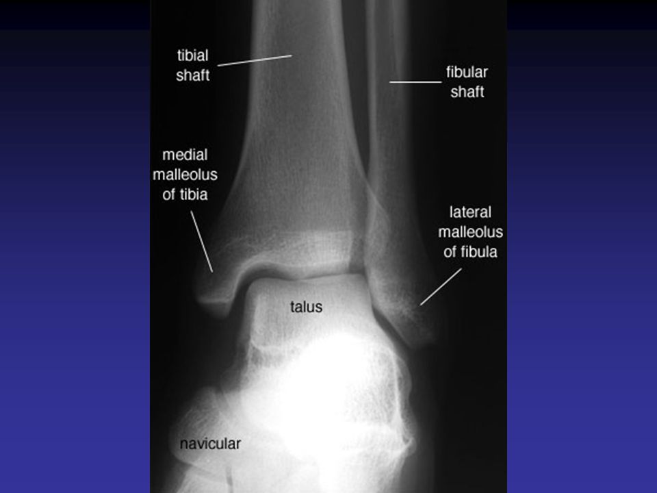
The most frequently used modality for evaluation of bone and joint disorder The radiologist should obtain at least two (2) views of the bone involved at 90° angles to each other with each view including two adjacent joints CONVENTIONAL RADIOGRAPHY: The most frequently used modality for evaluation of bone and joint disorder and particularly in trauma is conventional plain film radiography. The radiologist should obtain at least two (2) views of the bone involved at 900 angles to each other with each view including two adjacent joints. This decreases the risk of missing an associated fracture, subluxation, and/or dislocation at the site remote from the primary injury. In children, obtaining a radiograph of the normal and affected limb for comparison is also wise. Usually, the standard films compromised the arterio-posterior and lateral views. Occasionally, oblique and special views are necessary particularly in evaluating complex structures, such as the elbow, wrist, ankle, and pelvis. A weight bearing view may be of value for a dynamic evaluation of the joint space under the weight of the body.
 views of the bone involved at 90° angles to each other. with each view including two adjacent joints. CONVENTIONAL RADIOGRAPHY: The most frequently used modality for evaluation of bone and joint disorder and particularly in trauma is conventional plain film radiography. The radiologist should obtain at least two (2) views of the bone involved at 900 angles to each other with each view including two adjacent joints. This decreases the risk of missing an associated fracture, subluxation, and/or dislocation at the site remote from the primary injury. In children, obtaining a radiograph of the normal and affected limb for comparison is also wise. Usually, the standard films compromised the arterio-posterior and lateral views. Occasionally, oblique and special views are necessary particularly in evaluating complex structures, such as the elbow, wrist, ankle, and pelvis. A weight bearing view may be of value for a dynamic evaluation of the joint space under the weight of the body.")
7
AP & lat AP view 1, Lateral condyle of femur.
2, Femur.
3, Patella.
4, Medial condyle of femur.
5, Medial intercondylar tubercle of tibia.
6, Tibia.
7, Fibula. Lateral 1, Patella.
2, Tuberosity of tibia.
3, Tibia.
4, Femur.
5, Medial condyle of femur.
6, Fibula.

8
Additional views standard films compromised the anterio-posterior and lateral views. Occasionally, oblique and special views elbow, wrist, ankle, and pelvis In children, obtaining a radiograph of the normal and affected limb for comparison is also wise. Usually, the standard films compromised the arterio-posterior and lateral views. Occasionally, oblique and special views are necessary particularly in evaluating complex structures, such as the elbow, wrist, ankle, and pelvis. A weight bearing view may be of value for a dynamic evaluation of the joint space under the weight of the body. Cervical Spine X-ray, 3/4 (Left Neural Foramina). 1, Rib. 2, Clavicle. 3, Neural Foramina. 4, Pedicle. 5, Trachea.
. 1, Rib. 2, Clavicle. 3, Neural Foramina. 4, Pedicle. 5, Trachea.")
9
FLUOROSCOPY: Many radiologic procedures Arthrography Tenography
Versography Arteriography Percutaneous Bone or Soft Tissue Biopsy. FLUOROSCOPY: Fluoroscopy is a fundamental diagnostic tool for many radiologic procedures including Arthrography, Tenography, Versography, Arteriography and Percutaneous Bone or Soft Tissue Biopsy. Fluoroscopy combined with videotaping is useful in evaluating the _____ of joints. In arthrography, checking proper placement of the needle and to monitor the flow of contrast agent, and intra-operatively to assess reduction of a fracture or placement of hardware.

11
COMPUTED TOMOGRAPHY: COMPONENTS: -X ray source -Detectors
- Computer data processing system COMPUTED TOMOGRAPHY: CT is radiological modality containing an x-ray source, detectors and the computer data processing system. The essential component of a CT system include a circular scanning gantry which holds the x-ray tube, and imaging sensors, a table for a patient, an x-ray generator and a computerized data processing unit. The patient lies on the table and is placed inside the gantry. The x-ray tube is rotated around the patient while the computer collects the data and formulates an axial image or slice. Each cross sectional slice represents a thickness between 3mm in 1.5cm of body tissue.

12
Uses of CT Trauma Intraarticular abnormalities
Detection of small bony fragements CT is indispensable in the evaluation of many traumatic conditions in various bone and soft tissue tumors because of its cross sectional imaging capabilities.

13
Complex hip fractures

14
CT Vs. Xray Advantages: Disadvantages: Excellent contrast resolution.
Measures the tissue attenuation coefficient Obtain transaxial images Reformation Disadvantages: Radiation Inability to make a specific diagnosis

15
Uses -Tumors Delineates tumors extent Soft tissue extension.
Presence of Calcification Biopsy

16
Arthrography Arthrography is introduction of contrast agent positive contrast iodine iodide solution negative contrast, air or combination of both into the joint space. Advantages: Simple Effective Arthrography is introduction of contrast agent positive contrast iodine iodide solution negative contrast, air or combination of both into the joint space. Despite the evolution of new diagnostic imaging modalities such as CT and MRI, arthrography has retained its importance in daily radiological practice. The growing popularity of arthrography has been particularly due to advantages in its technique and interpretation. The fact that it is not a technically difficult procedure and is much simpler to interpret than Ultrasound, CT or MRI makes it very desirable for evaluation of various interarticulation. Although virtually every joint can be injected with contrast, the examination in the present time is mostly frequently performed in shoulder, breast, ankle and elbow. It is important to obtain preliminary films prior to any arthrographic procedure since contrast may obscure some joint abnormalities (osteochondro---) that can be easily detected on Conventional Radiographs. The examination of any of the joints, arthrography can be combined cith CT (CT arthrography) or with digitization of image (Digitized Subtraction Arthrography) or MR Arthrography. There are relatively few absolute contra indications to arthrography. Even hypersensitivity to iodine is a relative contra indication since in this case a single contrast study using only air can be performed.
 that can be easily detected on Conventional Radiographs. The examination of any of the joints, arthrography can be combined cith CT (CT arthrography) or with digitization of image (Digitized Subtraction Arthrography) or MR Arthrography. There are relatively few absolute contra indications to arthrography. Even hypersensitivity to iodine is a relative contra indication since in this case a single contrast study using only air can be performed.")
17
Arthrography Any joint Shoulder Ankle Elbow Knee
Although virtually every joint can be injected with contrast, the examination in the present time is mostly frequently performed in shoulder, breast, ankle and elbow. It is important to obtain preliminary films prior to any arthrographic procedure since contrast may obscure some joint abnormalities (osteochondro---) that can be easily detected on Conventional Radiographs. The examination of any of the joints, arthrography can be combined cith CT (CT arthrography) or with digitization of image (Digitized Subtraction Arthrography) or MR Arthrography. There are relatively few absolute contra indications to arthrography. Even hypersensitivity to iodine is a relative contra indication since in this case a single contrast study using only air can be performed.
 that can be easily detected on Conventional Radiographs. The examination of any of the joints, arthrography can be combined cith CT (CT arthrography) or with digitization of image (Digitized Subtraction Arthrography) or MR Arthrography. There are relatively few absolute contra indications to arthrography. Even hypersensitivity to iodine is a relative contra indication since in this case a single contrast study using only air can be performed.")
18
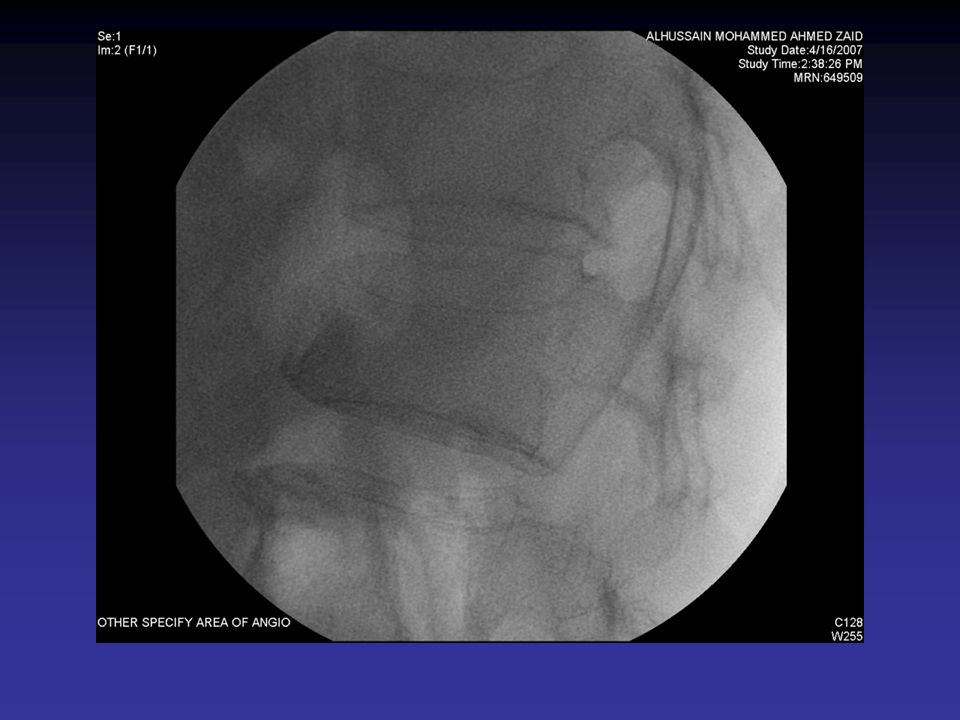
Angiography Advantages: Map-out bone lesions
Demonstrate the vascularity of the lesion. Demonstrate the vascular supply of a tumor Locate vessels suitable for pre operative intraarterial chemotherapy. Demonstrating the area suitable for open biopsy. ANGIOGRAPHY: The use of contrast material injected into selective branches of both arterial and venous circulation has aided greatly in assessing the involvement of the circulatory system in various condition and has provided a precise diagnostic method for local pathology. In evaluation of tumors, arteriography is used mainly to mop-out bone lesions demonstrate the vascularity of the lesion and assess the extent of the disease. It is also used to demonstrate the vascular supply of a tumor and to locate vessels suitable for pre operative intraarterial chemotherapy. It is very useful in demonstrating the area suitable for open biopsy, since the most vascular part of tumor contain the most aggressive component of the lesion. Occasionally, arteriography can be used to demonstrate abnormal tumor vessels. Arteriography is often extremely helpful in planning for limb salvage procedure since it demonstrate the regional vascular anatomy and thus, commit a plan to be drawn up for the tumor presection. It is also sometimes use to outline the major vessels prior to resection of a benign lesions. It can also be combined with interventional procedure such as embolization of hypervascular tumors prior to further treatment.

19
Post placement of two protective coils

21
ULTRASOUND: Rarely used Advantages: inexpensive
allows comparison with the opposite side, normal side uses no ionizing radiation, performed at bed side or in the operating room. It is a non invasive modality ULTRASOUND: Over the past several years, ultrasound has made an enormous impact in the field of radiology, however it is only rarely used its skeletal radiology. It has a various inherit advantages, it is relatively inexpensive, allows comparison with the opposite side, normal side, uses no ionizing radiation, and can be performed at bed side or in the operating room. It is a non invasive modality, relying upon the interaction of propagated soundwaves with tissue interface in the body whenever the direct pulsating of sound waves encounters an interface of tissue an acoustic impedence reflection or refraction occurs. The sound waves reflected back through the ultrasound transducer are recorded and converted into images. Applications of ultrasound in orthopaedics include evaluation of the rotator cuff, injuries to various tendons, e.g. the achilles tendons, and occasionally soft tissue tumors such as hemangioma. The most effective application however is in evaluation of the infant hip for which ultrasound has become the imaging modality of choice

22
Applications Evaluation of the rotator cuff
Injuries to various tendons, e.g. the achilles tendons. Evaluation of the infant hip for which ultrasound has become the imaging modality of choice

23
Shoulder US

24
SCINTIGRAPHY RADIONUCLIDE BONE SCAN
image the entire skeleton at once. It provides a metabolic picture. It is particularly helpful in condition such as fibrodysplasia, Langerhans Cell Histocytosis or metastatic cancer. SCINTIGRAPHY RADIONUCLIDE BONE SCAN: One major advantage of skeletal scintigraphy over all other imaging techniques is its ability to image the entire skeleton at once. It provides a metabolic picture. It is particularly helpful in condition such as fibrodysplasia, Langerhans Cell Histocytosis or metastatic cancer.

25
Prostate Mets

26
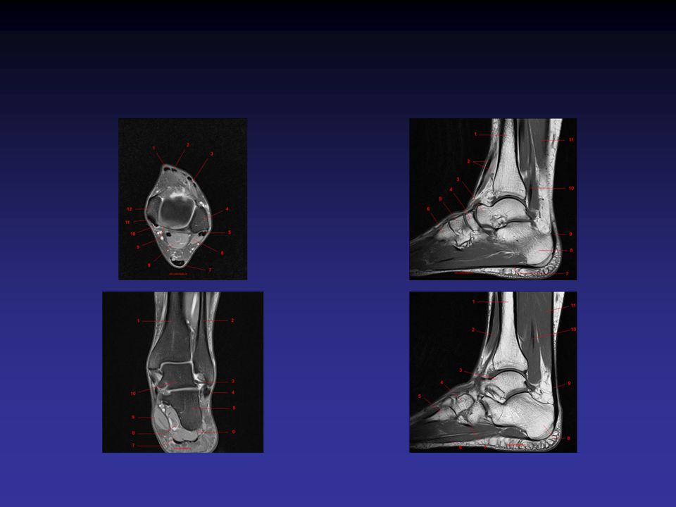
MRI Magnet RF coils Computer MAGNETIC RESONANCE IMAGING:
MRI is based upon the remission of an absorb radio frequency signal while the patient is in a strong magnetic field. An external magnetic field is usually generated by a magnet with field strength of 0.2 to 1.5 Tesla. The system includes a magnet, RF coils (Transmitter and receiver), gradient coils, and a computer display unit with digital storage facilities. The principle of MRI cannot be discussed in detail. The musculoskeletal system is ideally suited for evaluation by MRI since different tissue displayed different signal intensities on T1 & T2 weighted images. The images displayed may have a low signal intensity, intermediate signal intensity, or high signal intensity
, gradient coils, and a computer display unit with digital storage facilities. The principle of MRI cannot be discussed in detail. The musculoskeletal system is ideally suited for evaluation by MRI since different tissue displayed different signal intensities on T1 & T2 weighted images. The images displayed may have a low signal intensity, intermediate signal intensity, or high signal intensity.")
27
MRI The musculoskeletal system is ideally suited for evaluation by MRI since different tissue displayed different signal intensities on T1 & T2 weighted images. The images displayed may have a low signal intensity, intermediate signal intensity, or high signal intensity

28
MRI-uses Traumatic & non-traumatic conditions Bone Soft tissue
Contusions Microfractures Traumatic conditions of the bone, and soft tissue are particularly well suited to diagnose and evaluate by MRI. Some abnormalities such as bone contusions or trabecular micro fractures not seen on radiography and CT are well demonstrated by this technique. The use of intravenous gadolinium produces enhancement in MRI. Magnetic Resonance arthrography has become popular by recent years. Although MRI has many advantages and disadvantages exists as well, this include the typical contra indication of scanning of patients with cardiac pacemakers, cerebral aneurysm clips, and claustrophobia.

29
Relation to neurovascular bundle

30
Tumor composition

31
MRI Contraindications
ABSOLOUTE Patients with cardiac pacemakers Cerebral aneurysm clips RELATIVE: Claustrophobia.

32
Summary Imaging Techniques in Orthopaedics Conventional Radiography
Fluoroscopy Computed Tomography Arthrography Angiography Ultrasound Scintigraphy Magnetic Resonance Imaging

33
Radiologic Anatomy of the Musculoskeletal System
Upper limb

34
1

35
B A C

38
2

39
B A

41
3

42
C A D B

44
4

45
A B C

47
5

48
D A B C

50
6

51
D A B C

53
7

54
K J L I M F E H G C D A B

56
8

57
D E G F A E B C

59
Radiologic Anatomy of the Musculoskeletal System
Lower limb

60
9

61
A E E B C D

64
10

65
A C B D

68
11

69
B A C D

71
12

72
D A B E C

75
13

76
D E A B C

78
14

79
F G H I J D F E B C A

81
15

82
A C D B F E

Similar presentations


 Ultrasound Magnetic Resonance Imaging (MRI) Radioisotopes Studies.>")


