Download presentation
Presentation is loading. Please wait.

1
Peptic Ulcer disease

2
Anatomy Stomach Regions

3
Stomach – cont. Anatomy – cont. Layers of walls Serosa Muscularis
Submucosa Mucosa

4
Stomach – cont. Anatomy – cont. Glands in mucosa Cardiac glands
Gastric glands Chief cells Parietal cells Mucous neck cells gastrin

5
Gastric pit mucus neck & surface cells parietal cells (oxyntic)
Mucus & HCO3 parietal cells (oxyntic) H+ secretion & intrinsic factor Peptic cells (chief, zymogen) Pepsinogen secn
 H+ secretion & intrinsic factor. Peptic cells. (chief, zymogen) Pepsinogen secn.")
6
Functions of gastric secretions
Digestion of proteins ( pepsinogen & HCl) Protection of stomach ( HCO3- & mucus) Absorption of vitamin B12 ( intrinsic factor) Destroy bacteria & other micro-organisms (HCl) ~ 3 li per day
 Protection of stomach ( HCO3- & mucus) Absorption of vitamin B12 ( intrinsic factor) Destroy bacteria & other micro-organisms. (HCl) ~ 3 li per day.")
7
Peptic ulcer disease General consideration
Peptic ulcers result from the corrosive action of acid gastric juice Ulcers may occur in oesophagus, stomach,duodenum, jejunum or ileum from ectopic gastric mucosa Can be anywhere in GI tract exposed to acid-pepsin gastric juice Other factors also contribute H. pylori Mucosal bicarbonate secretion Stress Genetics

8
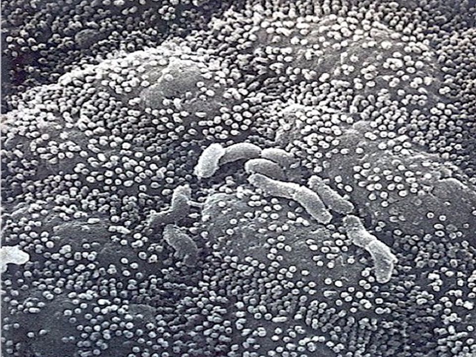
GI Pathology Helicobacter pylori

10
Peptic ulcer disease - cont.
Pathogenesis Two factors prevent stomach from digesting itself Gastric mucosal barrier First line of defense NSAIDS cause in changes mucosa that my facilitate its digestion by pepsin Destruction of barrier believed to be important factor in pathogenesis of gastric ulcers Results of back diffusion of H+ injuring underlying tissues Antrum more susceptible to back diffusion than fundus Duodenum resistant to ulceration due to Brunner’s glands which produce a highly alkaline secretion

11
Peptic ulcer disease - cont.
Epithelial barrier Depends of abundant vascular supply and continual, rapid regeneration of epithelial cells (~3 days)
")
12
Peptic ulcer disease - cont.
Other factors 10-12 % incidence in population Duodenal ulcers occur in much younger group than gastric years Males affected 3X as often as women Duodenal ulcers 10X as common as gastric >90% of duodenal ulcers are on anterior or posterior wall within 3 cm of pyloric ring 40-60% have family history

13
Peptic ulcer disease - cont.
Clinical features Principle feature is chronic, intermittent epigastric pain – typically relieved by food ~25% have bleeding (more common with duodenal) Other signs and symptoms Vomiting Red or “coffee-ground” emesis Nausea Anorexia Weight loss Pain-food-relief pattern may not be typical of gastric ulcers – food sometimes aggravates
 Other signs and symptoms. Vomiting. Red or coffee-ground emesis. Nausea. Anorexia. Weight loss. Pain-food-relief pattern may not be typical of gastric ulcers – food sometimes aggravates.")
14
Diagnostic procedures
Barium radiologic studies Gastric analysis of acid secretion Aspirate gastric juices with nasogastric tube Endoscopy Photography Biopsy Exfoliative cytology

15
Differential Diagnosis
Gallbladder disease Pancreatitis Functional indegestion Reflux oesphagitis

16
Peptic ulcer disease - cont.
Medical treatment Primary consideration is to inhibit or buffer acid to relieve symptoms and promote healing Antacids – increase pH so pepsin isn’t activated Dietary management – small frequent meals, avoid alcohol and caffeine Anticholinergics – inhibit vagal stimulation Antimicrobial therapy Physical and emotional rest Ulcers caused by H. pylori are successfully treated with antimicrobial agents, bismuth salts, and H2 blockers 65-95% eradication rates

17
10 Day Regimen clarithromycin 500 mg bid X 10
amoxicillin 1 gram bid X 10 omeprazole 20 mg bid X 10 in patients with current ulcer, continue omeprazole 20 mg/day for 18 days

18
kPeptic ulcer disease - cont.
Complications Hemorrhage Most frequent complication – 15-20% Most common in ulcers of the posterior wall of duodenal bulb due to proximity of arteries Symptoms depend on severity Anemia Occult blood in stool Black and tarry stool Hematemesis Shock Mortality up to 10% - higher for patients over 50

19
Peptic ulcer disease - cont.
Perforation Approximately 5% of all ulcers perforate - accounts for 65% of deaths from peptic ulcers Usually on anterior wall of duodenum or stomach Thought to be due to excess acid and often a result of NSAIDS Characteristic presentation Sudden onset of excruciating pain in upper abdomen – chemical peritonitis Patient fears to move or breath Abdomen becomes silent to auscultation and board like rigidity to palpation Treatment – immediate surgery

20
Peptic ulcer disease - cont.
Obstruction Obstruction of gastric outlet in ~5% of patients Due to inflammation and edema, pylorospasm or scarring More often with duodenal ulcers Symptoms Anorexia Nausea Bloating after eating Pain and vomiting when severe Treatment Restore fluids and electrolytes Decompress stomach with nasogastric tube Surgical correction - pyloroplasty

21
Peptic ulcer disease - cont.
Intractability Medical therapy fails to control symptoms adequately, resulting in frequent, rapid recurrences Typically surgery is recommended

22
Peptic ulcer disease - cont.
Surgical treatment – for patients who do not respond to therapy For duodenal ulcers aim is to permanently reduce stomach’s capacity to secrete acid and pepsin Vagotomy Cut vagal branches to stomach Eliminates cephalic phase Several techniques Antrectomy Removal of entire antrum Eliminates gastric phase Vagotomy plus antrectomy Eliminates both cephalic and gastric phases

23
Peptic ulcer disease - cont.
Partial gastrectomy Removal of distal 50-75% of stomach Gastric remnant anastamosed to duodenum (Billroth I) or jejunum (Billroth II) For gastric ulcers Usually partial gastrectomy and a gastroduodenal anastomosis Normally do not do vagotomy as patients have normal to low acid production
 or jejunum (Billroth II) For gastric ulcers. Usually partial gastrectomy and a gastroduodenal anastomosis. Normally do not do vagotomy as patients have normal to low acid production.")
24
Normal Stomach

25
Esophagus & Stomach Normal

26
Gastric Ulcer

27
Peptic ulcer - Endoscopy

28
Duodenal Peptic Ulcer

29
Gastric Ulcer

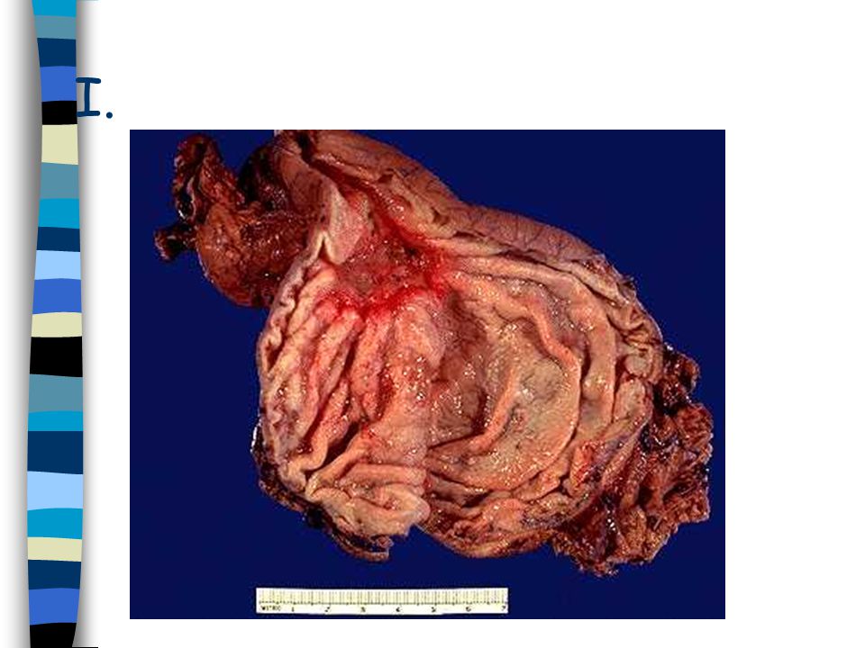
31
GI Pathology Duodenal Peptic Ulcers, Gross

32
GI Pathology Giant gastric ulcer

33
GI Pathology Gastric Peptic Ulcer
Gross Lesser curvature is the most common location in the stomach; greatest frequency is in the first part of the duodenum Less than three centimeters in diameter Round to oval in shape Punched-out area with clean base Margins are usually level with surrounding mucosa or slightly elevated due to edema; the mucosa is undermined at the edges

34
GI Pathology Gastric Peptic Ulcer
Up to 50% of those with gastric peptic ulcer have concurrent duodenal ulcer These ulcers typically occur at mucosal junctions exposed to acid and pepsin (e.g., body of stomach/antrum)
")
35
GI Pathology Gastric peptic ulcer
Associations: Chronic gastritis, Helicobacter pylori (50-60%) Peak incidence = 50's Location = Lesser curvature, antrum
 Peak incidence = 50 s. Location = Lesser curvature, antrum.")
36
GI Pathology Anatomy of the stomach

Similar presentations
 and Vomiting>")


>")
![Peptic Ulcer Disease Dr Maha Arafah. Objectives Upon completion of this lecture the students will : A] Understand the Pathophysiology of acute and chronic.](/13/3809458/big_thumb.jpg "Peptic Ulcer Disease Dr Maha Arafah. Objectives Upon completion of this lecture the students will : A] Understand the Pathophysiology of acute and chronic.>")


>")
 Dr. Gehan Mohamed Dr. Abdelaty Shawky.>")


