Download presentation
Presentation is loading. Please wait.

1
Muscle Physiology CHAPTER 6

2
Muscle Physiology Questions to ponder….. Can I turn fat into muscle if I start working out? Body fat and muscle are two completely different tissues. They have different structures and functions, they react to training in different ways and, simply put, one does not have the capability to turn into the other Can I increase the number of muscle cells I have by working out? While the number of muscles cells/fibers can not increase, each individual muscle fiber has the potential to increase in size, density and efficiency. These changes may occur together but not necessarily to the same degree, however, all will translate to an increase in strength. I keep doing abdominal crunches, why can I not get those 6-pack abs?

3
IComparison of different types of muscle tissue A. Skeletal Muscle A. Skeletal Muscle 1. Cells/fibers are long (some up to 1 ft), cylindrical and striated in appearance. They have multiple nuclei in each cell. 1. Cells/fibers are long (some up to 1 ft), cylindrical and striated in appearance. They have multiple nuclei in each cell. 2. The tissue is under mostly voluntary control. 2. The tissue is under mostly voluntary control. 3. The tissue is located in the body attached to bones and/or skin (ex. facial muscles) 3. The tissue is located in the body attached to bones and/or skin (ex. facial muscles) 4. Fiber contractions vary from slow to fast depending on type and location in the body. 4. Fiber contractions vary from slow to fast depending on type and location in the body. a. Slow oxidative fibers: posture muscles in the back, typically very red; fatigue slowly. a. Slow oxidative fibers: posture muscles in the back, typically very red; fatigue slowly. b. Fast oxidative fibers: sprinting and walking muscles; fatigue is moderate. b. Fast oxidative fibers: sprinting and walking muscles; fatigue is moderate. c. Fast glycolytic fibers: short-term intense muscles, ex. hitting a baseball; fatigue quickly. c. Fast glycolytic fibers: short-term intense muscles, ex. hitting a baseball; fatigue quickly. d. Some aptitude in sports is due to the ratio of these fibers in an individual. Ex. Marathon runners have more slow oxidative fibers for less fatigue and more endurance. d. Some aptitude in sports is due to the ratio of these fibers in an individual. Ex. Marathon runners have more slow oxidative fibers for less fatigue and more endurance.
, cylindrical and striated in appearance. They have multiple nuclei in each cell. 1. Cells/fibers are long (some up to 1 ft), cylindrical and striated in appearance. They have multiple nuclei in each cell. 2. The tissue is under mostly voluntary control. 2. The tissue is under mostly voluntary control. 3. The tissue is located in the body attached to bones and/or skin (ex. facial muscles) 3. The tissue is located in the body attached to bones and/or skin (ex. facial muscles) 4. Fiber contractions vary from slow to fast depending on type and location in the body. 4. Fiber contractions vary from slow to fast depending on type and location in the body. a. Slow oxidative fibers: posture muscles in the back, typically very red; fatigue slowly. a. Slow oxidative fibers: posture muscles in the back, typically very red; fatigue slowly. b. Fast oxidative fibers: sprinting and walking muscles; fatigue is moderate. b. Fast oxidative fibers: sprinting and walking muscles; fatigue is moderate. c. Fast glycolytic fibers: short-term intense muscles, ex. hitting a baseball; fatigue quickly. c. Fast glycolytic fibers: short-term intense muscles, ex. hitting a baseball; fatigue quickly. d. Some aptitude in sports is due to the ratio of these fibers in an individual. Ex. Marathon runners have more slow oxidative fibers for less fatigue and more endurance. d. Some aptitude in sports is due to the ratio of these fibers in an individual. Ex. Marathon runners have more slow oxidative fibers for less fatigue and more endurance..")
4
5. Fibers have very specific arrangement to enhance the strength and durability of the muscle tissue. 5. Fibers have very specific arrangement to enhance the strength and durability of the muscle tissue. a: Each fiber is surrounded by connective tissue – Endomysium a: Each fiber is surrounded by connective tissue – Endomysium b. Bundle of muscle fibers are surrounded by connective tissue – Perimysium (called a fascicle, can be large or small) b. Bundle of muscle fibers are surrounded by connective tissue – Perimysium (called a fascicle, can be large or small) c. Many fascicles are bound together by connective tissue – Epimysium (these layers blend together to form tendon). Extremely durable tissue for crossing joints and bony projections so that muscles are not torn. c. Many fascicles are bound together by connective tissue – Epimysium (these layers blend together to form tendon). Extremely durable tissue for crossing joints and bony projections so that muscles are not torn. d. Orientation of the bundles in the muscle are different for different muscles. Ex. fat belly of the biceps brachii vs. the flat parallel bundles of the latissimus dorsi. d. Orientation of the bundles in the muscle are different for different muscles. Ex. fat belly of the biceps brachii vs. the flat parallel bundles of the latissimus dorsi.
 b. Bundle of muscle fibers are surrounded by connective tissue – Perimysium (called a fascicle, can be large or small) c. Many fascicles are bound together by connective tissue – Epimysium (these layers blend together to form tendon). Extremely durable tissue for crossing joints and bony projections so that muscles are not torn. c. Many fascicles are bound together by connective tissue – Epimysium (these layers blend together to form tendon). Extremely durable tissue for crossing joints and bony projections so that muscles are not torn. d. Orientation of the bundles in the muscle are different for different muscles. Ex. fat belly of the biceps brachii vs. the flat parallel bundles of the latissimus dorsi. d. Orientation of the bundles in the muscle are different for different muscles. Ex. fat belly of the biceps brachii vs. the flat parallel bundles of the latissimus dorsi..")
5
B. Smooth Muscle 1. Cells/fibers are long spindle shaped, not striated and have one nucleus. 1. Cells/fibers are long spindle shaped, not striated and have one nucleus. 2. The tissue is under involuntary nervous control. 2. The tissue is under involuntary nervous control. 3. The tissue is located surrounding hollow organs (blood vessels,urinary bladder, esophagus, stomach, intestine, and respiratory passages) 3. The tissue is located surrounding hollow organs (blood vessels,urinary bladder, esophagus, stomach, intestine, and respiratory passages) 4. Composed of two layers with the fibers running perpendicular to each other. Helps muscle prevent muscle fatigue and change the size and shape of the organ. (ex. urinary bladder empty vs. full) 4. Composed of two layers with the fibers running perpendicular to each other. Helps muscle prevent muscle fatigue and change the size and shape of the organ. (ex. urinary bladder empty vs. full) 5. Fatigue very slowly, contraction is slow and steady.
 3. The tissue is located surrounding hollow organs (blood vessels,urinary bladder, esophagus, stomach, intestine, and respiratory passages) 4. Composed of two layers with the fibers running perpendicular to each other. Helps muscle prevent muscle fatigue and change the size and shape of the organ. (ex. urinary bladder empty vs. full) 4. Composed of two layers with the fibers running perpendicular to each other. Helps muscle prevent muscle fatigue and change the size and shape of the organ. (ex. urinary bladder empty vs. full) 5. Fatigue very slowly, contraction is slow and steady..")
6
c. Cardiac Muscle 1. Cells/fibers are long, cylindrical fibers that are striated and branched. 1. Cells/fibers are long, cylindrical fibers that are striated and branched. 2. Tissue is under involuntary nervous control. 2. Tissue is under involuntary nervous control. 3. Fibers are connected by intercalated discs to facilitate the passing of the contraction impulse through the tissue of the organ. Fibers are arranged in a spiral arrangement to help with coordinated heart contractions. 3. Fibers are connected by intercalated discs to facilitate the passing of the contraction impulse through the tissue of the organ. Fibers are arranged in a spiral arrangement to help with coordinated heart contractions. 4. Fibers are located only in the heart. 4. Fibers are located only in the heart.

7
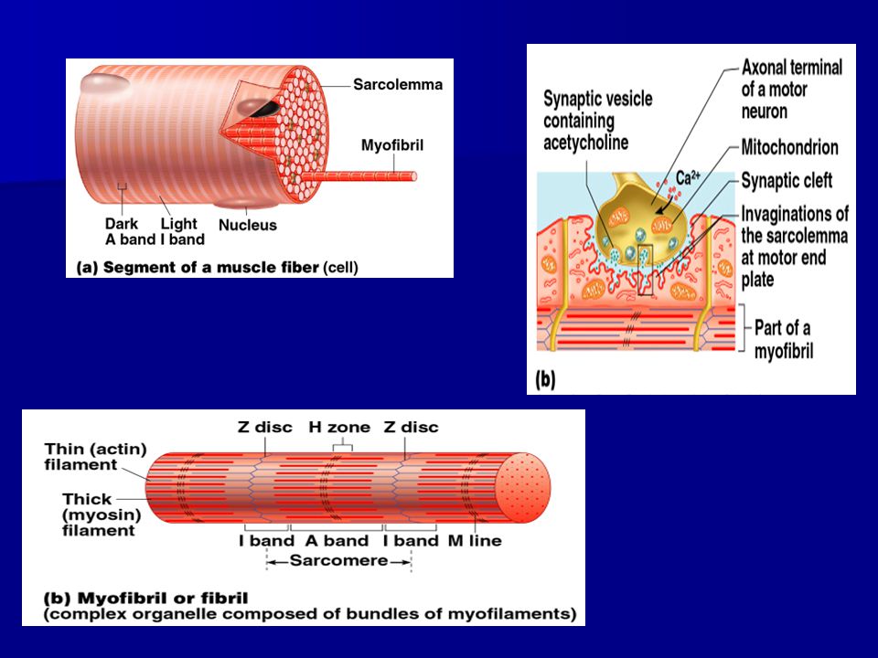
IIThe functions of muscles 1. Producing movement – facial expressions, movement of food stuffs through digestions and blood through the heart. 1. Producing movement – facial expressions, movement of food stuffs through digestions and blood through the heart. 2. Maintaining posture (skeletal only), very fatigue resistant muscles, ex.erector spinae; and stabilizing joints (holding the skeleton together) 2. Maintaining posture (skeletal only), very fatigue resistant muscles, ex.erector spinae; and stabilizing joints (holding the skeleton together) 3. Generating heat to maintain body temperature. Muscles make up 40% of body weight and ¾ of energy given off by ATP is heat. 3. Generating heat to maintain body temperature. Muscles make up 40% of body weight and ¾ of energy given off by ATP is heat. IIIHow the muscle fiber contracts – The Sliding Filament Theory a. Basic structure of sarcomere. a. Basic structure of sarcomere. 1. Sarcomere is the functional contracting unit of the muscle fiber. 1. Sarcomere is the functional contracting unit of the muscle fiber.
, very fatigue resistant muscles, ex.erector spinae; and stabilizing joints (holding the skeleton together) 2. Maintaining posture (skeletal only), very fatigue resistant muscles, ex.erector spinae; and stabilizing joints (holding the skeleton together) 3. Generating heat to maintain body temperature. Muscles make up 40% of body weight and ¾ of energy given off by ATP is heat. 3. Generating heat to maintain body temperature. Muscles make up 40% of body weight and ¾ of energy given off by ATP is heat. IIIHow the muscle fiber contracts – The Sliding Filament Theory a. Basic structure of sarcomere. a. Basic structure of sarcomere. 1. Sarcomere is the functional contracting unit of the muscle fiber. 1. Sarcomere is the functional contracting unit of the muscle fiber..")
8
Muscles are made of muscle fibers (cells) Muscles are made of muscle fibers (cells) Muscle fibers (cells) are made of myofibrils which contain the functioning units of contraction called sarcomeres Muscle fibers (cells) are made of myofibrils which contain the functioning units of contraction called sarcomeres
 Muscles are made of muscle fibers (cells) Muscle fibers (cells) are made of myofibrils which contain the functioning units of contraction called sarcomeres Muscle fibers (cells) are made of myofibrils which contain the functioning units of contraction called sarcomeres")
11
b. Functioning of the sarcomere. What you can do in 1000th of a second! b. Functioning of the sarcomere. What you can do in 1000th of a second!http://media.pearsoncmg.com/bc/bc_campbell_biology_6/cipl/ins/49/HTML/source/71.html 1. Nerve impulse reaches the synaptic cleft between the nerve and the muscle fiber (each fiber must be individually stimulated). This is called the neuromuscular junction. 1. Nerve impulse reaches the synaptic cleft between the nerve and the muscle fiber (each fiber must be individually stimulated). This is called the neuromuscular junction. 2. Signal causes Calcium (Ca) to be released from the sarcoplasmic reticulum. (as soon as nerve impulse is over Ca is reabsorbed) 2. Signal causes Calcium (Ca) to be released from the sarcoplasmic reticulum. (as soon as nerve impulse is over Ca is reabsorbed) 3. Calcium causes the protein complex on actin to move away from the binding site. 3. Calcium causes the protein complex on actin to move away from the binding site. 4. ATP activates the myosin head to form the cross bridge (myosin head attached to actin and pivots toward the H- zone. Causing a shortening of the sarcomere. 4. ATP activates the myosin head to form the cross bridge (myosin head attached to actin and pivots toward the H- zone. Causing a shortening of the sarcomere. 5. This action occurs simultaneously through out the entire muscle fiber and through out the entire muscle causing the muscle itself to contract/shorten. In order to maintain a contraction (ex. hold your arm out in front of you for several seconds) the nerve must constantly restimulate the muscle fiber. 5. This action occurs simultaneously through out the entire muscle fiber and through out the entire muscle causing the muscle itself to contract/shorten. In order to maintain a contraction (ex. hold your arm out in front of you for several seconds) the nerve must constantly restimulate the muscle fiber.
. This is called the neuromuscular junction. 1. Nerve impulse reaches the synaptic cleft between the nerve and the muscle fiber (each fiber must be individually stimulated). This is called the neuromuscular junction. 2. Signal causes Calcium (Ca) to be released from the sarcoplasmic reticulum. (as soon as nerve impulse is over Ca is reabsorbed) 2. Signal causes Calcium (Ca) to be released from the sarcoplasmic reticulum. (as soon as nerve impulse is over Ca is reabsorbed) 3. Calcium causes the protein complex on actin to move away from the binding site. 3. Calcium causes the protein complex on actin to move away from the binding site. 4. ATP activates the myosin head to form the cross bridge (myosin head attached to actin and pivots toward the H- zone. Causing a shortening of the sarcomere. 4. ATP activates the myosin head to form the cross bridge (myosin head attached to actin and pivots toward the H- zone. Causing a shortening of the sarcomere. 5. This action occurs simultaneously through out the entire muscle fiber and through out the entire muscle causing the muscle itself to contract/shorten. In order to maintain a contraction (ex. hold your arm out in front of you for several seconds) the nerve must constantly restimulate the muscle fiber. 5. This action occurs simultaneously through out the entire muscle fiber and through out the entire muscle causing the muscle itself to contract/shorten. In order to maintain a contraction (ex. hold your arm out in front of you for several seconds) the nerve must constantly restimulate the muscle fiber..")
12
c. Graded Responses and Various Muscle Contractions c. Graded Responses and Various Muscle Contractions 1. Strength of contraction: (Picking up a feather vs. picking up a large book): Dependent on the number of motor units stimulated within the muscle. Only a few motor units would be stimulated for the feather vs. many motor units for the book. Have you ever gone to pick something up that you thought would be heavy, when you lifted the object it was with great force and speed? 1. Strength of contraction: (Picking up a feather vs. picking up a large book): Dependent on the number of motor units stimulated within the muscle. Only a few motor units would be stimulated for the feather vs. many motor units for the book. Have you ever gone to pick something up that you thought would be heavy, when you lifted the object it was with great force and speed? 2. Length of contraction: Dependent on the frequency of stimulation, nerves stimulate the muscle cell so quickly it does not have a chance to completely relax and can sustain a contraction, this is called tetanus. 2. Length of contraction: Dependent on the frequency of stimulation, nerves stimulate the muscle cell so quickly it does not have a chance to completely relax and can sustain a contraction, this is called tetanus. 3. Muscle Twitch: Single, brief, jerky muscle contraction. Not normally how the nervous system stimulates a muscle. 3. Muscle Twitch: Single, brief, jerky muscle contraction. Not normally how the nervous system stimulates a muscle. 4. Muscle paralysis: Can be due to numerous factors such as: 1) inadequate nervous stimulation (severed nerve); 2) no ATP in the cell; 3) blocked receptor site on muscle cell or the actin filament (ex. poison dart frog poison); or 4) cross bridges are not broken 4. Muscle paralysis: Can be due to numerous factors such as: 1) inadequate nervous stimulation (severed nerve); 2) no ATP in the cell; 3) blocked receptor site on muscle cell or the actin filament (ex. poison dart frog poison); or 4) cross bridges are not broken
: Dependent on the number of motor units stimulated within the muscle. Only a few motor units would be stimulated for the feather vs. many motor units for the book. Have you ever gone to pick something up that you thought would be heavy, when you lifted the object it was with great force and speed. 1. Strength of contraction: (Picking up a feather vs. picking up a large book): Dependent on the number of motor units stimulated within the muscle. Only a few motor units would be stimulated for the feather vs. many motor units for the book. Have you ever gone to pick something up that you thought would be heavy, when you lifted the object it was with great force and speed. 2. Length of contraction: Dependent on the frequency of stimulation, nerves stimulate the muscle cell so quickly it does not have a chance to completely relax and can sustain a contraction, this is called tetanus. 2. Length of contraction: Dependent on the frequency of stimulation, nerves stimulate the muscle cell so quickly it does not have a chance to completely relax and can sustain a contraction, this is called tetanus. 3. Muscle Twitch: Single, brief, jerky muscle contraction. Not normally how the nervous system stimulates a muscle. 3. Muscle Twitch: Single, brief, jerky muscle contraction. Not normally how the nervous system stimulates a muscle. 4. Muscle paralysis: Can be due to numerous factors such as: 1) inadequate nervous stimulation (severed nerve); 2) no ATP in the cell; 3) blocked receptor site on muscle cell or the actin filament (ex. poison dart frog poison); or 4) cross bridges are not broken 4. Muscle paralysis: Can be due to numerous factors such as: 1) inadequate nervous stimulation (severed nerve); 2) no ATP in the cell; 3) blocked receptor site on muscle cell or the actin filament (ex. poison dart frog poison); or 4) cross bridges are not broken.")
13
5. Muscle Atrophy: Loss of muscle mass due to lack of activity either from lack of nervous stimulation (ex. spinal cord injuries) or extreme inactivity (ex. leg in a cast) 5. Muscle Atrophy: Loss of muscle mass due to lack of activity either from lack of nervous stimulation (ex. spinal cord injuries) or extreme inactivity (ex. leg in a cast) 6. Muscle Fatigue: Caused by oxygen debt, unable to contract even with stimulation. Lactic acid build up also alters the pH of the cell which makes the muscle contraction less efficient. Recovery occurs when you continue to breath deeply after exercise has stopped. (new explanation deals with athletes that experience fatigue after long intense training) 6. Muscle Fatigue: Caused by oxygen debt, unable to contract even with stimulation. Lactic acid build up also alters the pH of the cell which makes the muscle contraction less efficient. Recovery occurs when you continue to breath deeply after exercise has stopped. (new explanation deals with athletes that experience fatigue after long intense training) 7. Muscle Tone: Even when muscles are relaxed voluntarily, some of the motor units have been stimulated to contract in a systematic way though out the muscle. This keeps the muscle firm and ready for action. 7. Muscle Tone: Even when muscles are relaxed voluntarily, some of the motor units have been stimulated to contract in a systematic way though out the muscle. This keeps the muscle firm and ready for action. 8. Isotonic Contractions: Myofilaments contract when actin fibers slide towards each other shortening the sarcomere and therefore the muscle. The muscle itself shortens and movement occurs. Ex. bending your knee. 8. Isotonic Contractions: Myofilaments contract when actin fibers slide towards each other shortening the sarcomere and therefore the muscle. The muscle itself shortens and movement occurs. Ex. bending your knee. 9. Isometric Contractions: Contractions where the muscle does not shorten. Fibers do not slide over each other because there is an immovable object (ex. a wall) that prevents the muscle from contracting. Tension builds in the muscle but no movement occurs. Ex. pushing against a wall with your foot. 9. Isometric Contractions: Contractions where the muscle does not shorten. Fibers do not slide over each other because there is an immovable object (ex. a wall) that prevents the muscle from contracting. Tension builds in the muscle but no movement occurs. Ex. pushing against a wall with your foot.
 or extreme inactivity (ex. leg in a cast) 5. Muscle Atrophy: Loss of muscle mass due to lack of activity either from lack of nervous stimulation (ex. spinal cord injuries) or extreme inactivity (ex. leg in a cast) 6. Muscle Fatigue: Caused by oxygen debt, unable to contract even with stimulation. Lactic acid build up also alters the pH of the cell which makes the muscle contraction less efficient. Recovery occurs when you continue to breath deeply after exercise has stopped. (new explanation deals with athletes that experience fatigue after long intense training) 6. Muscle Fatigue: Caused by oxygen debt, unable to contract even with stimulation. Lactic acid build up also alters the pH of the cell which makes the muscle contraction less efficient. Recovery occurs when you continue to breath deeply after exercise has stopped. (new explanation deals with athletes that experience fatigue after long intense training) 7. Muscle Tone: Even when muscles are relaxed voluntarily, some of the motor units have been stimulated to contract in a systematic way though out the muscle. This keeps the muscle firm and ready for action. 7. Muscle Tone: Even when muscles are relaxed voluntarily, some of the motor units have been stimulated to contract in a systematic way though out the muscle. This keeps the muscle firm and ready for action. 8. Isotonic Contractions: Myofilaments contract when actin fibers slide towards each other shortening the sarcomere and therefore the muscle. The muscle itself shortens and movement occurs. Ex. bending your knee. 8. Isotonic Contractions: Myofilaments contract when actin fibers slide towards each other shortening the sarcomere and therefore the muscle. The muscle itself shortens and movement occurs. Ex. bending your knee. 9. Isometric Contractions: Contractions where the muscle does not shorten. Fibers do not slide over each other because there is an immovable object (ex. a wall) that prevents the muscle from contracting. Tension builds in the muscle but no movement occurs. Ex. pushing against a wall with your foot. 9. Isometric Contractions: Contractions where the muscle does not shorten. Fibers do not slide over each other because there is an immovable object (ex. a wall) that prevents the muscle from contracting. Tension builds in the muscle but no movement occurs. Ex. pushing against a wall with your foot..")
14
10. Muscle Cramp: Involuntarily and forcibly contracted muscle that will not relax. Cramps have multiple causes: 1)hyperexciteability of nerves caused by vigorous activity, injury, periods of inactivity, dehydration, body fluid changes, low blood calcium, potassium, or magnesium; 2) depletion of ATP in the cell and cross bridges can’t break (contractures); 3)Unintentional stimulation of muscles not necessary for the movement, ex. writers cramp. 10. Muscle Cramp: Involuntarily and forcibly contracted muscle that will not relax. Cramps have multiple causes: 1)hyperexciteability of nerves caused by vigorous activity, injury, periods of inactivity, dehydration, body fluid changes, low blood calcium, potassium, or magnesium; 2) depletion of ATP in the cell and cross bridges can’t break (contractures); 3)Unintentional stimulation of muscles not necessary for the movement, ex. writers cramp. d. Energy for muscle contractions 1. Muscle fibers typically keep 4 to 6 seconds worth of ATP stored. Therefore the cell must very quickly begin generating more ATP. 1. Muscle fibers typically keep 4 to 6 seconds worth of ATP stored. Therefore the cell must very quickly begin generating more ATP. 2. First, use of creatine phosphate to make more ATP. (about 20 seconds) adding the phosphate to ADP to create ATP 2. First, use of creatine phosphate to make more ATP. (about 20 seconds) adding the phosphate to ADP to create ATP 3. Aerobic respiration. Used during light exercise and rest. 95% of all muscle energy is generated this way. Requires oxygen. 3. Aerobic respiration. Used during light exercise and rest. 95% of all muscle energy is generated this way. Requires oxygen. 4. Anaerobic respiration. During more intense exercise (30 to 60 seconds) but results in the build up of lactic acid (byproduct) in the muscles which promotes muscle fatigue and soreness. 4. Anaerobic respiration. During more intense exercise (30 to 60 seconds) but results in the build up of lactic acid (byproduct) in the muscles which promotes muscle fatigue and soreness.
hyperexciteability of nerves caused by vigorous activity, injury, periods of inactivity, dehydration, body fluid changes, low blood calcium, potassium, or magnesium; 2) depletion of ATP in the cell and cross bridges can’t break (contractures); 3)Unintentional stimulation of muscles not necessary for the movement, ex. writers cramp. 10. Muscle Cramp: Involuntarily and forcibly contracted muscle that will not relax. Cramps have multiple causes: 1)hyperexciteability of nerves caused by vigorous activity, injury, periods of inactivity, dehydration, body fluid changes, low blood calcium, potassium, or magnesium; 2) depletion of ATP in the cell and cross bridges can’t break (contractures); 3)Unintentional stimulation of muscles not necessary for the movement, ex. writers cramp. d. Energy for muscle contractions 1. Muscle fibers typically keep 4 to 6 seconds worth of ATP stored. Therefore the cell must very quickly begin generating more ATP. 1. Muscle fibers typically keep 4 to 6 seconds worth of ATP stored. Therefore the cell must very quickly begin generating more ATP. 2. First, use of creatine phosphate to make more ATP. (about 20 seconds) adding the phosphate to ADP to create ATP 2. First, use of creatine phosphate to make more ATP. (about 20 seconds) adding the phosphate to ADP to create ATP 3. Aerobic respiration. Used during light exercise and rest. 95% of all muscle energy is generated this way. Requires oxygen. 3. Aerobic respiration. Used during light exercise and rest. 95% of all muscle energy is generated this way. Requires oxygen. 4. Anaerobic respiration. During more intense exercise (30 to 60 seconds) but results in the build up of lactic acid (byproduct) in the muscles which promotes muscle fatigue and soreness. 4. Anaerobic respiration. During more intense exercise (30 to 60 seconds) but results in the build up of lactic acid (byproduct) in the muscles which promotes muscle fatigue and soreness..")
Similar presentations


 Generate heat - body temp 3 types: Skeletal - moves bone, voluntary Smooth.>")


