Download presentation
Presentation is loading. Please wait.

1
THE ANATOMY OF THE LOWER LIMB
BY DR. AHMAD KAMIL SHAHWAN PH.D. GENERAL SURGERY

2
THE LOWER LIMB ANATOMY THE BONES OF THE LOWER LIMB : THE HIP BONE :
It is a large bone ,thick in some places & thin in others. It is formed of 3 bones : THE ILIUM , THE ISCHIUM & THE PUPIS which are fused together before birth. They also fused at the acetabulum. There is a large opening below the acetabulum called the obturator foramen

6
THE HIP BONE : THE ILIUM: is the upper expanded part of the hip bone ;it is the largest part & consists of body & a large flat wing called the ala of the ilium . It has 3 borders ;The upper border called the iliac crest & ant. & post. borders. The iliac crest lies between the ant .sup. Iliac spine & the post. Sup. Iliac spine. Its ant. 2/3 is thick & convex outward & has inner & outer lips with an intermediate rough area in between , while the post. 1/3 is thin & convex inward & has 2 sloping surfaces separated by bony ridge.A bony prominence called the tubercle of the iliac crest is on the outer lip 5 cm . behind the ant. Sup. Iliac spine.

9
THE HIP BONE : The ant. border begins at the ASIS below which there is a notch then ant. Inf. Iliac spine. The post. Border begins in the PSIS & below it there is post inf. Iliac spine then form the greater sciatic notch then be continuous with the post. border of the ischium. The ilium has 2 surfaces :the outer(=gluteal) surface & the inner (=pelvic) surface.
 surface & the inner (=pelvic) surface.")
10
THE HIP BONE : THE ISCHIUM : forms the lower & the post. part of hip bone &consists of body , tuberosity & one ramus. the body :the ant .border of the body is continuous below with the upper border of the ramus & both form part of the wall of the obturator foramen. The post. border of the body is continuous above with the post border of the ilium forming the lower part of the greater sciatic notch ,then project to form the ischial spine. , & then form the lesser sciatic notch before it form the ischial tuberosity .

11
THE HIP BONE : The ischial ramus is continuous in front with the inf. Ramus of the pubis. The ischial tuberosity is a very strong piece of bone which project from the inf. pole of the body of the ischium . It divided to 4 parts : 1- part give origin to semimembransus M. 2-part give origin to semitendenosus & long head of biceps Mm. 3-part give origin to adductor magnus M. 4-part which we sit on& does not give origin to any M.

12
THE HIP BONE : THE PUBIS :forms the lower & ant. Part of the hip bone& has a body & 2 rami. The body : is flat triangular part which articulate with its fellow at the symphysis pubis. The body has 3 borders : The upper border called the pubic crest & it ends laterlaly by a projection called the pubic tubercle. The lateral border is very sharp & form boundary of obturator foramen.

13
THE HIP BONE : The sup. Ramus is triangular in shape while the inf. Ramus starts at the symphysis pubis & run obliquely downwards & laterally to join the ischial ramus & form together the conjoint (ischio-pubic ) ramus. THE ARTICULATION OF THE HIP BONE: 1- above & behind with the sacrum to form the sacro-iliac joint. 2- below & in front with the other hip bone at symphysis pubis. 3- through the acetabulum with the head of the femur to form the hip joint.
 ramus. THE ARTICULATION OF THE HIP BONE: 1- above & behind with the sacrum to form the sacro-iliac joint. 2- below & in front with the other hip bone at symphysis pubis. 3- through the acetabulum with the head of the femur to form the hip joint.")
18
THE FEMUR It is the longest & strongest bone in the body
It is formed of upper end , shaft (body) & lower end. THE UPPER END :consists of the head , the neck ,the greater trochanter & the lesser trochanter . THE HEAD: is less than 2/3 of sphere & faces upwards forwards & medially. In life it is covered by a cartilage except with central depression called the fovea where the legamentum teres is attached.
 & lower end. THE UPPER END :consists of the head , the neck ,the greater trochanter & the lesser trochanter . THE HEAD: is less than 2/3 of sphere & faces upwards forwards & medially. In life it is covered by a cartilage except with central depression called the fovea where the legamentum teres is attached.")
21
THE FEMUR THE NECK : is 5 cm long & connect the head with the shaft .It forms an angle ( ) with the axis of the shaft . This angle is smaller ( i.e. more acute ) in the female (who has wide pelvis ) than in male .This angle is normally 160 in children. THE GREATER TROCHANTER: is a large quadrangular piece of bone lies at the lateral & upper part of the junction between the neck & the shaft. In its medial surface there is deep depression called the trochantric fossa .
 with the axis of the shaft . This angle is smaller ( i.e. more acute ) in the female (who has wide pelvis ) than in male .This angle is normally 160 in children. THE GREATER TROCHANTER: is a large quadrangular piece of bone lies at the lateral & upper part of the junction between the neck & the shaft. In its medial surface there is deep depression called the trochantric fossa .")
23
THE FEMUR THE LESSER TROCHANTER: is a small pyramidal projection.
The intertrochanteric line : connects the greater & lesser trochanters in front & continues below the lesser troch. as the spiral line on the upper part of the shaft . The intertrochanteric crest : is a rough ridge joins the 2 troch. behind. In the middle of the crest there is a bony prominence called the quadrate tubercle.

25
Blood Supply Of The Head of The Femur
1- Blood ascending upwards from the shaft along the cancellous bone. 2- Blood from the Vv. In the capsule of the hip joint. 3- Blood from the artery in the ligamentum teres.

27
THE SHAFT OF THE FEMUR It is cylindrical in shape & be flattened posteriorly & downward. It is very slightly curved( convex) anteriorly . Along the middle of the shaft posteriorly there is rough ridge called linea aspera with 2 lips (lat. & med.). The lateral lip of linea aspera superiorly join the gluteal tuberosity which extends upward to the base of greater troch. The medial lip of linea aspera passes above to form the spiral line & ends in the intertroch. Line.
. The lateral lip of linea aspera superiorly join the gluteal tuberosity which extends upward to the base of greater troch. The medial lip of linea aspera passes above to form the spiral line & ends in the intertroch. Line.")
28
THE SHAFT OF THE FEMUR The pactineal line arises from the lesser troch. & runs down parallel to the medial lip of linea aspera. In the lower 1/3 of the shaft the lat. & med. Lips diverge from each other & continue down as the lateral & medial supracondylar lines to the back of the lat. & med. Condyles respectively. leaving between them a flat triangular area called popliteal surface .The med. Supracondylar line ends below in the adductor tubercle.

30
THE LOWER END OF THE FEMUR
It consists of 2 condyles (med. & lat.) & 2 epicondyles( med. & lat.) . The condyles are large bony masses (the lat. Is larger) .Posteriorly the 2 cond. are separated from each other by a wide deep intercondylar fossa while anteriorly the 2 cond. fused to form the articular (patellar) surface. The most prominent parts of cond. is the epicondyles where between them posteriorly is the popliteal surface.
 & 2 epicondyles( med. & lat.) . The condyles are large bony masses (the lat. Is larger) .Posteriorly the 2 cond. are separated from each other by a wide deep intercondylar fossa while anteriorly the 2 cond. fused to form the articular (patellar) surface. The most prominent parts of cond. is the epicondyles where between them posteriorly is the popliteal surface.")
31
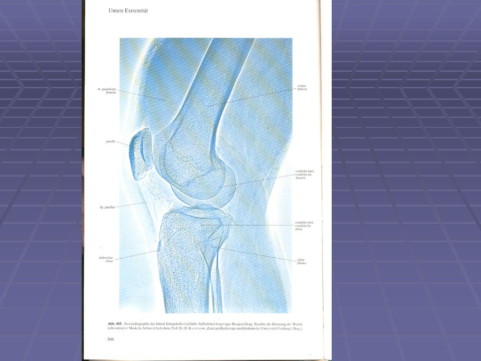
The joints of the femur 1- The head articulate with the acetabulum to form the hip joint. 2- The 2 femoral condyles articulate with the 2 tibial condyles in the knee joint . 3- The ant. surface of the lower end articulates with the upper 2/3 of the post. surface of the patella.

32
THE PATELLA The patella(=the knee cap) : It is a flat & the largest sesamoid bone in the body located in the tendon of quadriceps femoris M. in front of the lower end of the femur . It is triangular in shape with a base (upper border ) & an apex (rounded lower tip )& 2 borders (medial & lateral) & 2 surfaces (ant. & post.).
 : It is a flat & the largest sesamoid bone in the body located in the tendon of quadriceps femoris M. in front of the lower end of the femur . It is triangular in shape with a base (upper border ) & an apex (rounded lower tip )& 2 borders (medial & lateral) & 2 surfaces (ant. & post.).")
34
THE PATELLA The lower 1/3 of the post. surface is rough while its upper 2/3 is smooth & is called the articular surface as it articulates with the patellar surface of the femur . The vastus medialis & vastus lateralis Mm are attached to the medial & lateral borders of the patella respectively while the vastus intermedius & the rectus femoris are attached to its upper border ( the base ). The patellar ligament is attached to the apex. These attachments made it stable &rarely dislocated.
. The patellar ligament is attached to the apex. These attachments made it stable &rarely dislocated.")
37
THE BONES OF THE LEG They are THE TIBIA & THE FIBULA
THE TIBIA: It is the large , weight bearing ,medial bone of the leg .It articulates with the condyles of the femur & the head of the fibula above & with the talus & distal part of the fibula below. It consist of expanded upper end , shaft & smaller lower end . The upper end of the tibia :There are lateral & medial condyles of the tibia which articulate with the lat. & med. Condyles of the femur . The lat. & med. Menesci intervening . Between the 2 condyles is the intercondylar eminance.

38
THE TIBIA On the lateral aspect of the lateral condyle ;there is circular articular facet for the head of the fibula .

39
THE TIBIA The shaft of the tibia is triangular in cross section with 3 borders &3 surfaces ;Its ant. & med. borders with medial surface between them are subcutaneous ; the ant. border is prominent & form the shin of the leg . At the junction of the ant. border & the upper end is the tibial tuberosity which receives the attachment of ligamentum patellae . The med. border becomes rounded below where it becomes continuous with the medial malleolus.

40
THE TIBIA The lateral(=interosseous) border gives attachment to interosseous membrane . The upper post. Surface shows an oblique line called the soleal line for the attachment of soleus M .Below it there is vertical line which extend to the interosseous border . The lower end of the tibia is slightly expanded & on the inf. surface shows saddle shape articular surface for articulation with talus, the lower end is prolonged down ward & medially to form the medial malleolus.

44
THE TIBIA The lateral surface of medial malleolus articulate also with the talus , On the lat. Surface of the lower end there is a wide rough depression for articulation with the fibula . THE JOINTS OF THE TIBIA: 1- the upper end : 1-1-the upper surface of the tibial condyles with the femoral condyles in the knee joint. 1-2-the fibular facet articulate with the flat circular facet on the fibula in the sup. tibio-fibular joint. (synovial joint.) .
 .")
45
THE TIBIA 2- the lower end :
2-1- the inf. surface of its lower end & the smooth lateral surface of med malleolus articulate with the talus in the ankle joint. 2-2- the fibular notch articulate with the lower end of the fibula in the inf. tibio fibular joint.(a fibrous jt.),
,")
46
THE FIBULA It is a very thin, long bone on the lateral side of the tibia .It takes no part in the articualion in the knee joint ; but below it forms the lat. Malleolus of the ankle joint. It takes no part in transmission of body weight, but it provides attachments for Mm. It consists of upper end (head ), neck , shaft & lower end. The upper end of the fibula: 1- The head :It is the bulky upper end has aposterolateral projection called the styloid process in which the biceps tendon is inserted .
, neck , shaft & lower end. The upper end of the fibula: 1- The head :It is the bulky upper end has aposterolateral projection called the styloid process in which the biceps tendon is inserted .")
47
THE FIBULA The upper part of its medial surface has a circular flat articular process to articulate with the fibular facet on the lateral condyle of the tibia . 2- The neck :the constricted upper part . The shaft :Its long twisted bone covered all with Mm. except triangular area above the lat. malleolus which is subcutaneous .It has 3 surfaces & 3 borders . The lower end of the fibula :is called the lateral malleolus , Its flattened from side to side & more pointed & longer than the medial malleolus. Its lateral surface is subcutaneous & its medial smooth surface for articulation with the lateral surface of the talus .while its posterior surface has a shallow groove for the tendons of peroneus longus & brevis .

49
THE FIBULA The fibula has 3 functions :
1- gives origin to Mm. of the leg . 2- form part of the ankle joint . 3- the lower end form a pulley for the tendons of peroneal mm. The Mm. attached to the fibula: One M.,biceps, inserted in the head of the fibula. Three mm. arise from the medial surface : 1- Extensor digitorum longus :from the upper ¾. 2- Peroneus tertius :from the lower ¼ . 3- extensor hallucis longus :from the middle ½.

50
THE FIBULA Two Mm. arise from the lateral (peroneal) surface:
1-peroneus longus :from the upper 2/3 . 2-Peroneus brevis : from the lower 2/3 . Three Mm. arise from the post. (flexor )surface : 1- Soleus :from the back of the head & upper 1/3 2-Tibialis post. M. from the medial side of post surface . 3- Flexor hallucis longus: from the lat. side of post. Surface.
surface : 1- Soleus :from the back of the head & upper 1/3. 2-Tibialis post. M. from the medial side of post. surface . 3- Flexor hallucis longus: from the lat. side of post. Surface.")
51
THE FOOT The foot consists of : 1- the tarsal bones (7) .
2- the metatarsal bones (5) . 3- the toes =digits(5). TheTarsal bones : * the Calcaneus : is the largest ,the longest & the most posterior . *the Talus : lies on the top of the calcaneus . *the Navicular :lies between the talus & the cuneiform bones . *the 3 Cuneiform bones ( medial , intermediate & lateral ) lies between the navicular bone & the medial 3 metatarsls. *the cuboid lies between the calcaneus & the lateral 2 metatarsals.
 . 3- the toes =digits(5). TheTarsal bones : * the Calcaneus : is the largest ,the longest & the most posterior . *the Talus : lies on the top of the calcaneus . *the Navicular :lies between the talus & the 3 cuneiform bones . *the 3 Cuneiform bones ( medial , intermediate & lateral ) lies between the navicular bone & the medial 3 metatarsls. *the cuboid lies between the calcaneus & the lateral 2 metatarsals.")
52
THE FOOT The metatarsals: they are 5 bones , one for each digit .each consists of base , shaft & head . The medial 3 articulate with the 3 cuneiform bones & the lateral 2 with the cuboid bone . The toes :each toe has 3 phalanges except the big toe has 2.Each phalanx consists of base ,shaft & head .

54
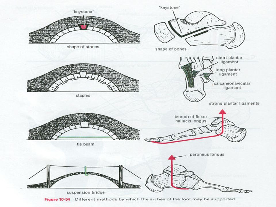
THE ARCHES OF THE FOOT The bones of the foot are arranged to form 2 longitudinal arches( medial & lateral) & one transverse arch. These are bony parts which do not come in contact with ground when the weight of the body is born on the foot during standing . The medial longitudinal arch form from calcaneus ,talus ,navicular ,3 cuneiforms & medial 3 metatarsals. Its apex is the talus . Its concerned with support of the weight of the body during standing.
 & one transverse arch. These are bony parts which do not come in contact with ground when the weight of the body is born on the foot during standing . The medial longitudinal arch form from calcaneus ,talus ,navicular ,3 cuneiforms & medial 3 metatarsals. Its apex is the talus . Its concerned with support of the weight of the body during standing.")
56
THE ARCHES OF THE FOOT The lateral longitudinal arch form from calcaneus ,cuboid,& lateral 2 metatarsals. Its lower than the medial arch. Its concerned with elastic propulsion during walking or running The transverse arch at the bases of metatarsals bone. In the flat foot ,there is falling of the arches . The factors maintain the arches : 1- the shape of the bones. 2- the ligaments of the foot & the planter aponeurosis. 3- the action of certain Mm.

60
THE RETINACULI OF THE LEG & FOOT
There are 5 retinaculi : 1- The superior extensor ret.: lies immediately above the ankle joint. 2- The inferior extensor ret. :like Y-shape below the sup. Ext. ret. 3- The superior peroneal ret.: lies over the peroneus longus & brevis tendons on the back of the lat. Malleolus.: 4- The inferior peroneal ret.: lies on the peroneal tendons on the lat. Surface of the calcaneus . 5- The flexor ret.: between the calcaneus & the medial malleolus.

62
THE MUSCLES OF THE FOOT There are 4 layers of Mm. & tendons in the sole separated by layers of facsia in which the planter vessels & nerves lie.

63
THE ARTERIES OF THE LOWER LIMB
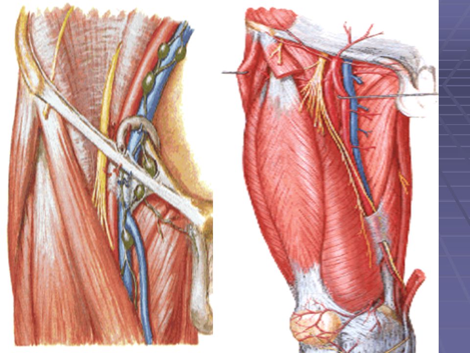
The femoral artery : Its is the direct continuation of the external iliac A. , It enters the thigh by passing behind the mid-inguinal point ( midway between the ASIS & the pubic tubercle). After running vertically downward , it enters the femoral triangle then in the adductor canal then pierce the adductor magnus M to enter to the popliteal fossa to be the popliteal A. The upper 10 cm of femoral A. is superficial & may easily injured or punctured by a needle. The femoral vein is medial to it while the femoral nerve is lateral to it ( VAN).
. After running vertically downward , it enters the femoral triangle then in the adductor canal then pierce the adductor magnus M to enter to the popliteal fossa to be the popliteal A. The upper 10 cm of femoral A. is superficial & may easily injured or punctured by a needle. The femoral vein is medial to it while the femoral nerve is lateral to it ( VAN).")
66
The femoral artery The branches of femoral A.:
1- Superficial branches : 1-1- superficial epigastric A. 1-2- superficial circumflex iliac A. 1-3- superficial external pudendal A. 2- Deep branches : 2-1- deep external pudednal A. 2-2- the profunda femoris A.

67
The femoral artery The profunda femoris A.: is the largest branch , arise from the upper part of fem. A. to pass behind it to end as the 4th perforating Aa. It gives of: 1- lateral circumflex femoral A. 2- medial circumflex femoral A. 3- four perforating Aa.

69
THE POPLITEAL ARTERY It begins at the opening in the adductor magnus as the continuation of the femoral A. & ends at the lower border of the popliteus M. by dividing into ant. & post. Tibial arteries. It is the deepest str. of the popl. Fossa. It lies on( from above –downward) :the popliteal surface of the femur , then the back of the capsule of the knee joint , then the fascia over the popliteus Mm.It gives of muscular & articular branches to form rich anastamosis around the knee joint.
 :the popliteal surface of the femur , then the back of the capsule of the knee joint , then the fascia over the popliteus Mm.It gives of muscular & articular branches to form rich anastamosis around the knee joint.")
71
THE ANTERIOR TIBIAL ARTERY
It begins at the lower border of the popliteus M. in the posterior comp. as the smaller terminal branch of the popl. A. & after running downwards in the ant. compt. of the leg to ends in front of the ankle joint by becoming the dorsalis pedis A. It gives off 1- ant.& post. Recurrent br. to anastamose around the knee joint . 2- ant.medial malleolar & ant. Lateral malleolar br. to anastamose around the ankle joint 3- muscular br. to M. of ant. compt of the leg .

72
THE DORSALIS PEDIS ARTERY
It begins in front of the ankle joint as a continuation of ant. tibial A. runs forward on the dorsum of the foot to pass in the 1st intermetatarsal space then reach the sole of the foot to anastamose with the end of the planter arch. It passes on the talus ,navicular & intermediate cuneiform bone & pass behind the inf. Ext. ret.

74
THE POSTERIOR TIBIAL ARTERY
It begins at the lower border of the popliteus M.as the larger terminal branch of the popliteal A.& after running downward in the post. cmpt. It ends by dividing to medial & lateral planter Aa. It is deep in the upper part of the leg while superficial in the lower part to be palpated midway between the medial malleolus & medial tubercle of the calcaneum , there it divided to med. & lat.planter Aa. which form the main blood supply of the foot.

75
THE POSTERIOR TIBIAL ARTERY
The peroneal A.: is a big branch arise just below the origin of the post. tibial A. passing downward & lateraly towards the fibula then vertically with the medial border of the fibula.in the lower part to end behind the inf.tibio-fibular joint& share in the anastamosis around the lat. Malloelus . It gives of a perforating br. which pierce the interosseus membrane & share in the same anastamosis.

76
The lateral planter A. : It begins under cover of flexor retinaculum . Passing toward the base of 5th metatarsal then pass medially across the foot to form the planter arch with the dorsalis pedis A. It gives off planter digital Aa. & planter metatarsal Aa. The medial planter A.: is a small branch arise under the flex. Ret. & supply the big toe.

77
THE LUMBAR PLEXUS It is formed in the Psoas major M. in front of the lower lumbar transverse processes by the anterior primary rami of the upper 4 lumbar nerves.( L1,L2, L3, L4). It gives 4 small & 2 big nerves : 1- the small branches : 1-1- ilio-hypogastric N (L1) . 1-2- ilio-inguinal N.(L1) . 1-3- Genito-femoral N. (L1&L2) . 1-4- Lateral coetaneous N .of the thigh(L2&L3) 2-The main branches : 2-1-Femoral N.(L2,L3,L4) dorsal divisions. 2-2-Obturator N .(L2,L3,L4) ventral divisions
. It gives 4 small & 2 big nerves : 1- the small branches : 1-1- ilio-hypogastric N (L1) ilio-inguinal N.(L1) Genito-femoral N. (L1&L2) Lateral coetaneous N .of the thigh(L2&L3) 2-The main branches : 2-1-Femoral N.(L2,L3,L4) dorsal divisions. 2-2-Obturator N .(L2,L3,L4) ventral divisions.")
78
THE LUMBAR PLEXUS The genito-femoral N. emerge from the ant. surface of the Psoas M , the obturator N. from its medial side, while the ilio-hypogastric,ilio-inguinal , lat.cut. N. of the thigh & femoral N . emerge from its lateral border. The obturator N. is the N. of the medial (adductor) compt. of the thigh. It supplies the adductor Mm, gracilis & obturator externus Mm.,both hip & knee joints & skin over the medial aspect of the thigh.
 compt. of the thigh. It supplies the adductor Mm, gracilis & obturator externus Mm.,both hip & knee joints & skin over the medial aspect of the thigh.")
80
The femoral N.: It is the N. of ant. (extensor) comp. of the thigh.
It enters the thigh behind the inguinal lig. lateral to femoral A. to enter the femoral triangle. Before it enters the thigh , it supplies the iliacus M. then after it enters the thigh it supplies the pectineus M. then it divided to : 1- ant. Group : which gives : 1-1- intermediate cut. N. of the thigh . 1-2-medial cut. N. of the thigh . 1-3-branch to sartorius . 2- post. Group : which gives : 2-1-saphenous N. 2-2- branches to quadriceps femoris M . 2-3- articular br. to knee joint .

81
The saphenous N : It runs on the lat. side of the femoral A. till it enters the adductor canal where it crosses in front of the femoral A. from lateral to medial. It gives an infra-patellar br. to join the patellar plexus then supply the medial side of the leg then supplies the medial side of the foot.

83
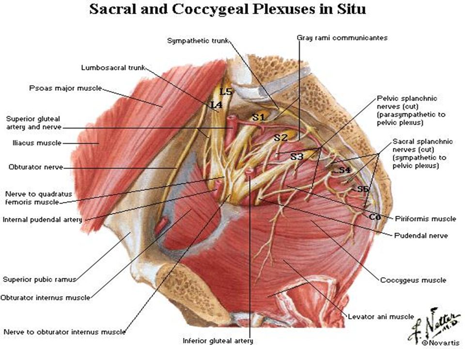
THE SACRAL PLUXES : It is formed by the lumbo-sacral trunk (L4,L5) & the anterior primary rami of S1 , S2 , S3 , S4 .
 & the anterior primary rami of S1 , S2 , S3 , S4 .")
84
THE BRANCHES. OF SACRAL PLUXES
1- From the post. surface : 1-1- The sup. gluteal N ( L4 ,L5 ,S1) 1-2- The inf. gluteal N( L5 ,S1 ,S2) 2- From the ant. Surface : 2-1-The N .to quadratus femoris (L4,L5,S1 ) 2-2-The N. to oburator internus (L5 ,S1 ,S2 ) 3- The cutaneous Nn.: 3-1- The perforating cutaneous N. (S2 & S3 ) 3-2- The post. cutaneous N. of the thigh (S1, S2 , S3 )
 1-2- The inf. gluteal N( L5 ,S1 ,S2) 2- From the ant. Surface : 2-1-The N .to quadratus femoris (L4,L5,S1 ) 2-2-The N. to oburator internus (L5 ,S1 ,S2 ) 3- The cutaneous Nn.: 3-1- The perforating cutaneous N. (S2 & S3 ) 3-2- The post. cutaneous N. of the thigh (S1, S2 , S3 )")
85
4- from the roots of the plexus :
4-1- Branches to piriformis ( S1 , S2 ) 4-2-- The pelvic splanchnic(parasympathetic) Nn.(S2 ,S 3, S4) 5- The two terminal branches : 5-1-the sciatic N. (L4,L5,S1,S2,S3 ) 5-2-The pudendal N.(S1,S2,S3,S4 )
 The pelvic splanchnic(parasympathetic) Nn.(S2 ,S 3, S4) 5- The two terminal branches : 5-1-the sciatic N. (L4,L5,S1,S2,S3 ) 5-2-The pudendal N.(S1,S2,S3,S4 )")
88
THE SCIATIC NERVE (L4,5+S1,2,3)
It is the largest N. in the body .It arise from the sacral plexus in the pelvis as flat & broad N. then be rounded as it traced downward & ends in the middle of the thigh by dividing to: 1- tibial (=medial popliteal )from ventral divisions of L4,5 & S1,2,3. 2- common peroneal(=lateral popliteal) N. from the dorsal divisions of L4,5 & S1,2. The sciatic N. leaves the pelvis through greater sciatic foramen below the piriformis M. to reach the gluteal region under cover of the gluteus maximus M. then downward in the back of the thigh where supply the biceps ,semitendenosus ,semimembrenesus & adductor magnus Mm.
from ventral divisions of L4,5 & S1,2,3. 2- common peroneal(=lateral popliteal) N. from the dorsal divisions of L4,5 & S1,2. The sciatic N. leaves the pelvis through greater sciatic foramen below the piriformis M. to reach the gluteal region under cover of the gluteus maximus M. then downward in the back of the thigh where supply the biceps ,semitendenosus ,semimembrenesus & adductor magnus Mm.")
90
THE SCIATIC NERVE (L4,5+S1,2,3

91
THE TIBIAL (MEDIAL POPLITEAL) N.(L4,5+S1,2,3)
It begins in the middle of the thigh as the largest terminal br. of sciatic N ,descend in the popliteal fossa to ends in the lower border of popliteus M by becoming the post. tibial N. which descends to the back of the leg. It lies superficial to popliteal Vv.& gives off: 1-the cutaneous br. Sural N. 2- the muscular br. to gastrocnemius , soleus ,popliteus & plantaris Mm. 3- the articular br. to the knee joint.

93
The sural N. descend over the the back of the calf then joinded with the sural communicatin br. Of lat. Popliteal N. to go behind & below the lateral malleolus( with the short saphenous vein) then along the lateral border of the foot to end by suppling the lateral side of the little toe.
 then along the lateral border of the foot to end by suppling the lateral side of the little toe..")
94
The posterior tibial n. It begins at the lower border of popliteus M as a continuation of tibial (medial popliteal) N.& pass downward to ends under the flexor retinaculum by dividing to medial & lateral planter Nn. It supplies: 1- muscular br. to soleus , tibialis post ,flexor digitorum longus & flexor hallucis longus, 2- medial calcaneal br. to the skin of the heel& medial side of the sole of the foot 3- articular br. to ankle joint .
 N.& pass downward to ends under the flexor retinaculum by dividing to medial & lateral planter Nn. It supplies: 1- muscular br. to soleus , tibialis post. ,flexor digitorum longus & flexor hallucis longus, 2- medial calcaneal br. to the skin of the heel& medial side of the sole of the foot. 3- articular br. to ankle joint .")
95
The medial planter N.: It arise under the flexor retinaculum as the larger terminal br. of post. Tibial N. to ends at the bases of metatarsal bones by dividing to 3 planter digital Nn. It supplies the flexor digitorum brevis , flexor hallucis brevis , abductor hallucis & 1st lumbrical Mm., also supplies the skin on the medial part of the sole & the medial 3 ½ toes by the planter digital Nn. The lateral planter N : It arise under the flexor retinaculum as the smaller terminal br. of post. Tibial N. to ends at the base of 5th metatarsal where supplies the skin of the lateral 1 ½ toes & some small Mm.

96
The common peroneal (lateral popliteal) N. L4,L5,S1,S2
The smaller terminal br. of sciatic N .,begins in the middle of the thigh then runs downward & laterally in the popliteal fossa to ends on the lateral aspect of the neck of the fibula by dividing to deep peroneal (ant. tibial) & the superficial peroneal ( musculo-cutaneous) Nn. It gives the lateral cutaneous N. of the calf & the sural communicating br.& articular br. to the knee joint.( i.e. no muscular br.).
 & the superficial peroneal ( musculo-cutaneous) Nn. It gives the lateral cutaneous N. of the calf & the sural communicating br.& articular br. to the knee joint.( i.e. no muscular br.).")
98
The anterior tibial N.(deep peroneal) N.
It begins on the lateral side of the neck of the fibula as the deep terminal br. of lat. Popliteal N. It runs downward in the ant.comp. of the leg & passes in front of the ankle joint under the inf. ext. retinaculum into the dorsum of the foot & ends by supplying the intertarsal & tarsometatarsal joints.It supplies all the Mm in the ant. comp. of the leg &small area of skin in the cleft between the 1st & 2nd toes.& the ankle joint .

99
The musculo-cutaneous(the superficial peroneal ) N.
It begins on the lateral side of the neck of the fibula as the superficial terminal br. of lat. Popliteal N. It runs downward in the lat. comp. of the leg & ends in the lower part of the leg by dividing to med. & lat. br. on the dorsum of the foot. It supplies the Mm of lat. comp. of the leg & the skin of the lower 2/3rd of the lateral aspect of the leg & the skin of dorsum of the foot& the toes except the cleft between the 1st & 2nd toes(ant.tibial N.) &lat. Side of little toe (sural N.).
 &lat. Side of little toe (sural N.).")
100
Segmental cutaneous supply of the lower limb
The skin of the lower limb is supplied by L1,2,3,4,5&S1,2,3. as fallows: L1,2,3 :front ,med. & lat. of the thigh . L4 :antero-medial aspect of the leg. L5 :ant. aspect of the leg & med.side of the foot S1 :lat. Side of the foot&lat. side of the leg. S2 :post. Surface of the leg & the thigh. S3 :lower&med. Part of gluteal region .

102
The veins of the lower limb:
The veins of the lower limb divided to superficial & deep veins according to the deep fascia. The superficial veins : which divided to great (long) saphenous vein & small (short ) saphenous vein. The great saphenous vein :It begins at the medial end of the dorsal venous plexus on the dorsum of the foot & ascends immediately in front of the medial malleolus where the saphenous N. lies in front of the vein. Then the vein ascend on the medial side of the leg then the post. parts of the medial condyles of the tibia & the femur to the groin where it pierces the deep fascia at the saphenous opening to enter the femoral vein.
 saphenous vein & small (short ) saphenous vein. The great saphenous vein :It begins at the medial end of the dorsal venous plexus on the dorsum of the foot & ascends immediately in front of the medial malleolus where the saphenous N. lies in front of the vein. Then the vein ascend on the medial side of the leg then the post. parts of the medial condyles of the tibia & the femur to the groin where it pierces the deep fascia at the saphenous opening to enter the femoral vein.")
103
The upper part of great saph. V
The upper part of great saph. V. receives superficial veins from the thigh , external genetalia & ant. Abdominal wall The great saph. Vein contain many valves below the knee & 2 valves just before entering the femoral vein . The small saphenous vein :It begins behind the lat. malleolus & drains the lat. side of the venous plexus on the dorsum of the foot .It ascends over the back of the calf to the popliteal fossa where it perforates the deep fascia & ends in the popliteal vein.

107
The deep veins of the lower limb:
The venae comitantes of the ant. & post. tibial Aa. unite together at the lower border of popliteus M. to form the popliteal vein which after ascending in the popliteal fossa ,it receives the small saph. vein ,then enters the opening in the adductor magnus M. to form the femoral vein. The femoral vein : It begins at the opening in the adductor magnus M. as a continuation of popliteal vein then pass in the adductor canal the femoral triangle & ends behind the inguinal ligament to become the external iliac vein. It is behind the femoral A. in the apex of femoral triangle to be medial to it in the base of the triangle.

108
The femoral vein receive :
1- deep external pudendal vein . 2- profunda femoris vein . 3- medial & lateral circumflex femoral veins. 4- great saphenous vein . 5- muscular veins .

110
The femoral triangle It is found in the upper1/3rd of the front of the thigh. From above: the inguinal lig. laterally :medial border of sartorius M. Medially :medial border of adductor longus M. The roof :skin , superficial fascia & deep fascia. The floor : 4 Mm.( from med. to lat.):adductor longus , pectineus, psoas major & iliacus.
:adductor longus , pectineus, psoas major & iliacus.")
112
The femoral triangle contents:
1- The femoral artery & its branches. 2- The femoral vein& its tributaries. 3- The femoral sheath( & the femoral canal ). 4- The femoral nerve & its branches . 5- The deep inguinal lymph nodes(3-4) along the medial side of femoral vein . 6- The femoral br. of genito-femoral N. 7- The lat. cut N. of the thigh.
. 4- The femoral nerve & its branches . 5- The deep inguinal lymph nodes(3-4) along the medial side of femoral vein . 6- The femoral br. of genito-femoral N. 7- The lat. cut N. of the thigh.")
113
The femoral sheath & the femoral canal
The femoral A. & V. enter the femoral triangle from behind the inguinal lig. within a sheath of fascia called the femoral sheath which descends from the lower abdomen to surround the upper part of the femoral Vv. The femoral sheath is funnel- shape ,4 cm long. It divided to : 1- lateral comp. contains femoral A. 2- Intermediate comp. contains the femoral vein. 3- medial comp.: a small vertically placed gap called the femoral canal contains a lymph node & loose areolar tissue.It opens in the abdomenal cavity by an opening called The femoral ring whish is normally closed by condensation of extra-peritoneal tissue called the femoral septum. The canal is closed below by the blending of its wall.

Similar presentations


.>")


>")


