Download presentation
Presentation is loading. Please wait.

1
Shark Dissection Study photos

22
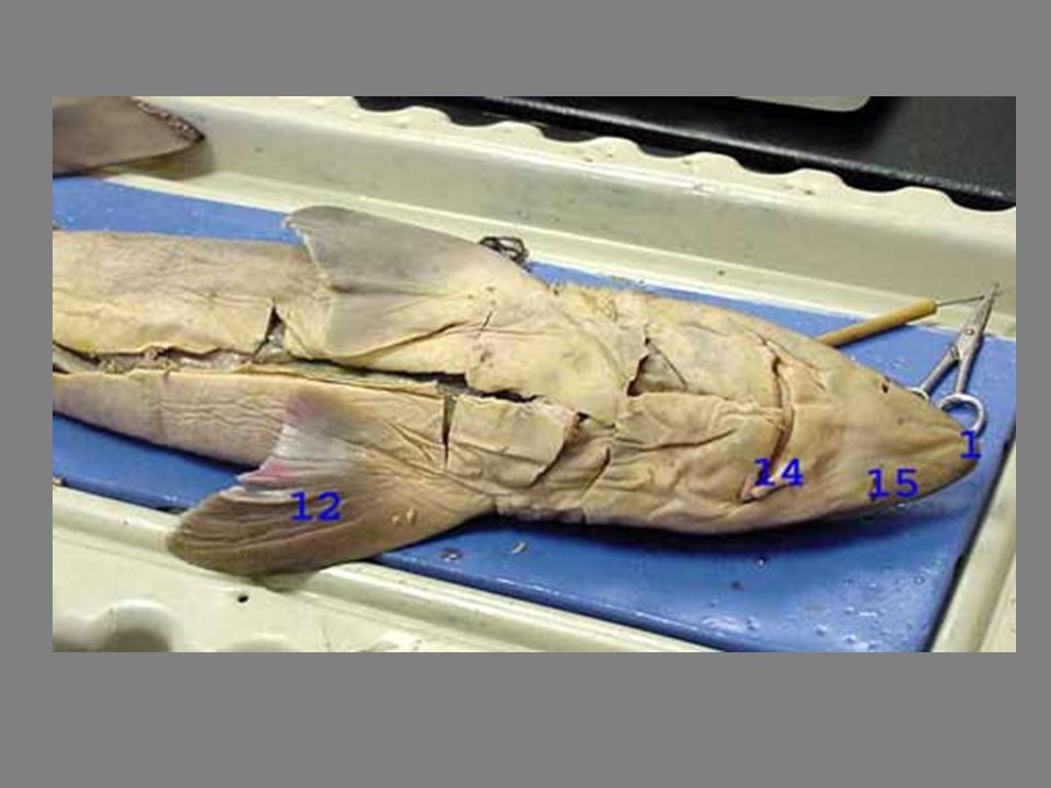
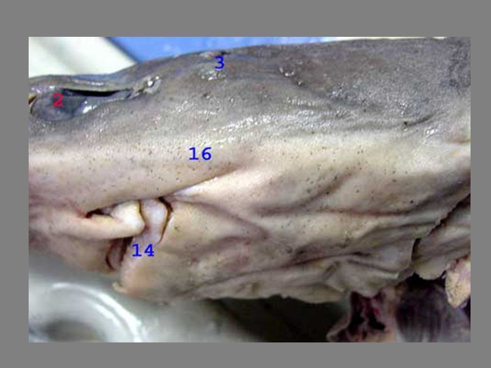
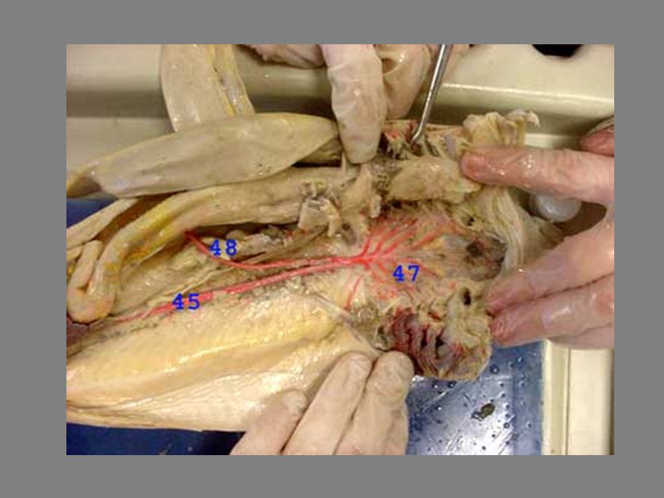
Shark Structures 1. Rostrum 2. Eye 3. Spiracle 4. Lateral Line
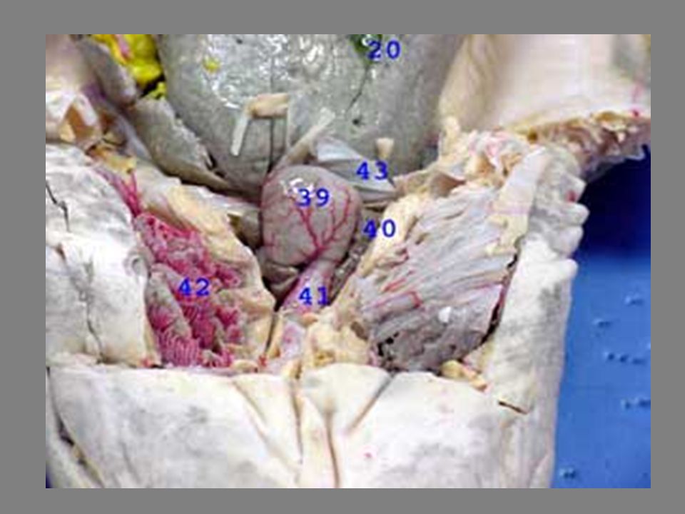
5. Fin Spine 6. Anterior Dorsal Fin 7. Posterior Dorsal Fin 8. Dorsal Lobe of Caudal Fin 9. Ventral Lobe of Caudal Fin 10. Clasper 11. Pelvic Fin 12. Pectoral Fin 13. External Gill Slits 14. Mouth 15. Nostril 16. Ampulla of Lorenzinii 17. Medial Lobe of Liver 18. Left Lobe of Liver 19. Right Lobe of Liver 20. Gallbladder

23
Shark Structures 21. Stomach 22. Duodenum 23. Ilium 24. Spiral Valve
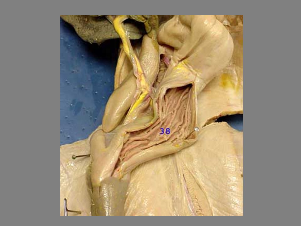
25. Colon 26. Cloaca 27. Urogenital Papilla 28. Ventral Lobe of Pancreas 29. Dorsal Lobe of Pancreas 30. Spleen 31. Kidney 32. Rectal Gland 33. Bile Duct 34. Right Testis 35. Left Testis 36. Sinus Venosus 37. Sperm Sac 38. Rugae in Stomach 39. Ventricle

24
Shark Structures 40. Atrium 41. Conus Arteriosus 42. Gills
43. Transverse Septum 44. Ductus Deferens ( Sperm Duct) 45. Dorsal Aorta 46. Ovary 47. Efferent Branchial Arteries 48. Celiac Artery 49. Internal Carotid Artery 50. Anterior Mesenteric Artery 51. Lienogastric Artery 52. Posterior Mesenteric Artery 53. Iliac Artery Pictures Courtesy: © Lori Starwalt. 22nd, February, All rights reserved.
 45. Dorsal Aorta. 46. Ovary. 47. Efferent Branchial Arteries. 48. Celiac Artery. 49. Internal Carotid Artery. 50. Anterior Mesenteric Artery. 51. Lienogastric Artery. 52. Posterior Mesenteric Artery. 53. Iliac Artery. Pictures Courtesy: © Lori Starwalt. 22nd, February, All rights reserved.")
25
Buccal Cavity Colon Dorsal Aorta External Gill Slits External Nares Gall Bladder Gill Rays Internal Gill Slits Kidney Labial Groove Liver (left, median, right lobes) Oviduct Pancreas Pectoral Fin Pelvic Fin Rectal Gland Small Intestine (duodenum, ileum) Spiracle Spleen Stomach (cardiac, pyloric) Tongue Upper & Lower Jaws
 Oviduct. Pancreas. Pectoral Fin. Pelvic Fin. Rectal Gland. Small Intestine (duodenum, ileum) Spiracle. Spleen. Stomach (cardiac, pyloric) Tongue. Upper & Lower Jaws.")
Similar presentations


>")

 Dissection: Anatomy and Physiology>")


