Download presentation
Presentation is loading. Please wait.

1
Unit 3: Biological Psychology
WHS AP Psychology Unit 3: Biological Psychology Essential Task 3-5a.Describe the subdivisions and functions of the Central Nervous System A. Brain i. Brain Stem Medulla, Pons, Reticular Formation, Cerebellum, and the Thalamus ii. Limbic System Hypothalamus, Amygdala, and the Hippocampus iii. Cerebral Cortex (Left and Right Hemispheres and the corpus callosum) Occipital Lobe, Parietal Lobe, Temporal Lobe, and the Frontal Lobe Primary Motor Cortex and Primary Sensory Cortex Wernicke's Area and Broca's Area B. Spinal Cord Logo Green is R=8 G=138 B= Blue is R= 0 G=110 B=184 Border Grey is R=74 G=69 B=64
 Occipital Lobe, Parietal Lobe, Temporal Lobe, and the Frontal Lobe. Primary Motor Cortex and Primary Sensory Cortex. Wernicke s Area and Broca s Area. B. Spinal Cord. Logo Green is R=8 G=138 B=76 Blue is R= 0 G=110 B=184. Border Grey is R=74 G=69 B=64.")
2
Biological Psychology
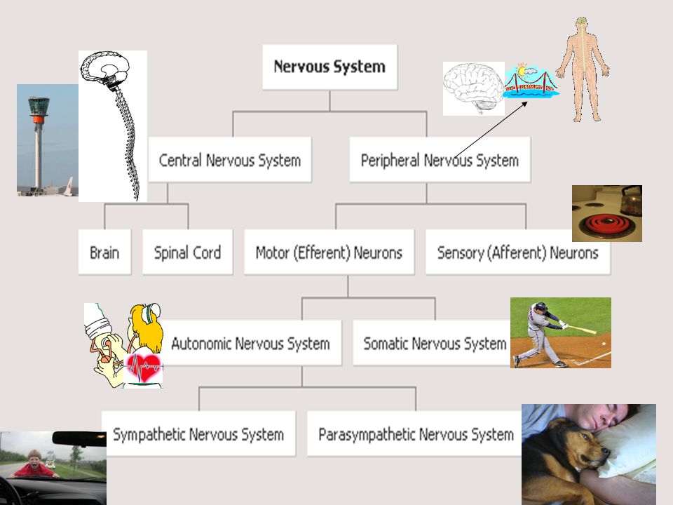
Nervous System Central Nervous System Brain Brain Imaging Peripheral Nervous System Building Blocks Genetics Evolutionary Endocrine System Neurotransmitters Somatic Autonomic Sympathetic Parasympathetic Biological Psychology Spinal Cord Neurons Sensory Motor We are here

4
Essential Task 3-5: CNS A. Brain i. Brain Stem
Outline A. Brain i. Brain Stem Medulla, Pons, Reticular Formation, Cerebellum, and the Thalamus ii. Limbic System Hypothalamus, Amygdala, and the Hippocampus iii. Cerebral Cortex (Left and Right Hemispheres and the corpus callosum) Occipital Lobe, Parietal Lobe, Temporal Lobe, and the Frontal Lobe Primary Motor Cortex and Primary Sensory Cortex Wernicke's Area and Broca's Area B. Spinal Cord
 Occipital Lobe, Parietal Lobe, Temporal Lobe, and the Frontal Lobe Primary Motor Cortex and Primary Sensory Cortex Wernicke s Area and Broca s Area. B. Spinal Cord.")
5
The Brain Stem (Automatic Functions)
Brain Structure Primary Function Secondary Function Medulla Respiration, blood pressure, heart rate Vomiting Pons Puts you to sleep Reticular Formation Attention, regulates awareness Cerebellum Balance and coordination Thalamus Directs sensory information to the rest of the brain

6
Older Brain Structures
The Brainstem is the oldest part of the brain, beginning where the spinal cord swells and enters the skull. It is responsible for automatic survival functions. OBJECTIVE 12| Describe the components of the brainstem and summarize the functions of the brainstem, thalamus and cerebellum.

7
Brain Stem The Medulla [muh-DUL-uh] is the base of the brainstem
It controls autonomic functions and relays nerve signals between the brain and spinal cord. respiration blood pressure heart rate reflex arcs vomiting
![Brain Stem The Medulla [muh-DUL-uh] is the base of the brainstem](http://slideplayer.com/slide/4280553/14/images/7/Brain+Stem+The+Medulla+%5Bmuh-DUL-uh%5D+is+the+base+of+the+brainstem.jpg "It controls autonomic functions and relays nerve signals between the brain and spinal cord. respiration. blood pressure. heart rate. reflex arcs. vomiting.")
8
Brain Stem Pons and inside that the (Reticular Formation) is a nerve network in the brainstem that plays an important role in controlling arousal. It is involved in motor control and sensory analysis... for example, information from the ear first enters the brain in the pons. It has parts that are important for the level of consciousness and for sleep. The Reticular Formation controls: Attention Cardiac Reflexes Motor Functions Regulates Awareness Relays Nerve Signals to the Cerebral Cortex Sleep

9
Brain Stem The Medulla [muh-DUL-uh] is the base of the brainstem that controls heartbeat and breathing. Reticular Formation is a nerve network in the brainstem that plays an important role in controlling arousal.
![Brain Stem The Medulla [muh-DUL-uh] is the base of the brainstem that controls heartbeat and breathing.](http://slideplayer.com/slide/4280553/14/images/9/Brain+Stem+The+Medulla+%5Bmuh-DUL-uh%5D+is+the+base+of+the+brainstem+that+controls+heartbeat+and+breathing..jpg "Reticular Formation is a nerve network in the brainstem that plays an important role in controlling arousal.")
10
Brain Stem The Thalamus [THAL-uh-muss] is the brain’s sensory switchboard, located on top of the brainstem. It directs messages to the sensory areas in the cortex and transmits replies to the cerebellum and medulla.

11
Cerebellum The “little brain” attached to the rear of the brainstem. It helps coordinate voluntary movements and balance.

12
Limbic System (Emotion Center)
Brain Structure Primary Function Secondary Function Hypothalamus Drives: Hunger, Thirst, Sex Temperature control Amygdala Fight or Flight Hippocampus STM to LTM

13
The Limbic System The Limbic System is a doughnut-shaped system of neural structures at the border of the brainstem and cerebrum, associated with emotions such as fear, aggression and drives for food and sex. It includes the hippocampus, amygdala, and hypothalamus. OBJECTIVE 13| Describe the structures and functions of the limbic system, and explain how one of these structures controls the pituitary gland.

14
Amygdala The Amygdala [ah-MIG-dah-la] consists of two almond-shaped neural clusters linked to the emotions of fear and anger.
![Amygdala The Amygdala [ah-MIG-dah-la] consists of two almond-shaped neural clusters linked to the emotions of fear and anger.](http://slideplayer.com/slide/4280553/14/images/14/Amygdala+The+Amygdala+%5Bah-MIG-dah-la%5D+consists+of+two+almond-shaped+neural+clusters+linked+to+the+emotions+of+fear+and+anger..jpg "Amygdala The Amygdala [ah-MIG-dah-la] consists of two almond-shaped neural clusters linked to the emotions of fear and anger.")
15
Hypothalamus The Hypothalamus lies below (hypo) the thalamus. It directs several maintenance activities like eating, drinking, body temperature, and control of emotions. It helps govern the endocrine system via the pituitary gland.
 the thalamus. It directs several maintenance activities like eating, drinking, body temperature, and control of emotions. It helps govern the endocrine system via the pituitary gland.")
16
Reward Center Rats cross an electrified grid for self-stimulation when electrodes are placed in the reward (hypothalamus) center (top picture). When the limbic system is manipulated, a rat will navigate fields or climb up a tree (bottom picture). Sanjiv Talwar, SUNY Downstate
 center (top picture). When the limbic system is manipulated, a rat will navigate fields or climb up a tree (bottom picture). Sanjiv Talwar, SUNY Downstate.")
17
Cerebral Cortex Brain Structure Primary Function Secondary Function
Occipital Lobe Visual Processing Parietal Lobe Spatial Reasoning Frontal Lobe Decision Making Temporal Lobe Auditory sensory information Motor Cortex Movement Sensory Cortex Sensation Wernicke’s Area Understanding Speech Broca’s Area Producing Speech

18
The Cerebral Cortex The intricate fabric of interconnected neural cells that covers the cerebral hemispheres. It is the body’s ultimate control and information processing center. OBJECTIVE 14| Define cerebral cortex and explain its importance fro the human brain.

19
Structure of the Cortex
Each brain hemisphere is divided into four lobes that are separated by prominent fissures. These lobes are the frontal lobe (forehead), parietal lobe (top to rear head), occipital lobe (back head) and temporal lobe (side of head). OBJECTIVE 15| Identify the four lobes of the cerebral cortex.
, parietal lobe (top to rear head), occipital lobe (back head) and temporal lobe (side of head). OBJECTIVE 15| Identify the four lobes of the cerebral cortex.")
20
Functions of the Cortex
The Motor Cortex is the area at the rear of the frontal lobes that control voluntary movements. The Sensory Cortex (parietal cortex) receives information from skin surface and sense organs. OBJECTIVE 16| Summarize some of the findings on the functions of the motor cortex and the sensory cortex, and discuss the importance of the association areas.
 receives information from skin surface and sense organs. OBJECTIVE 16| Summarize some of the findings on the functions of the motor cortex and the sensory cortex, and discuss the importance of the association areas.")
21
Visual Function The functional MRI scan shows the visual cortex is active as the subject looks at faces.

22
Auditory Function The functional MRI scan shows the auditory cortex is active in patients who hallucinate.

23
Association Areas More intelligent animals have increased “uncommitted” or association areas of the cortex.

24
Language Aphasia is an impairment of language, usually caused by left hemisphere damage either to Broca’s area (impaired speaking) or to Wernicke’s area (impaired understanding). OBJECTIVE 17| Describe the five brain areas that would be involved if you read this sentence aloud.
 or to Wernicke’s area (impaired understanding). OBJECTIVE 17| Describe the five brain areas that would be involved if you read this sentence aloud.")
25
Specialization & Integration
Brain activity when hearing, seeing, and speaking words

26
Can you make a purple circle with a cross in the middle?

27
Fun with your Hemispheres
Rotate your dominant hand in one direction while at the same time rotating the opposite foot in the other direction. No problem since controlled by two hemispheres Now, rotate your dominant hand in one direction while at the same time rotating the foot on the same side in the other direction.

28
Our brain is divided into two hemispheres.
Our Divided Brain Our brain is divided into two hemispheres. The left hemisphere processes reading, writing, speaking, mathematics, and comprehension skills. In the 1960s, it was termed as the dominant brain. OBJECTIVE 19| Describe split-brain research, and explain how it helps us to understand the functions of our left and right hemispheres.

29
Hemispheric Specialization
Corpus Callosum Fibers that connect the two hemispheres Allow close communication between left and right hemisphere Each hemisphere appears to specialize in certain functions (See Worksheet)
")
30
The Wagner Preference Inventory
(a) left, logical (b) left, verbal (c) right, manipulative/spatial (d) right, creative
 left, logical. (b) left, verbal. (c) right, manipulative/spatial. (d) right, creative.")
31
Hemispheric Specialization
People with intact brains also show left-right hemispheric differences in mental abilities. A number of brain scan studies show normal individuals engage their right brain when completing a perceptual task and their left brain when carrying out a linguistic task.

32
Splitting the Brain A procedure in which the two hemispheres of the brain are isolated by cutting the connecting fibers (mainly those of the corpus callosum) between them. Corpus Callosum
 between them. Corpus Callosum.")
33
Split Brain Patients With the corpus callosum severed, objects (apple) presented in the right visual field can be named. Objects (pencil) in the left visual field cannot.
 presented in the right visual field can be named. Objects (pencil) in the left visual field cannot.")
34
Divided Consciousness

35
The Spinal Cord Complex cable of nerves that connects brain to rest of the body Carries motor impulses from the brain to internal organs and muscles Carries sensory information from extremities and internal organs to the brain 400,000 people a year in US either partial or complete paralysis.

36
The Spinal Cord The spinal cord controls some protective reflex movements without any input from the brain

Similar presentations


 as well.>")


. The more complex.>")


