Download presentation
Presentation is loading. Please wait.

1
به نام خدایی که در این نزدیکی است

2
Dr.Mokhtare Iran University of Medical Sciences
History taking and physical exam in gastrointestinal and pancreatic & hepatobiliary diseases Dr.Mokhtare Iran University of Medical Sciences

6
1(راست) (چپ)
 2(چپ)")
27
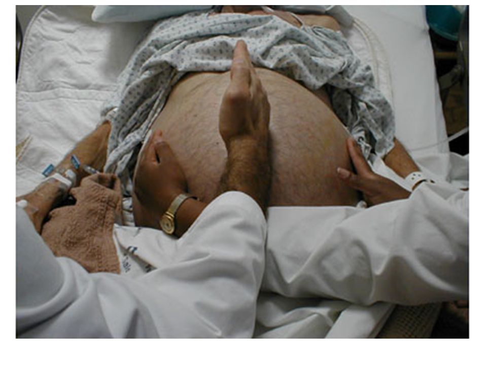
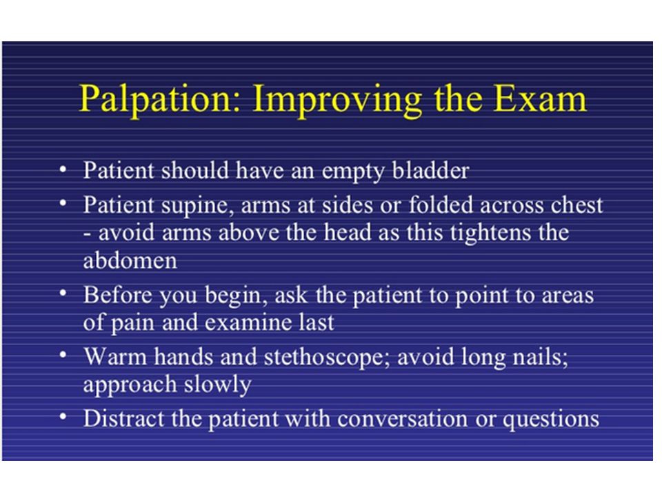
Physical examination Palpation of the aorta
to the left of the midline normal: < 3-4 cm >6 cm: aortic aneurysm transmitted pulsations: pancreatic or gastric tumor, pseudocyst of the pancreas

36
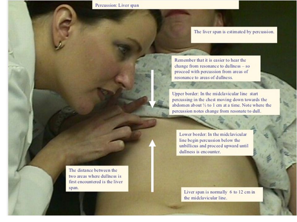
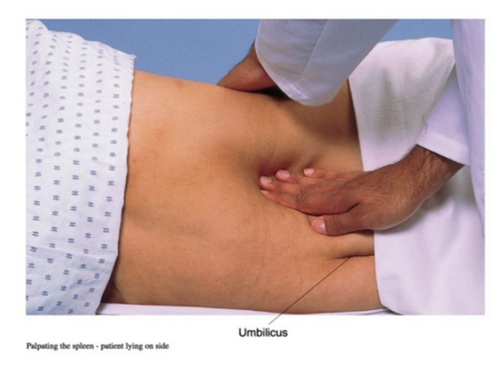
Physical examination Palpation of the liver and spleen
Characteristics: 1. size 2. surface 3. edge 4. consistency 5. tenderness (6. liver pulsation)
")
47
Physical examination Palpation of the gallbladder
Curvoisier’s sign - painless enlargement of the gallbladder due to cancer of the head of the pancreas Murphy’s sign - RUQ pain aggravated by inspiration - acute cholecystitis

49
History of Present Illness answers questions of ..
When the problem began, what and where the symptoms are, what makes the symptoms worse or better.

50
History of Present Illness
Ask about the nature of the symptoms (for pain, is it sharp or dull, localized or generalized).
.")
51
Chronological description of the development of the patient's present illness from the first sign and/or symptom 10 15 Abdominal pain Fever and chills jaundice

52
History of Present Illness (PAIN)
Location Quality Severity Duration Timing Context Modifying factors Associated signs and symptoms.

53
55-yr-old Men With Abdominal Pain
History of present illness LIQOR AAA

54
L Location of the symptom (forehead, wrist...)
")
56
I Intensity of the symptom (scale 1-10, 6/10)
")
57
Q Quality of the symptom (burning, pulsating pain...)
")
58
O Onset of the symptom + precipitating factors

59
R Radiation of the symptom (to left shoulder and arm)
")
60
A Associated symptom ( palpitations, shortness of breath)
")
61
A Alleviating factors (sitting with my chest on my knees)
")
62
A Aggravating factors (effort, smoking, large meals)
")
63
History taking in abdominal diseases

64
History taking Family history Colon cancer Gallstones

65
History taking Factors, habits and previous diseases
Diet Drugs Alcohol Smoking Transfusion Iv. drug abuse Lifestyle

66
History taking - summary
Abdominal pain Dysphagia Nausea and vomiting Anorexia and unexpected weight loss Abdominal gas Abdominal distension Diarrhea Constipation Gastrointestinal bleeding Jaundice

67
History taking Abdominal pain
Localisation Type Severity Chronology Aggravating or relieving factors Associated symptoms Radiation of pain

68
Diffuse abdominal pain
Peritonitis Intestinal obstruction Irritable bowel syndrome Tense ascites

69
Acute abdomen Peritonitis Appendicitis Bowel or gastric perforation
Gallbladder perforation Intestinal obstruction (ileus) Mesenterial ischaemia Extrauterine pregnancy (ectopic pregnancy) Acute necrotising pancreatitis Biliary colic Renal colic
 Mesenterial ischaemia. Extrauterine pregnancy (ectopic pregnancy) Acute necrotising pancreatitis. Biliary colic. Renal colic.")
70
History taking Other causes abdominal pain
Diabetic ketoacidosis Hyperthyroidism Acute intermittent porphyria Hypercalcemia, hyperkalemia Vasculitis Pneumonia Sickle cell crisis Herpes zoster

71
Radiation of pain Ulcer disease: to the back
Biliary pain: to the back, right scapula, right shoulder Pancreatic: band-like, to the back Kidney, ureter: to the genitalia, groin Splenic: left shoulder

72
History taking Substernal pain
Esophageal pain Radiation : back Type:burning, spasmodic Aggravating factors: body position, eating Relieving factors: antacid Associated symptoms: dysphagia,regurgitation Cardiac pain Radiation: left Type: pressing, constricting Aggravating factors: physical activity, stress Relieving factors: nitrates Associated symptoms: dyspnea, sweating

73
History taking Substernal pain
Esophageal pain Radiation : back Type:burning, spasmodic Aggravating factors: body position, eating Relieving factors: antacid Associated symptoms: dysphagia,regurgitation Cardiac pain Radiation: left Type: pressing, constricting Aggravating factors: physical activity, stress Relieving factors: nitrates Associated symptoms: dyspnea, sweating

74
History taking Weight loss
Is it associated with anorexia? Chronology Severity (significant:> 5% of body weight) Underlying diseases Causes: general disorders: diabetes, hyperthyroidism, chr.infections,malignancy, medications behavioral disorders: anorexia nervosa, depression GI disorders: malignancy, malabsorption, hepatic, biliary, pancreatic diseases
 Underlying diseases. Causes: general disorders: diabetes, hyperthyroidism, chr.infections,malignancy, medications. behavioral disorders: anorexia nervosa, depression. GI disorders: malignancy, malabsorption, hepatic, biliary, pancreatic diseases.")
75
History taking Nausea and vomiting
Organic, functional or psychogenic? connection with meals accompanied by weight loss Content of the vomit Factors: taste, smell, color, pH Subtypes: acid : reflux disease, duodenal ulcer bile: bilio-pancreatic diseases undigested food: obstruction of the upper GI feces (miserere): bowel obstruction (ileus) blood: ie. ulcer, tumor, eso.varix
: bowel obstruction. (ileus) blood: ie. ulcer, tumor, eso.varix.")
76
History taking Abdominal gas
Belching, bloating (meteorism), flatulence Causes Aerophagia (habitual, poor dentition, inadequate chewing, rapid eating) GI motor dysfunction or obstruction Malabsorption, maldigestion Bacterial overgrowth
, flatulence. Causes. Aerophagia (habitual, poor dentition, inadequate. chewing, rapid eating) GI motor dysfunction or obstruction. Malabsorption, maldigestion. Bacterial overgrowth.")
77
History taking Bowel movement
Constipation Chronic or recent onset Causes Decreased fluid and/or food intake Functional (irritable bowel syndrome) Medications Hypothyroidism Fecal impaction Rectal or colon cancer Chronic debilitating disease
 Medications. Hypothyroidism. Fecal impaction. Rectal or colon cancer. Chronic debilitating disease.")
78
History taking GI bleeding
Classification Hematemesis fresh blood - coffee ground Melena Hematochezia - blood on the stool - blood mixed with the stool Occult bleeding

79
History taking Jaundice
Observe it in bright, natural light First time you can observe on the sclerae

80
History taking Jaundice
Important anamnestic factors Color of the skin: overproduction: lemon obstructive: dark-yellow, greenish Color of the stool: overproduction: dark, greenish (pleiochromic) obstructive: hypocholic, acholic Color of the urine: overproduction: cherry-red obstructive: dark, brown Associated symptoms: anemia, pain, fever, hepatomegaly, splenomegaly, ascites
 obstructive: hypocholic, acholic. Color of the urine: overproduction: cherry-red. obstructive: dark, brown. Associated symptoms: anemia, pain, fever, hepatomegaly, splenomegaly, ascites.")
81
Dysphagia Dysphagia—difficulty with swallowing—refers to problems with the transit of food or liquid from the mouth to the hypopharynx or through the esophagus. Severe dysphagia can compromise nutrition, cause aspiration, and reduce quality of life. Aphagia denotes complete esophageal obstruction, most commonly encountered in the acute setting of a food bolus or foreign body impaction. Odynophagia refers to painful swallowing, typically resulting from mucosal ulceration within the oropharynx or esophagus. It commonly is accompanied by dysphagia, but the converse is not true. Globus pharyngeus is a foreign body sensation localized in the neck that does not interfere with swallowing and sometimes is relieved by swallowing. Transfer dysphagia frequently results in nasal regurgitation and pulmonary aspiration during swallowing and is characteristic of oropharyngeal dysphagia. Phagophobia (fear of swallowing) and refusal to swallow may be psychogenic or related to anticipatory anxiety about food bolus obstruction, odynophagia, or aspiration
 and refusal to swallow may be psychogenic or related to anticipatory anxiety about food bolus obstruction, odynophagia, or aspiration.")
82
Oropharyngeal Dysphagia
Oropharyngeal dysphagia may be due to neurologic, muscular, structural, iatrogenic, infectious, and metabolic causes. Iatrogenic causes include surgery and radiation, often in the setting of head and neck cancer. Neurogenic dysphagia resulting from cerebrovascular accidents, Parkinson's disease, and amyotrophic lateral sclerosis is a major source of morbidity related to aspiration and malnutrition. Oropharyngeal structural lesions causing dysphagia include Zenker's diverticulum, cricopharyngeal bar, and neoplasia. Zenker's diverticulum typically is encountered in elderly patients, with an estimated prevalence between 1:1000 and 1:10,000. In addition to dysphagia, patients may present with regurgitation of particulate food debris, aspiration, and halitosis. A cricopharyngeal bar, appearing as a prominent indentation behind al the lower third of the cricoid cartilage, However, a cricopharyngeal bar is a common radiographic finding, and most patients with transient cricopharyngeal bars are asymptomatic, making it important to rule out alternative etiologies of dysphagia before treatment. Furthermore, cricopharyngeal bars may be secondary to other neuromuscular disorders.

83
Esophageal Dysphagia The adult esophagus measures 18–26 cm in length and is anatomically divided into the cervical esophagus, extending from the pharyngoesophageal junction to the suprasternal notch, and the thoracic esophagus, which continues to the diaphragmatic hiatus. When distended, the esophageal lumen has internal dimensions of about 2 cm in the anteroposterior plane and 3 cm in the lateral plane. Solid food dysphagia becomes common when the lumen is narrowed to <13 mm but also can occur with larger diameters in the setting of poorly masticated food or motor dysfunction. Circumferential lesions are more likely to cause dysphagia than are lesions that involve only a partial circumference of the esophageal wall. The most common structural causes of dysphagia are Schatzki's rings, eosinophilic esophagitis, and peptic strictures. Dysphagia also occurs in the setting of gastroesophageal reflux disease without a stricture. Diseases affecting smooth muscle involve both the thoracic esophagus and the LES. A dominant manifestation of this, absent peristalsis, refers to either the complete absence of swallow-induced contraction or the presence of nonperistaltic, disordered contractions. Absent peristalsis and failure of deglutitive LES relaxation are the defining features of achalasia. In diffuse esophageal spasm (DES), LES function is normal, with the disordered motility restricted to the esophageal body. Absent peristalsis combined with severe weakness of the LES is a nonspecific pattern commonly found in patients with scleroderma.
, LES function is normal, with the disordered motility restricted to the esophageal body. Absent peristalsis combined with severe weakness of the LES is a nonspecific pattern commonly found in patients with scleroderma.")
85
History Key elements of the history are the localization of dysphagia, the circumstances in which dysphagia is experienced, other symptoms associated with dysphagia, and progression. Dysphagia that localizes to the chest is esophageal in origin. Nasal regurgitation and tracheobronchial aspiration with swallowing are hallmarks of oropharyngeal dysphagia or a tracheoesophageal fistula. The presence of hoarseness may be another important diagnostic clue. When hoarseness precedes dysphagia, the primary lesion is usually laryngeal; hoarseness that occurs after the development of dysphagia may result from compromise of the recurrent laryngeal nerve by a malignancy. The type of food causing dysphagia is a crucial detail. Intermittent dysphagia that occurs only with solid food implies structural dysphagia, whereas constant dysphagia with both liquids and solids strongly suggests a motor abnormality. Dysphagia that is progressive over the course of weeks to months raises concern for neoplasia. Episodic dysphagia to solids that is unchanged over years indicates a benign disease process such as a Schatzki's ring or eosinophilic esophagitis. Food impaction with a prolonged inability to pass an ingested bolus even with ingestion of liquid is typical of a structural dysphagia. Chest pain frequently accompanies dysphagia whether it is related to motor disorders, structural disorders, or reflux disease. A prolonged history of heartburn preceding the onset of dysphagia is suggestive of peptic stricture and, less commonly, esophageal adenocarcinoma. A history of prolonged nasogastric intubation, esophageal or head and neck surgery, ingestion of caustic agents or pills, previous radiation or chemotherapy, or associated mucocutaneous diseases may help isolate the cause of dysphagia. With accompanying odynophagia, which usually is indicative of ulceration, infectious or pill-induced esophagitis should be suspected. In patients with AIDS or other immunocompromised states, esophagitis due to opportunistic infections such as Candida, herpes simplex virus, or cytomegalovirus and to tumors such as Kaposi's sarcoma and lymphoma should be considered. A strong history of atopy increases concerns for eosinophilic esophagitis.

86
Physical Examination Physical examination is important in the evaluation of oral and pharyngeal dysphagia because dysphagia is usually only one of many manifestations of a more global disease process. Signs of bulbar or pseudobulbar palsy, including dysarthria, dysphonia, ptosis, tongue atrophy, and hyperactive jaw jerk, in addition to evidence of generalized neuromuscular disease, should be elicited. The neck should be examined for thyromegaly. A careful inspection of the mouth and pharynx should disclose lesions that may interfere with passage of food. Physical examination is less helpful in the evaluation of esophageal dysphagia as most relevant pathology is restricted to the esophagus. The notable exception is skin disease. Changes in the skin may suggest a diagnosis of scleroderma or mucocutaneous diseases such as pemphigoid and epidermol-ysis bullosa, all of which can involve the esophagus.

87
Vomiting&Nausea Nausea is the subjective feeling of a need to vomit. Vomiting (emesis) is the oral expulsion of gastrointestinal contents resulting from contractions of gut and thoracoabdominal wall musculature. Vomiting is contrasted with regurgitation, the effortless passage of gastric contents into the mouth. Rumination is the repeated regurgitation of stomach contents, which may be rechewed and reswallowed. In contrast to vomiting, these phenomena often exhibit volitional control. Indigestion is a nonspecific term that encompasses a variety of upper abdominal complaints including nausea, vomiting, heartburn, regurgitation, and dyspepsia (the presence of symptoms thought to originate in the gastroduodenal region). Some individuals with dyspepsia report predominantly epigastric burning, gnawing discomfort, or pain. Others with dyspepsia experience a constellation of symptoms including postprandial fullness, early satiety (an inability to complete a meal due to premature fullness), bloating, eructation (belching), and anorexia.
 is the oral expulsion of gastrointestinal contents resulting from contractions of gut and thoracoabdominal wall musculature. Vomiting is contrasted with regurgitation, the effortless passage of gastric contents into the mouth. Rumination is the repeated regurgitation of stomach contents, which may be rechewed and reswallowed. In contrast to vomiting, these phenomena often exhibit volitional control. Indigestion is a nonspecific term that encompasses a variety of upper abdominal complaints including nausea, vomiting, heartburn, regurgitation, and dyspepsia (the presence of symptoms thought to originate in the gastroduodenal region). Some individuals with dyspepsia report predominantly epigastric burning, gnawing discomfort, or pain. Others with dyspepsia experience a constellation of symptoms including postprandial fullness, early satiety (an inability to complete a meal due to premature fullness), bloating, eructation (belching), and anorexia.")
89
History and Physical Examination
The history helps define the etiology of unexplained nausea and vomiting. Drugs, toxins, and gastrointestinal infections commonly cause acute symptoms, whereas established illnesses evoke chronic complaints. Pyloric obstruction and gastroparesis produce vomiting within one hour of eating, whereas emesis from intestinal obstruction occurs later. In severe cases of gastroparesis, the vomitus may contain food residue ingested hours or days previously. Hematemesis raises suspicion of an ulcer, malignancy, or Mallory-Weiss tear, whereas feculent emesis is noted with distal intestinal or colonic obstruction. Bilious vomiting excludes gastric obstruction, while emesis of undigested food is consistent with a Zenker's diverticulum or achalasia. Relief of abdominal pain by emesis characterizes intestinal obstruction, whereas vomiting has no effect on pancreatitis or cholecystitis pain. Pronounced weight loss raises concern about malignancy or obstruction. Fevers suggest inflammation; an intracranial source is considered if there are headaches or visual field changes. Vertigo or tinnitus indicates labyrinthine disease. The physical examination complements information from the history. Orthostatic hypotension and reduced skin turgor indicate intravascular fluid loss. Pulmonary abnormalities raise concern for aspiration of vomitus. Abdominal auscultation may reveal absent bowel sounds with ileus. High-pitched rushes suggest bowel obstruction, while a succussion splash upon abrupt lateral movement of the patient is found with gastroparesis or pyloric obstruction. Tenderness or involuntary guarding raises suspicion of inflammation, whereas fecal blood suggests mucosal injury from ulcer, ischemia, or tumor. Neurologic disease presents with papilledema, visual field loss, or focal neural abnormalities. Neoplasm is suggested by palpation of masses or adenopathy.

90
Indigestion The most common causes of indigestion are gastroesophageal reflux and functional dyspepsia. Other cases are a consequence of a more serious organic illness

91
History and Physical Examination
Care of the patient with indigestion requires a thorough interview. GERD classically produces heartburn, a substernal warmth in the epigastrium that moves toward the neck. Heartburn often is exacerbated by meals and may awaken the patient. Associated symptoms include regurgitation of acid or nonacidic fluid and water brash, the reflex release of salty salivary secretions into the mouth. Atypical symptoms include pharyngitis, asthma, cough, bronchitis, hoarseness, and chest pain that mimics angina. Some patients with acid reflux on esophageal pH testing do not report heartburn, but note abdominal pain or other symptoms. Some patients with dyspepsia report a predominance of epigastric pain or burning that is intermittent and not generalized or localized to other regions. Others experience a postprandial distress syndrome characterized by fullness occurring after normal-sized meals and early satiety that prevents completion of regular meals, with associated bloating, belching, or nausea. Functional dyspepsia overlaps with other functional disorders such as IBS. The physical exam with GERD and functional dyspepsia usually is normal. In atypical GERD, pharyngeal erythema and wheezing may be noted. Recurrent acid regurgitation may cause poor dentition. Functional dyspeptics may report epigastric tenderness or distention. Discrimination between functional and organic causes of indigestion mandates exclusion of selected historic and examination features.Other alarming features include unexplained weight loss, recurrent vomiting, occult or gross gastrointestinal bleeding, jaundice, a palpable mass or adenopathy, and a family history of gastrointestinal malignancy.

93
Diarrhea Diarrhea is loosely defined as passage of abnormally liquid or unformed stools at an increased frequency. For adults on a typical Western diet, stool weight >200 g/d can generally be considered diarrheal. Diarrhea may be further defined as acute if <2 weeks, persistent if 2–4 weeks, and chronic if >4 weeks . Two common conditions, usually associated with the passage of stool totaling <200 g/d, must be distinguished from diarrhea, because diagnostic and therapeutic algorithms differ. Pseudodiarrhea, or the frequent passage of small volumes of stool, is often associated with rectal urgency and accompanies IBS or proctitis. Fecal incontinence is the involuntary discharge of rectal contents and is most often caused by neuromuscular disorders or structural anorectal problems. Diarrhea and urgency, especially if severe, may aggravate or cause incontinence. Overflow diarrhea may occur in nursing home patients due to fecal impaction that is readily detectable by rectal examination. A careful history and physical examination generally allow these conditions to be discriminated from true diarrhea.

94
Acute Diarrhea More than 90% of cases of acute diarrhea are caused by infectious agents; these cases are often accompanied by vomiting, fever, and abdominal pain. The remaining 10% or so are caused by medications, toxic ingestions, ischemia, and other conditions medications : antibiotics, cardiac antidysrhythmics, antihypertensives, NSAIDs, certain antidepressants, chemotherapeutic agents, bronchodilators, antacids, and laxatives. Occlusive or nonocclusive ischemic colitis typically occurs in persons >50 years; often presents as acute lower abdominal pain preceding watery, then bloody diarrhea; and generally results in acute inflammatory changes in the sigmoid or left colon while sparing the rectum. Acute diarrhea may accompany colonic diverticulitis and graft-versus-host disease. Acute diarrhea, often associated with systemic compromise, can follow ingestion of toxins including organophosphate insecticides; amanita and other mushrooms; arsenic; and preformed environmental toxins in seafood. Conditions causing chronic diarrhea can also be confused with acute diarrhea early in their course:IBD.

95
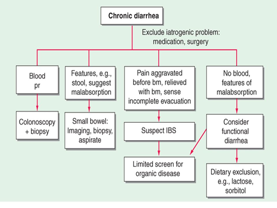
Chronic Diarrhea Diarrhea lasting >4 weeks warrants evaluation to exclude serious underlying pathology and most of the causes of chronic diarrhea are noninfectious. The classification of chronic diarrhea by pathophysiologic mechanism facilitates a rational approach to management, though many diseases cause diarrhea by more than one mechanism

97
Secretory Causes : due to derangements in fluid and electrolyte transport across the enterocolonic mucosa. They are characterized clinically by watery, large-volume fecal outputs that are typically painless and persist with fasting. Because there is no malabsorbed solute, stool osmolality is accounted for by normal endogenous electrolytes with no fecal osmotic gap.

98
Medications Side effects of drugs and toxins are the most common secretory causes of chronic diarrhea. Chronic ethanol consumption may cause a secretory-type diarrhea due to enterocyte injury with impaired sodium and water absorption as well as rapid transit and other alterations. Inadvertent ingestion of certain environmental toxins (e.g., arsenic) may lead to chronic rather than acute forms of diarrhea. Certain bacterial infections may occasionally persist and be associated with a secretory-type diarrhea
 may lead to chronic rather than acute forms of diarrhea. Certain bacterial infections may occasionally persist and be associated with a secretory-type diarrhea.")
99
Bowel Resection, Mucosal Disease, or Enterocolic Fistula
because of inadequate surface for reabsorption of secreted fluids and electrolytes. Unlike other secretory diarrheas, this subset of conditions tends to worsen with eating. With disease (e.g., Crohn's ileitis) or resection of <100 cm of terminal ileum, dihydroxy bile acids may escape absorption and stimulate colonic secretion (cholorrheic diarrhea). This mechanism may contribute to so-called idiopathic secretory diarrhea, in which bile acids are functionally malabsorbed from a normal-appearing terminal ileum. This idiopathic bile acid malabsorption may account for an average of 40% of unexplained chronic diarrhea.
 or resection of <100 cm of terminal ileum, dihydroxy bile acids may escape absorption and stimulate colonic secretion (cholorrheic diarrhea). This mechanism may contribute to so-called idiopathic secretory diarrhea, in which bile acids are functionally malabsorbed from a normal-appearing terminal ileum. This idiopathic bile acid malabsorption may account for an average of 40% of unexplained chronic diarrhea.")
100
Hormones Metastatic gastrointestinal carcinoid tumors Gastrinoma, one of the most common neuroendocrine tumors, most typically presents with refractory peptic ulcers, but diarrhea occurs in up to one-third of cases and may be the only clinical manifestation in 10%. The watery diarrhea hypokalemia achlorhydria syndrome, also called pancreatic cholera, is due to a non- cell pancreatic adenoma, referred to as a VIPoma, Medullary carcinoma of the thyroid. Prominent diarrhea is often associated with metastatic disease and poor prognosis. Systemic mastocytosis, which may be associated with the skin lesion urticaria pigmentosa, may cause diarrhea that is either secretory . Large colorectal villous adenomas may rarely be associated with a secretory diarrhea that may cause hypokalemia, can be inhibited by NSAIDs, and are apparently mediated by prostaglandins.

101
Osmotic Causes : when ingested, poorly absorbable, osmotically active solutes draw enough fluid into the lumen to exceed the reabsorptive capacity of the colon. Fecal water output increases in proportion to such a solute load. Osmotic diarrhea characteristically ceases with fasting or with discontinuation of the causative agent.

102
Carbohydrate Malabsorption
Carbohydrate malabsorption due to acquired or congenital defects in brush-border disaccharidases and other enzymes leads to osmotic diarrhea with a low pH. One of the most common causes is lactase deficiency.Most patients learn to avoid milk products without requiring treatment with enzyme supplements. Some sugars, such as sorbitol, lactulose, or fructose, are frequently malabsorbed, and diarrhea ensues with ingestion of medications, gum, or candies sweetened with these poorly or incompletely absorbed sugars

103
Steatorrheal Causes greasy, foul-smelling, difficult-to-flush diarrhea often associated with weight loss and nutritional deficiencies due to concomitant malabsorption of amino acids and vitamins. Increased fecal output is caused by the osmotic effects of fatty acids, especially after bacterial hydroxylation, and, to a lesser extent, by the neutral fat. Quantitatively, steatorrhea is defined as stool fat exceeding the normal 7 g/d; rapid-transit diarrhea may result in fecal fat up to 14 g/d; daily fecal fat averages 15–25 g with small intestinal diseases and is often >32 g with pancreatic exocrine insufficiency. Intraluminal maldigestion, mucosal malabsorption, or lymphatic obstruction may produce steatorrhea

104
Inflammatory Causes : generally accompanied by pain, fever, bleeding, or other manifestations of inflammation. The mechanism of diarrhea may not only be exudation but, depending on lesion site, may include fat malabsorption, disrupted fluid/electrolyte absorption, and hypersecretion or hypermotility from release of cytokines and other inflammatory mediators. The unifying feature on stool analysis is the presence of leukocytes or leukocyte-derived proteins such as calprotectin. With severe inflammation, exudative protein loss can lead to anasarca (generalized edema). Any middle-aged or older person with chronic inflammatory-type diarrhea, especially with blood, should be carefully evaluated to exclude a colorectal tumor.
. Any middle-aged or older person with chronic inflammatory-type diarrhea, especially with blood, should be carefully evaluated to exclude a colorectal tumor.")
105
Idiopathic Inflammatory Bowel Disease
Crohn's disease and chronic ulcerative colitis, are among adults and range in severity from mild to fulminant and life-threatening. They may be associated with uveitis, polyarthralgias, cholestatic liver disease (primary sclerosing cholangitis), and skin lesions (erythema nodosum, pyoderma gangrenosum). Microscopic colitis, including both lymphocytic and collagenous colitis, is an increasingly recognized cause of chronic watery diarrhea, especially in middle-aged women and those on NSAIDs, statins, PPIs, and SSRIs; biopsy of a normal-appearing colon is required for histologic diagnosis. It may coexist with symptoms suggesting IBS or with celiac sprue. It typically responds well to anti-inflammatory drugs (e.g., bismuth), to the opioid agonist loperamide, or to budesonide
, and skin lesions (erythema nodosum, pyoderma gangrenosum). Microscopic colitis, including both lymphocytic and collagenous colitis, is an increasingly recognized cause of chronic watery diarrhea, especially in middle-aged women and those on NSAIDs, statins, PPIs, and SSRIs; biopsy of a normal-appearing colon is required for histologic diagnosis. It may coexist with symptoms suggesting IBS or with celiac sprue. It typically responds well to anti-inflammatory drugs (e.g., bismuth), to the opioid agonist loperamide, or to budesonide.")
106
Dysmotility Causes Rapid transit may accompany many diarrheas as a secondary or contributing phenomenon, but primary dysmotility is an unusual etiology of true diarrhea. Stool features often suggest a secretory diarrhea, but mild steatorrhea of up to 14 g of fat per day can be produced by maldigestion from rapid transit alone. Hyperthyroidism, carcinoid syndrome, and certain drugs (e.g., prostaglandins, prokinetic agents) may produce hypermotility with resultant diarrhea. Primary visceral neuromyopathies or idiopathic acquired intestinal pseudoobstruction may lead to stasis with secondary bacterial overgrowth causing diarrhea. Diabetic diarrhea, often accompanied by peripheral and generalized autonomic neuropathies, may occur in part because of intestinal dysmotility. The exceedingly common IBS (10% point prevalence, 1–2% per year incidence) is characterized by disturbed intestinal and colonic motor and sensory responses to various stimuli. Symptoms of stool frequency typically cease at night, alternate with periods of constipation, are accompanied by abdominal pain relieved with defecation, and rarely result in weight loss
 may produce hypermotility with resultant diarrhea. Primary visceral neuromyopathies or idiopathic acquired intestinal pseudoobstruction may lead to stasis with secondary bacterial overgrowth causing diarrhea. Diabetic diarrhea, often accompanied by peripheral and generalized autonomic neuropathies, may occur in part because of intestinal dysmotility. The exceedingly common IBS (10% point prevalence, 1–2% per year incidence) is characterized by disturbed intestinal and colonic motor and sensory responses to various stimuli. Symptoms of stool frequency typically cease at night, alternate with periods of constipation, are accompanied by abdominal pain relieved with defecation, and rarely result in weight loss")
107
Approach to the Patient: Chronic Diarrhea
As such :a careful history and physical examination .The history, physical examination , and routine blood studies should attempt to characterize the mechanism of diarrhea, identify diagnostically helpful associations, and assess the patient's fluid/electrolyte and nutritional status. Patients should be questioned about the onset, duration, pattern, aggravating (especially diet) and relieving factors, and stool characteristics of their diarrhea. The presence or absence of fecal incontinence, fever, weight loss, pain, certain exposures (travel, medications, contacts with diarrhea), and common extraintestinal manifestations (skin changes, arthralgias, oral aphthous ulcers) should be noted. A family history of IBD or sprue may indicate those possibilities. Physical findings may offer clues such as a thyroid mass, wheezing, heart murmurs, edema, hepatomegaly, abdominal masses, lymphadenopathy, mucocutaneous abnormalities, perianal fistulas, or anal sphincter laxity. Peripheral blood leukocytosis, elevated ESR, or CRP suggests inflammation; anemia reflects blood loss or nutritional deficiencies; or eosinophilia may occur with parasitoses, neoplasia, collagen-vascular disease, allergy, or eosinophilic gastroenteritis. Blood chemistries may demonstrate electrolyte, hepatic, or other metabolic disturbances. Measuring tissue transglutaminase antibodies may help detect celiac disease.
 and relieving factors, and stool characteristics of their diarrhea. The presence or absence of fecal incontinence, fever, weight loss, pain, certain exposures (travel, medications, contacts with diarrhea), and common extraintestinal manifestations (skin changes, arthralgias, oral aphthous ulcers) should be noted. A family history of IBD or sprue may indicate those possibilities. Physical findings may offer clues such as a thyroid mass, wheezing, heart murmurs, edema, hepatomegaly, abdominal masses, lymphadenopathy, mucocutaneous abnormalities, perianal fistulas, or anal sphincter laxity. Peripheral blood leukocytosis, elevated ESR, or CRP suggests inflammation; anemia reflects blood loss or nutritional deficiencies; or eosinophilia may occur with parasitoses, neoplasia, collagen-vascular disease, allergy, or eosinophilic gastroenteritis. Blood chemistries may demonstrate electrolyte, hepatic, or other metabolic disturbances. Measuring tissue transglutaminase antibodies may help detect celiac disease.")
110
Constipation Constipation is a common complaint in clinical practice and usually refers to persistent, difficult, infrequent, or seemingly incomplete defecation. Because of the wide range of normal bowel habits, constipation is difficult to define precisely. Most persons have at least three bowel movements per week; however, low stool frequency alone is not the sole criterion for the diagnosis of constipation. Many constipated patients have a normal frequency of defecation but complain of excessive straining, hard stools, lower abdominal fullness, or a sense of incomplete evacuation. The individual patient's symptoms must be analyzed in detail to ascertain what is meant by "constipation" or "difficulty" with defecation. The perception of hard stools or excessive straining is more difficult to assess objectively, and the need for enemas or digital disimpaction is a clinically useful way to corroborate the patient's perceptions of difficult defecation. Psychosocial or cultural factors may also be important. A person whose parents attached great importance to daily defecation will become greatly concerned.

111
Causes

113
Gastrointestinal Bleeding
Bleeding from the gastrointestinal (GI) tract may present in five ways. Hematemesis is vomitus of red blood or "coffee-grounds" material. Melena is black, tarry, foul-smelling stool. Hematochezia is the passage of bright red or maroon blood from the rectum. Occult GIB may be identified in the absence of overt bleeding by a fecal occult blood test or the presence of iron deficiency. Finally, patients may present only with symptoms of blood loss or anemia such as lightheadedness, syncope, angina, or dyspnea
 tract may present in five ways. Hematemesis is vomitus of red blood or coffee-grounds material. Melena is black, tarry, foul-smelling stool. Hematochezia is the passage of bright red or maroon blood from the rectum. Occult GIB may be identified in the absence of overt bleeding by a fecal occult blood test or the presence of iron deficiency. Finally, patients may present only with symptoms of blood loss or anemia such as lightheadedness, syncope, angina, or dyspnea.")
114
Small-Intestinal Sources of Bleeding
Fortunately, small-intestinal bleeding is uncommon. The most common causes in adults are vascular ectasias, tumors (e.g., adenocarcinoma, leiomyoma, lymphoma, benign polyps, carcinoid, metastases, and lipoma), and NSAID-induced erosions and ulcers. Other less common causes in adults include Crohn's disease, infection, ischemia, vasculitis, small-bowel varices, diverticula, Meckel's diverticulum, duplication cysts, and intussusception. Meckel's diverticulum is the most common cause of significant lower GIB (LGIB) in children, decreasing in frequency as a cause of bleeding with age. In adults <40–50 years, small-bowel tumors often account for obscure GIB; in patients >50–60 years, vascular ectasias and NSAID-induced lesions are more commonly responsible.
, and NSAID-induced erosions and ulcers. Other less common causes in adults include Crohn s disease, infection, ischemia, vasculitis, small-bowel varices, diverticula, Meckel s diverticulum, duplication cysts, and intussusception. Meckel s diverticulum is the most common cause of significant lower GIB (LGIB) in children, decreasing in frequency as a cause of bleeding with age. In adults <40–50 years, small-bowel tumors often account for obscure GIB; in patients >50–60 years, vascular ectasias and NSAID-induced lesions are more commonly responsible.")
115
Colonic Sources of Bleeding
Hemorrhoids are probably the most common cause of LGIB; anal fissures also cause minor bleeding and pain. If these local anal processes, which rarely require hospitalization, are excluded, the most common causes of LGIB in adults are diverticula, vascular ectasias (especially in the proximal colon of patients >70 years), neoplasms (primarily adenocarcinoma), and colitis—most commonly infectious or idiopathic inflammatory bowel disease, but occasionally ischemic or radiation-induced. Uncommon causes include post-polypectomy bleeding, solitary rectal ulcer syndrome, NSAID-induced ulcers or colitis, trauma, varices (most commonly rectal), lymphoid nodular hyperplasia, vasculitis, and aortocolic fistulas. In children and adolescents, the most common colonic causes of significant GIB are inflammatory bowel disease and juvenile polyps. Diverticular bleeding is abrupt in onset, usually painless, sometimes massive, and often from the right colon; minor and occult bleeding is not characteristic.
, neoplasms (primarily adenocarcinoma), and colitis—most commonly infectious or idiopathic inflammatory bowel disease, but occasionally ischemic or radiation-induced. Uncommon causes include post-polypectomy bleeding, solitary rectal ulcer syndrome, NSAID-induced ulcers or colitis, trauma, varices (most commonly rectal), lymphoid nodular hyperplasia, vasculitis, and aortocolic fistulas. In children and adolescents, the most common colonic causes of significant GIB are inflammatory bowel disease and juvenile polyps. Diverticular bleeding is abrupt in onset, usually painless, sometimes massive, and often from the right colon; minor and occult bleeding is not characteristic.")
116
Differentiation of Upper from Lower GIB
Hematemesis indicates an upper GI source of bleeding (above the ligament of Treitz). Melena indicates that blood has been present in the GI tract for at least 14 h (and as long as 3–5 days). The more proximal the bleeding site, the more likely melena will occur. Hematochezia usually represents a lower GI source of bleeding, although an upper GI lesion may bleed so briskly that blood does not remain in the bowel long enough for melena to develop. When hematochezia is the presenting symptom of UGIB, it is associated with hemodynamic instability and dropping hemoglobin. Bleeding lesions of the small bowel may present as melena or hematochezia. Other clues to UGIB include hyperactive bowel sounds and an elevated blood urea nitrogen level (due to volume depletion and blood proteins absorbed in the small intestin.
. Melena indicates that blood has been present in the GI tract for at least 14 h (and as long as 3–5 days). The more proximal the bleeding site, the more likely melena will occur. Hematochezia usually represents a lower GI source of bleeding, although an upper GI lesion may bleed so briskly that blood does not remain in the bowel long enough for melena to develop. When hematochezia is the presenting symptom of UGIB, it is associated with hemodynamic instability and dropping hemoglobin. Bleeding lesions of the small bowel may present as melena or hematochezia. Other clues to UGIB include hyperactive bowel sounds and an elevated blood urea nitrogen level (due to volume depletion and blood proteins absorbed in the small intestin.")
117
Icter

119
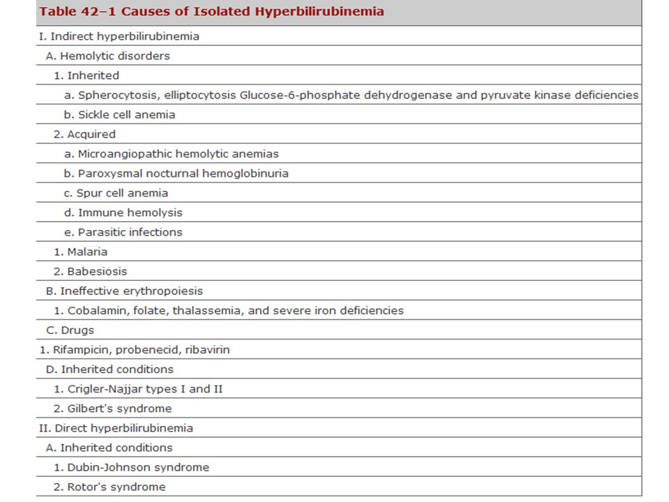
Elevation of Serum Bilirubin with Other Liver Test Abnormalities
The remainder of this chapter will focus on the evaluation of the patient with a conjugated hyperbilirubinemia in the setting of other liver test abnormalities. This group of patients can be divided into those with a primary hepatocellular process and those with intra- or extrahepatic cholestasis. This differentiation is made on the basis of the history and physical examination as well as the pattern of liver test abnormalities

120
History A complete medical history is perhaps the single most important part of the evaluation of the patient with unexplained jaundice. Important considerations ;the use of or exposure to any chemical or medication, either physician-prescribed, over-the-counter, complementary or alternative medicines such as herbal and vitamin preparations, or other drugs such as anabolic steroids& possible parenteral exposures, including transfusions, intravenous and intranasal drug use, tattoos, and sexual activity& recent travel history; exposure to people with jaundice; exposure to possibly contaminated foods; occupational exposure to hepatotoxins; alcohol consumption; the duration of jaundice; and the presence of any accompanying symptoms such as arthralgias, myalgias, rash, anorexia, weight loss, abdominal pain, fever, pruritus, and changes in the urine and stool. While none of these latter symptoms are specific for any one condition, they can suggest a particular diagnosis. A history of arthralgias and myalgias predating jaundice suggests hepatitis, either viral or drug-related. Jaundice associated with the sudden onset of severe right upper quadrant pain and shaking chills suggests choledocholithiasis and ascending cholangitis.

121
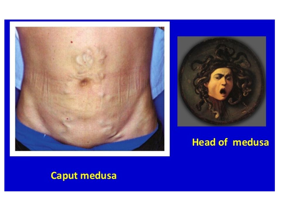
Physical Examination The general assessment include the patient's nutritional status. Temporal and proximal muscle wasting suggests long-standing diseases such as pancreatic cancer or cirrhosis. Stigmata of chronic liver disease, including spider nevi, palmar erythema, gynecomastia, caput medusae, Dupuytren's contractures, parotid gland enlargement, and testicular atrophy are commonly seen in advanced alcoholic cirrhosis and occasionally in other types of cirrhosis. An enlarged left supraclavicular node (Virchow's node) or periumbilical nodule (Sister Mary Joseph's nodule) suggests an abdominal malignancy. Jugular venous distention, a sign of right-sided heart failure, suggests hepatic congestion. Right pleural effusion, in the absence of clinically apparent ascites, may be seen in advanced cirrhosis. The abdominal examination should focus on the size and consistency of the liver, whether the spleen is palpable and hence enlarged, and whether there is ascites present. A grossly enlarged nodular liver or an obvious abdominal mass suggests malignancy. An enlarged tender liver could be viral or alcoholic hepatitis; an infiltrative process such as amyloid; or, less often, an acutely congested liver secondary to right-sided heart failure. Severe right upper quadrant tenderness with respiratory arrest on inspiration (Murphy's sign) suggests cholecystitis or, occasionally, ascending cholangitis. Ascites in the presence of jaundice suggests either cirrhosis or malignancy with peritoneal spread
 or periumbilical nodule (Sister Mary Joseph s nodule) suggests an abdominal malignancy. Jugular venous distention, a sign of right-sided heart failure, suggests hepatic congestion. Right pleural effusion, in the absence of clinically apparent ascites, may be seen in advanced cirrhosis. The abdominal examination should focus on the size and consistency of the liver, whether the spleen is palpable and hence enlarged, and whether there is ascites present. A grossly enlarged nodular liver or an obvious abdominal mass suggests malignancy. An enlarged tender liver could be viral or alcoholic hepatitis; an infiltrative process such as amyloid; or, less often, an acutely congested liver secondary to right-sided heart failure. Severe right upper quadrant tenderness with respiratory arrest on inspiration (Murphy s sign) suggests cholecystitis or, occasionally, ascending cholangitis. Ascites in the presence of jaundice suggests either cirrhosis or malignancy with peritoneal spread")
125
Abdominal Swelling Abdominal swelling is a manifestation of numerous diseases. Patients may complain of bloating or abdominal fullness and may note increasing abdominal girth on the basis of increased clothing or belt size. Abdominal discomfort is often reported, but pain is less frequent. When abdominal pain does accompany swelling, it is frequently the result of an intraabdominal infection, peritonitis, or pancreatitis. Patients with abdominal distention from ascites (fluid in the abdomen) may report the new onset of an inguinal or umbilical hernia. Dyspnea may result from pressure against the diaphragm and the inability to expand the lungs fully. The causes of abdominal swelling can be remembered conveniently by the six Fs: flatus, fat, fluid, fetus, feces, or a "fatal growth" (often a neoplasm).
 may report the new onset of an inguinal or umbilical hernia. Dyspnea may result from pressure against the diaphragm and the inability to expand the lungs fully. The causes of abdominal swelling can be remembered conveniently by the six Fs: flatus, fat, fluid, fetus, feces, or a fatal growth (often a neoplasm).")
126
Flatus Abdominal swelling may be the result of increased intestinal gas. The normal small intestine contains approximately 200 mL of gas made up of nitrogen, oxygen, carbon dioxide, hydrogen, and methane. Nitrogen and oxygen are consumed (swallowed), whereas carbon dioxide, hydrogen, and methane are produced intraluminally by bacterial fermentation. Increased intestinal gas can occur in a number of conditions. Aerophagia, the swallowing of air, can result in increased amounts of oxygen and nitrogen in the small intestine and lead to abdominal swelling. Aerophagia typically results from gulping food; chewing gum; smoking; or as a response to anxiety, which leads to repetitive belching. In some cases, increased intestinal gas is the result of bacterial metabolism of excess fermentable substances such as lactose and other oligosaccharides that can lead to production of hydrogen, carbon dioxide, or methane. In many cases, the precise cause of abdominal distention cannot be determined. increased lumbar lordosis accounts for apparent abdominal distention
, whereas carbon dioxide, hydrogen, and methane are produced intraluminally by bacterial fermentation. Increased intestinal gas can occur in a number of conditions. Aerophagia, the swallowing of air, can result in increased amounts of oxygen and nitrogen in the small intestine and lead to abdominal swelling. Aerophagia typically results from gulping food; chewing gum; smoking; or as a response to anxiety, which leads to repetitive belching. In some cases, increased intestinal gas is the result of bacterial metabolism of excess fermentable substances such as lactose and other oligosaccharides that can lead to production of hydrogen, carbon dioxide, or methane. In many cases, the precise cause of abdominal distention cannot be determined. increased lumbar lordosis accounts for apparent abdominal distention.")
127
Fat Weight gain with an increase in abdominal fat can result in an increase in abdominal girth and can be perceived as abdominal swelling. Abdominal fat may be the result of an imbalance between calorie intake and energy expenditure associated with a poor diet and sedentary lifestyle and also can be a manifestation of certain diseases such as Cushing's syndrome. Excess abdominal fat has been associated with an increased risk of insulin resistance and cardiovascular disease.

128
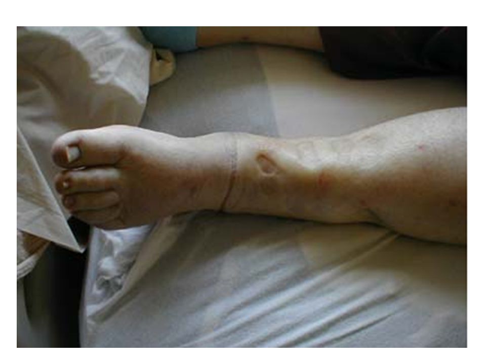
Fluid Fluid within the abdominal cavity, or ascites, often results in abdominal distention

130
Fetus Pregnancy results in increased abdominal girth. Typically, an increase in abdominal size is first noted at 12 to 14 weeks of gestation, when the uterus moves from the pelvis into the abdomen. Abdominal distention may be seen before this point as a result of fluid retention and relaxation of the abdominal muscles.

131
Feces Increased stool in the colon, in the setting of severe constipation or intestinal obstruction, also leads to increased abdominal girth. These conditions often are accompanied by abdominal pain, nausea, and vomiting and can be diagnosed by imaging studies

132
Fatal Growth An abdominal mass can result in abdominal swelling. Enlargement of the intraabdominal organs, specifically the liver (hepatomegaly) or spleen (splenomegaly) or an abdominal aortic aneurysm, can result in abdominal distention. Bladder distention also may result in abdominal swelling. In addition, malignancies, abscesses, or cysts can grow to sizes that lead to increased abdominal girth.
 or spleen (splenomegaly) or an abdominal aortic aneurysm, can result in abdominal distention. Bladder distention also may result in abdominal swelling. In addition, malignancies, abscesses, or cysts can grow to sizes that lead to increased abdominal girth.")
133
History and Physical Examination
Determining the etiology of abdominal swelling begins with history-taking and a physical examination. Patients questioned symptoms suggestive of malignancy, including weight loss, night sweats, and anorexia. Inability to pass stool or flatus together with nausea or vomiting suggest bowel obstruction, severe constipation, or an ileus (lack of peristalsis). Increased eructation and flatus may point toward aerophagia or increased intestinal production of gas. Patients should be questioned about risk factors for or symptoms of chronic liver disease, including excessive alcohol use and jaundice, which suggest ascites. Patients should also be asked about other symptoms of medical conditions, including heart failure and tuberculosis, which may cause ascites.
. Increased eructation and flatus may point toward aerophagia or increased intestinal production of gas. Patients should be questioned about risk factors for or symptoms of chronic liver disease, including excessive alcohol use and jaundice, which suggest ascites. Patients should also be asked about other symptoms of medical conditions, including heart failure and tuberculosis, which may cause ascites.")
134
Physical Examination Physical examination assess for signs of systemic disease. The presence of lymphadenopathy, especially supraclavicular lymphadenopathy (Virchow's node), suggests metastatic abdominal malignancy. Care also should be taken during the cardiac examination to evaluate for elevation of JVP; Kussmaul's sign (elevation of the JVP during inspiration); or a pericardial knock, which may be seen in heart failure or constrictive pericarditis, as well as a murmur of tricuspid regurgitation. Spider angiomas, palmar erythema, dilated superficial veins around the umbilicus (caput medusae), and gynecomastia suggest chronic liver disease.
, suggests metastatic abdominal malignancy. Care also should be taken during the cardiac examination to evaluate for elevation of JVP; Kussmaul s sign (elevation of the JVP during inspiration); or a pericardial knock, which may be seen in heart failure or constrictive pericarditis, as well as a murmur of tricuspid regurgitation. Spider angiomas, palmar erythema, dilated superficial veins around the umbilicus (caput medusae), and gynecomastia suggest chronic liver disease.")
135
Physical Examination The abdominal examination begin with inspection for the presence of uneven distention or an obvious mass. Auscultation should follow. The absence of bowel sounds or the presence of high-pitched localized bowel sounds point toward an ileus or intestinal obstruction. An umbilical venous hum may suggest the presence of portal hypertension, and a harsh bruit over the liver in hepatocellular carcinoma or alcoholic hepatitis. Abdominal swelling caused by intestinal gas can be differentiated from swelling caused by fluid or a solid mass by percussion; an abdomen filled with gas is tympanic, whereas an abdomen containing a mass or fluid is dull to percussion. The absence of abdominal dullness, however, does not exclude ascites, because a minimum of 1500 mL of ascites is required for detection on physical examination. Finally, the abdomen should be palpation to assess for tenderness, a mass, enlargement of the spleen or liver, or presence of a nodular liver suggesting cirrhosis or tumor. Light palpation of the liver may detect pulsations suggesting retrograde vascular flow from the heart in patients with right-sided heart failure, particularly tricuspid regurgitation.

136
Physical examination Inspection
Configurations of the abdomen in the level or above or below the chest apple-type : visceral obesity - cardiovascular risk pear-type : gluteal obesity Abdominal skin striae : white, pink hernias veins : caput Medusae visible peristalsis visible pulsations scars

137
Physical examination Abdominal distension
Localised Hepatomegaly Splenomegaly Polycystic kidney Gastric distension Inflammatory mass Tumor Obstructed bladder Hernia Generalised Obesity Pregnancy Ascites Bowel obstruction - ileus Big ovarian cyst Peritonitis

141
Physical examination Auscultation
Bowel sounds above the umbilicus or in the RUQ normal: 5-35/min, clicks and gurgles altered: absent: paralytic ileus hyperperistalsis: diarrhea, mechanical bowel obstruction Bruits arterial aortic, renal, iliac arteries Friction rubs spleen, liver, peritonitis Succussion splash normal: above the stomach pathologic: gastric or bowel obstruction

147
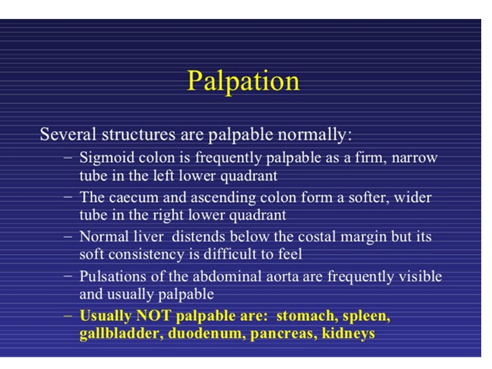
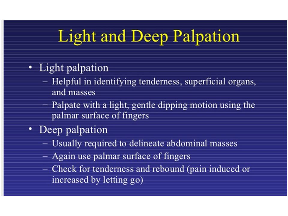
Physical examination Palpation
Characteristics of an abdominal mass 1. location 2. size 3. shape 4. consistency 5. surface 6. tenderness 7. movable or fixed 8. shifting by respiration

149
Physical examination Rectal digital examination
Perianal diseases fistulas, masses Anal alterations hemorrhoids, fisssuras, masses Rectal alterations polyp, neoplasm, ulcer Prostate gland Douglas’s space Stool on the glove

150
Causes Cirrhosis accounts for 84% of ascites. Cardiac ascites, peritoneal carcinomatosis, and "mixed" ascites resulting from cirrhosis and a second disease account for 10 to 15% of cases. Less common causes of ascites include massive hepatic metastasis, infection (tuberculosis, Chlamydia), pancreatitis, and renal disease (nephrotic syndrome). Rare causes of ascites include hypothyroidism and familial Mediterranean fever.
, pancreatitis, and renal disease (nephrotic syndrome). Rare causes of ascites include hypothyroidism and familial Mediterranean fever.")
151
Evaluation Once the presence of ascites has been confirmed, the etiology of the ascites is best determined by paracentesis. Paracentesis is a bedside procedure in which a needle or small catheter is passed transcutaneously to extract ascitic fluid from the peritoneum. The lower quadrants are the most frequent sites for paracentesis. Occasionally, an infraumbilical approach is used. The left lower quadrant is preferred because of the greater depth of ascites and thinner abdominal wall. Paracentesis is a safe procedure even in patients with coagulopathy; complications, including abdominal wall hematomas, hypotension, hepatorenal syndrome, and infection, are infrequent.

Similar presentations


>")
 The McGraw-Hill Companies, Inc. Permission required for reproduction or display. 23-1 Chapter 23 Abdominal and Gastrointestinal Disorders.>")


 and dys (with difficulty). It specifically.>")


