Download presentation
Presentation is loading. Please wait.

1
Fractures and dislocations around the elbow in adults
Prepared by Dr. Jamal Maqram

2
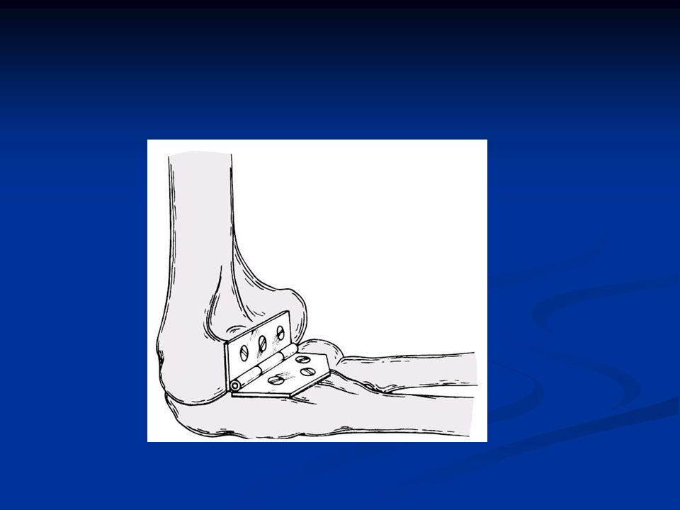
Anatomy The elbow joint is acomplex hinge occur b/n trochlea and capitulum of humerus and trochlear notch of ulna and radial head Movement: flex135 ext 0-5 . Appear in slight valgus {carrying angle}m 5deg/ f10-15. Stability : depend on shape of joint collateral ligaments, capsule& muscles around it .

5
Fractures of distal humerus
Mech.of injury: -high energy except in osteoporotic falling on flexed elbow > 90 degree. classification [ A O ] : divided into: - type A: extraarticular - typeB: intraarticular unicondylar frct .[one condyle sheared off and the still in contact with the shaft. - typeC: intraarticular bicondylar [no one in contact with the shaft] . has subgroups:- simpleTorY - extraarticular comminution - intraarticular comminution

6
Mechanism of injury of distal humerus#

7
classification [ A O ] of distal humerus#
![classification [ A O ] of distal humerus#](http://slideplayer.com/slide/4062431/13/images/7/classification+%5B+A+O+%5D+of+distal+humerus%23.jpg "classification [ A O ] of distal humerus#")
8
other classification of distal humerus#
Some classified them into : - Supracondylar # . - Intracondylar # . - transcondylar # - Chondyles[med.and lat.]# - Articular surface[capitulum and trochlea]# -Epicondyles #

9
Diagnosis of distal humerus#
C.P: pain , swelling etc….. Careful neurovascular assessment : (median & ulnar n. brachial a.) x-ray : APV & LAT.V gentle traction x-ray help in: - - accurate Dx -classification - pre-op. planning - C. T
 x-ray : APV & LAT.V. gentle traction x-ray help in: - - accurate Dx. -classification. - pre-op. planning. - C. T.")
10
X-ray APV Gentle traction x-ray

11
Treatment of distal humerus#
I. Conservative: -(rare) for undisplaced # p.o.p in 90 flexion for 6-8 w . -weekly x-ray II. Surgical is the treatment of choice. : because fracture usually unstable III.Alternative:
 for undisplaced #. -p.o.p in 90 flexion for 6-8 w . -weekly x-ray. II. Surgical is the treatment of choice. : because fracture usually unstable. III.Alternative:")
12
Treatment of distal humerus#
III.Alternative: indicated for: 1.severely commin.# . -2. severely soft tissue damage. -3.Patient bad condition. - 4.lack of expertise &facilities. - 5. severely osteoporotic(contraversed).
.")
13
III.Alternative treatment
Types : 1.bag of bones:arm held in a collar &cuff or p.o.p flexion>90. active motion encouraged if pat.well exercise continue after # healed. we get motion ranged :(45—90). 2.Olecnon traction. 3.Ilizarof ext. fix. (hinged type). 4.Total elbow arthroplasty. (eldery&less active pat)
. 2.Olecnon traction. 3.Ilizarof ext. fix. (hinged type). 4.Total elbow arthroplasty. (eldery&less active pat)")
14
Olecnon traction. Ilizarof ext. fix. (hinged )
")
15
Ilizarof ext. fix. (hinged ) Total elbow arthroplasty
 Total elbow arthroplasty")
16
Surgical treatment It include:
- pre operative planning: careful reading of x-ray C.T - prepare for the worst before op. - internal fixation: * it should be early(24-48h)except open #, accurate & rigid to give good stability& permit early motion
except open #, accurate & rigid to give good stability& permit early motion.")
17
Follow int.fixation O.R.I.F depend on the type of fracture:
1. Clsed #: .Uncomm#:screws,K.W(crossed or tension band). .Commin#:contoured plate(single or double). It is the best strong stability. .2. Open #: acc.to Gastilo: -GI&II O.R.I.F early. -GIII– dibridment .&delay O.R.I.F
. .Commin#:contoured plate(single or double). It is the best strong stability. .2. Open #: acc.to Gastilo: -GI&II O.R.I.F early. -GIII– dibridment .&delay O.R.I.F.")
18
Follow int.fixation Technique:
-position: prone, lateral. (help for bone graft) supine (in multitraumtic pat.) - incision: posterior 5cm distal olecranon up to10—12cm above. - isolate ulner n. - Approach -Campbell Transolecranon. -The medial triceps-elevating exposure for elbow arthroplasty
 supine (in multitraumtic pat.) - incision: posterior 5cm distal olecranon up to10—12cm above. - isolate ulner n. - Approach -Campbell -Transolecranon. -The medial triceps-elevating exposure for elbow arthroplasty.")
19
Prone position lateral position

20
Approaches of intercondylar #
I. Campbell app. advantages: 1- it is the only soft tissue approach to the elbow that expose all the articular surfaces of the joint, after the ulnar nerve has been isolated no large vessels or nerves lie in the area of the incision. II Transolecranon app. that provides an even better exposure of the articular surface but does not give exposure as far proximally as the Campbell app. - disadvantage: non union transolecranon #

21
Campbell App. Transolecranon APP

22
Follow int.fixation Steps of reduction of intercondylar # :-
Reduction &fix. Of condyles : Reduction &fix. Of epicondylar ridge : to the proximal fragment. (it form a buttress to which condyle later attached) Reduction &fix. Of reassembled condyles: to metaphysis with : screws, K.W or plates.
 3. Reduction &fix. Of reassembled condyles: to metaphysis with : screws, K.W or plates.")
24
Follow int.fixation Screws: if #line not extend far proximally.
K.W: if #line extend more proximally. Contoured plates(single or double) or Y shape: 1/3 tubular p. in the medial edge of med.pillar. Reconstructive p. in post. Aspect of lat.pillar. Good stability.
 or Y shape: 1/3 tubular p. in the medial edge of med.pillar. Reconstructive p. in post. Aspect of lat.pillar. Good stability.")
25
Tension band wire

26
Position of plates in distal humerus#

27
caution: not to encourage screw in olecranon or
coronoid fossae or penetrate trochlear surface. trochlea is spool in shape

28
After treatment Light post. Splint.
When wound healing is satisfactory7—10d Remove p.o.p periodically &gentle active exercise started. after 3 w p.o.p removed and the arm is supported by a sling with active motion as pain permit vigorous motion contraindicated

29
Transchondylar # often is grouped with suprachond#. but requires special considerations b/s usually extends to articular surface . Quite unstable unite slowly if treated conservatively . -so treated with percutaneous pins , lag screw (through small incision without opening the frac.), or canulated screw . if Was intraarticular and not fixed properly can be complicated by avascular necrosis . -Displaced # O.R.I.F.
, or canulated screw . if Was intraarticular and not fixed properly can be complicated by avascular necrosis . -Displaced # O.R.I.F.")
30
Undisplaced transcond# A vascular necrosis

31
Displaced transcondylar #

32
Side swipe fracture -occure in arm protruded from window of car and struck with other car . - fracture always open . Vary from GI ---- GIII - the most combination of this fracture consist of: * open distal 1/3 # of olecranon . * anterior dislocation of redial head & distal fragment of ulna . * comminuted distal humerous fracture . &other

33
Treated by : reduction of dislocation ,O.R. I. F . of olecranon # & ext. fix. To stabilize the all complex. Primary goal: care of open wound &restoration of elbow joint. Always complicated by infection, non union severe myositis ossificans arthroplasty

34
Complication of intercondylar # of distal humerus#
I. Early: neurovascular injury. II. Late : -Failure of fixation. -Non union & malunion. - Non union of olecranon osteotomy. -Infection. -Nerve palsy. -Hetrotopic ossification.

35
Failure of fixation. Nonunion Hetrotopic ossification

36
Fracture of capitulum - Mech. Of injuiry: F.O.S.H---- head of radius impacted to capitulum ----fracture classification: - type I: large fragment of bone and articular surface (involve trochlea) are fractured. -type II: small shell of bone and articular surface (not involve trochlia). - type III: comminuted #.
 are fractured. -type II: small shell of bone and articular surface (not involve trochlia). - type III: comminuted #.")
37
Classification of capitulum Fracture

38
Fracture of capitulum Treatment: ( through lat. Approach)
Diagnosis:- x-ray :lateral view (diagnostic). & A.P.V Deff. diag.: from # of radial head but the later rarely to displaced anteriorlly (so any # fragment ant.to lat. Condyle is capitulum fragment till prove otherwise.) C.T scan Treatment: ( through lat. Approach) Type I : O.R.I. F with small AO screw or Herbert's screw ( from post. to ant.) Type II&III: excision . -After treatment: like intercondylar # .
. & A.P.V. Deff. diag.: from # of radial head but the later rarely to displaced anteriorlly (so any # fragment ant.to lat. Condyle is capitulum fragment till prove otherwise.) C.T scan. Treatment: ( through lat. Approach) Type I : O.R.I. F with small AO screw or Herbert s screw ( from post. to ant.) Type II&III: excision . -After treatment: like intercondylar # .")
39
Fracture of capitulum Lat.V APV

40
Treatment of capitulum Fracture
Screw countersinked posteriorly. Not damage articular surface anteriorly.

41
Epicondylar fractures
Med.& Lat. Epic. # are rare in adult. Mech. Of injury: direct blow. Treatment: -lat. Epic Usually conservative :p.o.p for 3w. followed by supportive motion. -Med Epic. - Undisplaced: p.o.p. - displaced>1cm:O.R.I.F. -if med.epi. displaced to joint in: (rare in adult). 1.close Red:vulgus of elbow, arm supination&ext. of wrest. 2.open Red.
. 1.close Red:vulgus of elbow, arm supination&ext. of wrest. 2.open Red.")
42
Olecranon fractures Mech. Of inj. : direct: blow on elbow.
indirect: falling on partially flexed elbow with indirect force generated by triceps ___ avulsion. classification: type I :# of proximal 1/3. type II:# of middle 1/3 . type III :#of distal 1/3.it may be associated with ant. displacement of radius.

43
Classification of Olecranon fractures
I II III

44
Follow olecranon fracture
Other classification: [Colton] according to: displacement and the anatomy of the fracture, thus give guidance as to the appropriate type of fixation : I.Nondisplaced and stable II.Displaced fractures - Avulsion fractures - oblique fractures . - Transverse fractures - Isolated comminuted fractures - Fracture/dislocations

45
classification of olecranon fracture [Colton]
![classification of olecranon fracture [Colton]](http://slideplayer.com/slide/4062431/13/images/45/classification+of+olecranon+fracture+%5BColton%5D.jpg "classification of olecranon fracture [Colton]")
46
Treatment of olecranon fracture
I.Nondisplaced and Stable: 1.if the fractures displacement <2 mm exhibit no change in position with gentle flexion to 90 degrees or with extension against gravity treated by: p.o.p in 90 degrees of flexion for 3 to 4 w -followed by protected range of motion avoiding flexion past 90 degrees until bone healing is complete radiographically usually around 6 to 8 weeks In the elderly patient , motion may be initiated earlier than 3 weeks if the patient can tolerate it. -Control x-Ray after 5-7d. -P.o.p in full extension avoided b/s lead to stiffness

47
Nondisplaced and Stable

48
Treatment of olecranon fracture
II.Displaced Fractures: O.R.I.F is the treatment of choice. The goals of treatment are: 1.Maintain power of elbow extension. 2.Restore congruity of the articular surface. 3.Restore stability of the elbow. 4.Prevent stiffness of the joint. 5.Allow the patient to do early motion

49
Follow olecranon fracture
1.Avulsion #: -tension band wire. (T.BW) - if fragment small--- excision . 2.Transverse #: a. Without comminution: tension band wire is suitable - if fragment is big----- cancellous screw 6.5mm -if fragment is small --- K.W. b. with comminution: contoured plate with or without bone graft (T.B.W cause compression at # site & narrowing of trochlear notch.)
 - if fragment small--- excision . 2.Transverse #: a. Without comminution: tension band wire is suitable. - if fragment is big----- cancellous screw 6.5mm. -if fragment is small --- K.W. b. with comminution: contoured plate with or without bone graft (T.B.W cause compression at # site & narrowing of trochlear notch.)")
50
Avulsion #: small fragment

51
Transverse # without comminution

52
Follow olecranon fracture
3.Oblique #: a. without comminu .:(T.BW may displace#) 1. plate :reconstructiv(thick),1/3tubular(fatigue)or contoured limited contact dynamic compression LCDC it permit greater angulation of screws &has low profile. 2. some indicate T.B.W with Interfragmentery screw. b. with comminu.: plate with bone graft.
 1. plate :reconstructiv(thick),1/3tubular(fatigue)or contoured limited contact dynamic compression LCDC. it permit greater angulation of screws &has low profile. 2. some indicate T.B.W with Interfragmentery screw. b. with comminu.: plate with bone graft.")
53
Oblique # without comminu. Oblique # with T. B
Oblique # without comminu Oblique # with T.B.W with Interfragmentery screw comminution

54
contoured limited contact dynamic compression plate LCDC

55
-If no association with previous excision.
4.Isolated comminu# results from direct trauma. There are multiple fracture planes, &crushing of many fragments may be associated with fractures of the distal end of the humerus, the radial & ulnar shafts, and the radial head. -If no association with previous excision. & not in distal 1/3 of olecranon -if association occur (excision unsuitable) combination of plate & tension band wire.
--- combination of plate & tension band wire.")
56
Excision of proximal fragment:
-used only if there is proximal# & the remnant distal part form stable base for trochlea. Advantages: The possibility of non union is eliminated. The possibility of traumatic artheritis is menimiz due to irregular articular surface. Indication: severely comminuted fractures in which open reduction and internal fixation are not Possible. -non articular # Non union after failed O.R.I.F . -when reduction is delayed 10—14d. -in type III open# or if local soft tissue damaged . Contraindication: in distal 1/3 olecranon# joint instbility

57
Technique excision of proximal fragment

58
After excision of proximal fragment
- p.o.p in flexion 70 deg. For 3w. - gentle motion when wound heal permit 7—10d. -avoid forceful movement (ext. or flex.) for 3 month.
 for 3 month.")
59
Note : up to 80% olecranon can be excised safely.
If mid portion of olecranon is very comminuted while the proximal 1/3 intact ,excision of comminuted area as wedge& reconstitute a large olecranon notch then fixed with plate or tension band wire.

60
Follow olecranon fracture
5.Fracture-Dislocation Fracture-dislocations present a challenging problem because of the combination of severe bone and soft tissue damage . ORIF with restoration of alignment and stability of the ulna is the goal .This can be achieved by intramedullary wires or a long screw to ulnar canal. . Often plate is required in spite of such soft tissue damage Primary excision of the olecranon# must be carefully considered joint instability

61
Treatment of olecranon fracture

62
After treatment of olecranon fracture
P.o.p at 90degree for 3—4w. When wound heal permit, (7-10) gentle exercise. Periodic removing of p.o.p. Maximal function not return before 6—12m.
 gentle exercise. Periodic removing of p.o.p. Maximal function not return before 6—12m.")
63
Complication of olecranon fracture
the most common complication are: -nonunion Limitation of motion (esp. extension) Subcutaneous pain due to fixation devices.
. -Subcutaneous pain due to fixation devices.")
64
Coronoid fracture It indicate severe trauma to elbow.
Mech. of inj Struck of trochlea in coronoid. -avulsion (less common). Classification: type I: simple avulsion of tip. type II: involve <50%. type III :involve >50%. Treatment: typeI&II: heavy suture to the proximal of ulna. typeIII :I.F with screw.
. Classification: type I: simple avulsion of tip. type II: involve <50%. type III :involve >50%. Treatment: typeI&II: heavy suture to the proximal of ulna. typeIII :I.F with screw.")
65
Coronoid fracture Classification Treatment

66
Fracture of radial head
It is common in adult. Mech.of inj. :F.O.S.H while arm pronated, head impacted in capitulum. Classification: of radial head: Mason type I: undisplaced. type II: displaced. typeIII: Comminuted. type IV:# associated with post. Elbow dislocation & coroniod #.

67
Classification: of radial head: Mason

68
Fracture of radial head
Treatment: I. conservative: for : type I. -type II : - if # in<1/3 of head circum in outer part. - or get 70% of pronation & supination.

69
Fracture of radial head
2. surgical [Excision of radial head] is the treatment of choice Indication: a. typeIII# b. If head become oval in shape. c. if>1/3 of head circumflex involved d. .fracture lie in the inner side. e. those with loss fragments in the joint. f. neck# with enough angulation that interfere with rotation.

70
Excision of radial head
Technique: -excision should be early 24—48h. -incision:5cm below radial head up to lat. condyle. -pass b/n E.C.U&E.D or E.C.U&anconeus. -excision: transverse just proximal biceptal tuber. -anular lig should be excised. & debris removed After treatment: p.o.p in90 deg.for 1w then converted to sling till 3w. Within this interval start gentle active motion.

71
Site of excision of radial head

72
Note: if# segment is large, isolated& uncomminuted fixed with : mini O
Note: if# segment is large, isolated& uncomminuted fixed with : mini O.A, Herbert or Accutrac screw.

73
# of radial head & neck associated with elbow dislocation & coronoid #(type IV):
1 - If coronoid#undisp excision early. 2- If coronoid#disp. but not commin----- O.R.I.F of coronoid #&excision of head at the same time. 3 - If coronoid# was commin. &difficult to fix it wait 3-6m till# healed then excision In this time some indicate radial prosthesis to maintain joint stability.

74
Fracture of radial neck
Radial neck classified same as# of head. treatment: -conservative: undisp. or minimally displaced. -surgical: excision of head for severely displaced. if joint unstable small T plate. or small cortical screw in oblique # .

75
small T plate. small cortical screw in oblique #

76
if # irreducible--- radial head arthroplsty.
Radial head & neck # with dislocation of distal radio ulnar j. (Essex- Lopresti fracture dislocation). Mech. Of inj.:F.O.S.H cause disruption of distal radio ulner j &tearing of interosseus memb.---radial migration Diagnosis: pain at the wrest associated with displaced radial head or neck #. it should be early .once migration has occurred ,late reconstructive is unsatisfactory. Treatment :O.R.I.F of proximal radial #+pinning of distal R.U.J In supination. pin removed after 3—6 w. if # irreducible--- radial head arthroplsty.
. Mech. Of inj.:F.O.S.H cause disruption of distal radio ulner j &tearing of interosseus memb.---radial migration. Diagnosis: pain at the wrest associated with displaced radial head or neck #. it should be early .once migration has occurred ,late reconstructive is unsatisfactory. Treatment :O.R.I.F of proximal radial #+pinning of distal R.U.J. In supination. pin removed after 3—6 w. if # irreducible--- radial head arthroplsty.")
77
Essex- Lopresti fracture dislocation

78
Dislocation of elbow joint
Form 20% of joint dislocation (after shoulder& finger) classification: posterior [most common 80%] -ant. - med. - lat. - divergent. [rare]. posterior or post.lat. dislocation : mech of inj. :FOSH while elbow extended. Diagnosis: -C.P it may ass. with neurovascular inj (median & ulnar n. &brachial a.) - X—Ray.
 classification: posterior [most common 80%] -ant. - med. - lat. - divergent. [rare]. posterior or post.lat. dislocation : mech of inj. :FOSH while elbow extended. Diagnosis: -C.P it may ass. with neurovascular inj. (median & ulnar n. &brachial a.) - X—Ray.")
79
classification elbow Dislocation

80
posterior elbow dislocation

81
Treatment of posterior dislocation:
I. Un complicated: - close reduction: traction &counter traction of slightly flexed elbow, correction of lateral displacement.& olecranon Pressure. - traction with hyperext. to unlock olecranon. from distal humerus. dangers is entrapment of median n. & trauma to brachialis. - reduction in prone position if no assistant .

82
Reduction Reduction in supine position in prone position

83
Lock of olecranon Entrapment of in distal humerus median n

84
Treatment of posterior dislocation:
II. Complicated: associated with : 1 -coronoid# radial head #. 3 -olecranon # medial epicondylar# Dislocation with coronoid #:treated as before. 2. Dislocation with radial head #: -a. we try to preserve radial head especially if associated with coronoid# or medial lig.- by O.R. I.F. -b. if# irreducible:-*stitch of med. lig.& pronater mass& p.o.p for 3-4w then excision. *- or early excision &immobilization for 3-4w but if the joint unstable --- temporal arthroplasty .

85
Dislocation of elbow +radial head# + displaced coronoid# treated improperly with early excision of radial head &no coronoid I.F Dislocation of elbow radial head# displaced coronoid# improper treatment after 5days

86
Complication of Dislocation of elbow joint
1. Stiffness& post traumatic arthritis . 2. Neurovascular injury. 3. Hetrotopic calcification .(severe inj. long immobilization, aggressive passive motion) . treatment:- NSAID& Radiotherapy but ineffective. -Resection of calcification but delayed till month. -Early resection is contra indicated. -passive motion also avoided.
 . treatment:- NSAID& Radiotherapy but ineffective. -Resection of calcification but delayed till 12 month. -Early resection is contra indicated. -passive motion also avoided.")
87
4. Recurrent instability due to: a. weak collat. Lig. b
4.Recurrent instability due to: a .weak collat. Lig b. residual articcular defect in trochlea or trochlear notch c. ununited coronoid# d. unhealed ant. Capsule.

88
Treatment: b/s the cause not clear, so number of surgical procedures was tried :
1. Block of tibial bone put on coronoid. 2.Transfere of biceps tendon to coronoid. 3.Creation of cruciating lig. from triceps &bifceps . 4. Collateral lig. Reconstruction.

89
THE END Dr. Jamal Maqram

90
MoKazem.com هذه المحاضرة هي من سلسلة محاضرات تم إعدادها و تقديمها من قبل الأطباء المقيمين في شعبة الجراحة العظمية في مشفى دمشق, تحت إشراف د. بشار ميرعلي. الموقع غير مسؤول عن الأخطاء الواردة في هذه المحاضرة. This lecture is one of a series of lectures were prepared and presented by residents in the department of orthopedics in Damascus hospital, under the supervision of Dr. Bashar Mirali. This site is not responsible of any mistake may exist in this lecture. Dr. Muayad Kadhim د. مؤيد كاظم

Similar presentations


, F.R.C.S.(C )>")


 Lecture 3 Myology of the Elbow.>")


