Download presentation
Presentation is loading. Please wait.

1
INTEGUMENTARY SYSTEM PN 124 BACTERIAL AND FUNGAL INFECTIONS

2
Objectives Discuss s/s of 8 infectious disorders of the skin; bacterial and fungal Define the nursing management of the client with infectious disorders of the skin Discuss common diagnostic tests used as diagnostic tools for integumentary disorders

3
CELLULITIS, Bacterial infection
Etiology/Pathophysiology -infection is potentially serious. -not contagious -can be spread by direct contact with an open area from a person that has an infection. -causes in adults: group A streptococci and Staphylococcus aureus.

4
CELLULITIS -Hemophilus influenzae type B is more common in children.
-increase the risk for cellulitis: -venous insufficiency or stasis -diabetes mellitus -lymph edema -surgery -malnutrition -substance abuse -treatment with steroids or cancer chemotherapy

5
RISKS FOR CELLULITIS - presence of another infection
- compromised immune function due to human immunodeficiency virus (HIV) - autoimmune diseases, such as lupus erythematosus
 - autoimmune diseases, such as lupus. erythematosus.")
6
CELLULITIS -Develops as an edematous, erythematous area of skin
-hot and tender -bacteria enters through a break in the skin -can be from a cut, scratch, insect bite, etc -common areas are the lower extremities. -usually is a superficial infection -may spread and become life-threatening

8
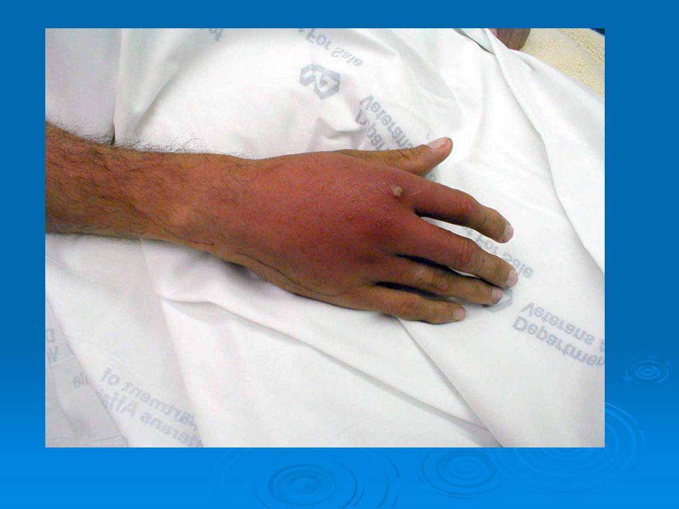
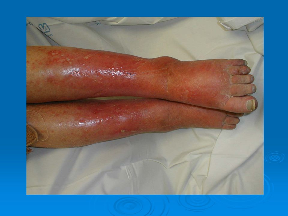
CELLULITIS

9
CELLULITIS CLINICAL MANIFESTATIONS:
-affected areas of the skin/underlying subcutaneous tissues -erythematous, tender, warm, edematous. -fever

11
CELLULITIS -s/s are caused by the bacteria, and the
body’s attempts to stop the infection. -skin appears pitted, like an orange peel. -area of redness spreads and small red spots appear -vesicles may form and burst

12
CELLULITIS -nearby lymph nodes may become enlarged and tender. (lymphadenitis) -edema secondary to the infected area occludes the lymphatic vessels in the skin. -most patients only feel mildly ill, - but some have fever, chills, headache, tachycardia, confusion, hypotension.

13
ERYSIPELAS A specific acute, inflammatory disease
-caused by a beta- hemolytic streptococci -characterized by hot, red, edematous and sharply defined eruptions

15
ASSESSMENT SUBJECTIVE: - fatigue - tenderness - pain
- limited movement of the involved extremity , - feeling of general malaise.

16
ASSESSMENT OBJECTIVE: -Inspection of the skin - erythema - edema
- areas that are warm to the touch. -Vesicles may be present. -Elevated temperature. -Tachycardia -Leukocytosis.

17
DIAGNOSTIC TESTS Cultures - identifies the causative bacteria
-from the blood, purulent exudate, or tissue specimens -Gram stain -determines the appropriate antibiotic therapy. Complete blood count (CBC). Inspection of the area
. Inspection of the area.")
18
DIAGNOSTIC TESTS -Tests done to differentiate cellulitis from
deep vein thrombosis. - ( they both have similar s/s) - X-ray, ultrasound, computed tomography or magnetic resonance imaging (MRI) - determines the extent of inflammation -identifies abscess formations
 - X-ray, ultrasound, computed tomography. or magnetic resonance imaging (MRI) - determines the extent of inflammation. -identifies abscess formations.")
19
MEDICAL MANAGEMENT Antibiotic treatment
- effect against streptococci and staphylococci - 10 day course -can be either oral or IV depending on severity

20
Nursing Diagnosis Deficient knowledge, related to the cause and the spread of the disease. Pain related to edema

21
NURSING INTERVENTIONS
-Treat s/s and to prevent the spread of the infection. -Administer the antibiotic -Assess pain; administer an analgesic if necessary -Warm, moist dressings applied to the affected area may relieve discomfort. -Monitor fluid intake and nutritional status.

22
NURSING INTERVENTIONS
-Keep the affected part immobile - helps reduce the edema -Stress the importance of taking the entire prescription of antibiotics. -Monitor for secondary diseases, such as yeast infections

23
PROGNOSIS Cure is possible with 7-10 days of treatment.
Cellulitis may be more severe in people with chronic diseases and those who are susceptible to infection, such as the immunocompromised. Complications: sepsis, meningitis, and lymphangitis.

24
Bacterial Disorders of the Skin
Impetigo contagiosa Etiology/pathophysiology Staphylococcus aureus or streptococci, or a mixed bacterial invasion of the skin. Common in children.

25
IMPETIGO Clinical manifestations/assessment Lesions begin as macules
- develop into pustule vesicles. Pustules rupture -form honey-colored exudate. -under the exudate is smooth, red skin. Affects exposed areas -face, hands, arms, and legs. Highly contagious— -direct or indirect contact Low-grade fever; leukocytosis t

26
Nursing assessment SUBJECTIVE DATA: -Ask about pruritis.
-Ask about pain and malaise. -Ask about the spreading of the disease to different body parts -Ask about other diseases present.

27
IMPETIGO OBJECTIVE DATA: -Focal erythema. -Pruritic areas.
-Honey-colored crust over dried lesions. -Smooth, red skin under the crust. -Low-grade fever. -Leukocytosis. -Positive culture for streptococcus or staphylococcus aureus. -Purulent exudate.

28
Diagnostic Tests Medical management -Culture of exudate from lesions
-Antiseptic soap (Betadine of Hibiclens) to remove crusted exudate and clean area -Topical cream, ointment or lotion -Antibiotics, oral or IV (Penicillin) -Keep area clean and dry
 to remove crusted exudate and clean. area. -Topical cream, ointment or lotion. -Antibiotics, oral or IV (Penicillin) -Keep area clean and dry.")
29
Folliculitis, furuncles, carbuncles, and felons
Etiology/pathophysiology Folliculitis Infected hair follicle (generally from Staphylococcus aureus). Furuncle (boil) Infection deep in hair follicle; involves surrounding tissue. Carbuncle Cluster of furuncles. Felons Infected soft tissue under and around an area.
. Furuncle (boil) Infection deep in hair follicle; involves surrounding tissue. Carbuncle. Cluster of furuncles. Felons. Infected soft tissue under and around an area.")
30
Folliculitis, furuncles, carbuncles, and felons
Clinical manifestations/assessment -Pustule -Edema -Erythema -Pain -Pruritus -Shiny, point up Carbuncle-the center will turn yellow.

31
Folliculitis Furuncles

32
Carbuncle Felon

33
ASSESSMENT SUBJECTIVE: -patient’s symptoms.
-family history of diabetes mellitus. -wearing of improperly fitting clothes.

34
ASSESSMENT OBJECTIVE: -erythema an -edema of the involved area.
-often overweight -may use poor body hygiene practices.

35
NURSING DIAGNOSES Impaired skin integrity, related to exudate from wound Pain, related to edema

36
DIAGNOSTIC TESTS Diagnostic tests Physical exam Culture of drainage
Health history

37
MEDICAL MANAGEMENT -Goal - prevent the spread of the infection.
-Patients in the hospital are isolated - using wound and secretion precautions.

38
Folliculitis, furuncles carbuncles and felons
Medical management/nursing interventions -Warm soaks 2-3 times per day -promote suppuration -Once the lesion ruptures, -hot soaks are discontinued -prevents damage to the surrounding skin and the spread of infection.

39
-medical asepsis. -topical antibiotic cream or ointment -surgical incision and drainage -immobilize affected area to prevent pain -elevate affected area to decrease the edema.

40
PATIENT TEACHING -Patient should not touch the exudate.
-Meticulous hand washing -BEFORE and AFTER contact with the lesions. -Hygiene practices should be demonstrated and return demonstrations done by the family and the patient.

41
-Whole family needs individual toilet
items and bath linens -bacteriostatic soap and shampoo. -Demonstrate proper disposal of contaminated articles.

42
YEAST INFECTIONS

43
FUNGAL INFECTIONS OF THE SKIN

44
FUNGAL INFECTIONS -Dermatophytoses
-Superficial infections of the skin. -Common types are: -tinea capitis -tinea corporis -tinea cruris -tinea pedis

45
TINEA CAPITIS

46
TINEA CORPORIS -Ringworm of the body.
-Body parts that have little or no hair.

47
TINEA CORPORIS

48
TINEA CRURIS -Jock itch. -Found in the groin area.

49
TINEA PEDIS -Most common of all fungal infections. -Athlete’s foot.
-Between the toes of people whose feet perspire heavily. -Contaminated swimming pools and public bathroom facilities

50
SIGNS AND SYMPTOMS TINEA CAPITIS: -erythematous.
-round lesion with pustules around the edges -temporary alopecia -infected hairs will turn blue-green under a Wood’s light.

51
SIGNS AND SYMPTOMS -TINEA CORPORIS:
-flat lesions that are clear in the center with erythematous borders. -scaliness -pruritis is severe.

52
SIGNS AND SYMPTOMS 3. TINEA CRURIS:
Has brownish-red lesions that migrate out from the groin area. Pruritis is a symptom. Scratching is done to relieve the itching. As a result, skin excoriation is present.

53
SIGNS AND SYMPTOMS TINEA PEDIS:
This fungal infection produces more skin maceration than the others. Fissures and vesicles are commonly seen around and below the toes, with occasional discoloration of the infected area.

54
ASSESSMENT SUBJECTIVE DATA: -extreme itching
-tenderness from excoriation of the area

55
ASSESSMENT OBJECTIVE DATA: -TINEA CAPITIS: -inspection
-round, scaled lesion -purulent vesicles around the edges of the scalp. -erythema alopecia to the surrounding area

56
ASSESSMENT OBJECTIVE DATA: -TINEA CORPORIS:
-flat lesions with clear centers and red borders on non-hairy body parts.

57
ASSESSMENT TINEA CRURIS:
-groin-brown to red lesions that spread outward. -skin excoriation from scratching. TINEA PEDIS: -fissures between the toes and soft skin. -vesicular lesions -thick toenails.

58
DIAGNOSTIC TESTS -visual inspection.
-Wood’s lamp-diagnose tinea capitis. -thorough health history

59
MEDICAL MANAGEMENT -topical or oral antifungal agents. -Griseofulvin (oral) -topical drugs do not penetrate the hair bulb -antifungal soaps and shampoos

60
Antifungal agents-Tinactin, Lotrimin, or Desenex
-2-6 weeks. See AHN p. 77. for a list of drugs that are used.

61
NURSING DIAGNOSES Impaired skin integrity, related to increased moisture and pruritis

62
NURSING INTERVENTIONS
-Protect the involved area from trauma and irritation -keep the area clean and dry. -Apply medications -warm compresses -Tinea pedis -warm soaks (usually Burrow’s solution) -topical antifungal agents.
 -topical antifungal agents.")
63
NURSING INTERVENTIONS
-Clean and dry the feet thoroughly -completely dry the toes -Wear sandal-like shoes/go barefoot -prevents moisture in the toes -Footwear needs to be of an absorbent material -socks, stockings, etc. -Wear loose-fitting clothing.

64
TEACHING 1. Teach proper skin care and comfort measures to relieve itching. 2. The nurse needs to review the meds. and procedures to be done at home by the pt. 3. The nurse should remind the pt. that it may take months for fungal disorders to be cured. 4. Clarify any misconceptions about athlete’s foot. 5. Teach the pt. the process of this disease.

65
FOOT CARE

66
FOOT CARE

67
PARASITIC DISEASES OF THE SKIN
Pediculosis Etiology/pathophysiology -Lice infestation

68
Pediculosis Three types of lice -Head lice (capitis)
-Attaches to the hair shaft and lays eggs -Body lice (corpis) -Found around the neck, waist, and thighs -Found in seams of clothing -Pubic lice (crabs) -Looks like a crab with pinchers -Found in pubic areas
 -Found around the neck, waist, and thighs. -Found in seams of clothing. -Pubic lice (crabs) -Looks like a crab with pinchers. -Found in pubic areas.")
69
Pediculosis Clinical manifestations/assessment Diagnostic tests
-Nits and/or lice on involved area -Pinpoint raised, red macules -Pinpoint hemorrhages -Severe pruritis -Excoriation Diagnostic tests -Physical exam

70
Pediculosis Medical management/nursing intervention
-Lindane (Kwell); Pyrethrins (RID) -Cool compresses -Corticosteroid ointment -Assess all contacts -Wash linens and clothes in hot water -Properly clean furniture or non-washable materials
; Pyrethrins (RID) -Cool compresses. -Corticosteroid ointment. -Assess all contacts. -Wash linens and clothes in hot water. -Properly clean furniture or non-washable. materials.")
71
Scabies

72
Scabies Etiology/Pathophysiology -Sarcoptes scabiei (itch mite)
-Mites lay eggs under the skin -Transmitted by prolonged contact with infected area

73
Scabies Clinical manifestations/assessment
-Wavy, brown, threadlike lines on the body -Pruritis -Excoriation

74
Scabies Diagnostic tests -Microscopic examination of infected skin
-Scratch test Medical management/nursing interventions -Lindane (Kwell), Pyrethrins (RID), Crotamiton (Eurax), 4-8% solution of sulfur in petrolatum -Treat all family members -Wash linens and clothing in hot water
, Pyrethrins (RID), Crotamiton. (Eurax), 4-8% solution of sulfur in petrolatum. -Treat all family members. -Wash linens and clothing in hot water.")
Similar presentations