Download presentation
Presentation is loading. Please wait.

1
به نام ایزد دانا

2
Gastrointestinal Bleeding

3
Epidemiology Gastrointestinal (GI) bleeding is a relatively common problem encountered in emergency medicine that requires early consultation and often admission The overall mortality of GI bleeding is approximately 10% Diagnostic modalities have improved much more than therapeutic techniques
 bleeding is a relatively common problem encountered in emergency medicine that requires early consultation and often admission. The overall mortality of GI bleeding is approximately 10% Diagnostic modalities have improved much more than therapeutic techniques.")
4
GI bleeding is often easy to identify when there is clear evidence of vomiting blood or passing blood in the stool, but it may present subtly with signs and symptoms of hypovolemia, such as dizziness, weakness, or syncope

5
The approach to GI bleeding depends on whether the hemorrhage is located in the proximal or distal segments of the GI tract (i.e., upper or lower GI bleeding) These segments are defined by the ligament of Treitz in the fourth section of the duodenum

6
In the United States, upper GI bleeding (UGIB) affects 50 to 150 people per 100,000 population each year and results in 250,000 admissions at an estimated annual cost of almost $1 billion Lower GI bleeding (LGIB) affects a smaller portion of patients and results in proportionally fewer hospital admissions than UGIB
 affects a smaller portion of patients and results in proportionally fewer hospital admissions than UGIB.")
7
GI bleeding can occur in individuals of any age, but most commonly affects people in their 40s through 70s (mean age 59 years) Most deaths caused by GI bleeding occur in patients older than age 60 years UGIB is more common in men than women (2:1), whereas LGIB is more common in women
, whereas LGIB is more common in women.")
8
Significant UGIB requiring admission is more common in adults, whereas LGIB requiring admission is more common in children

9
Differential Considerations

10
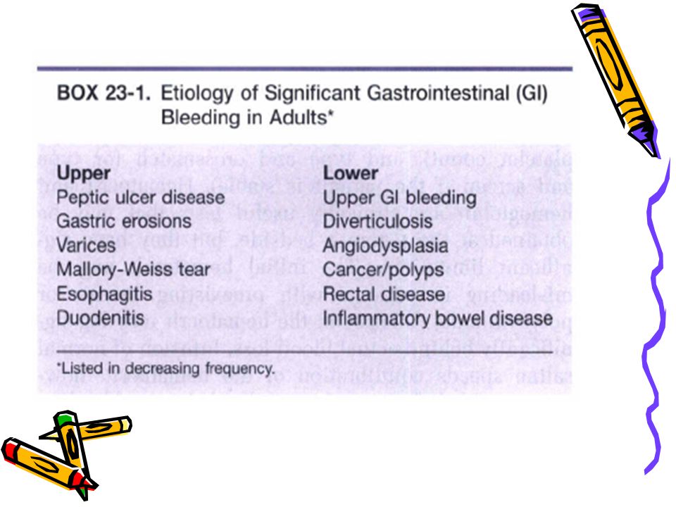
Peptic ulcer disease, gastric erosions, and varices account for approximately three fourths of adult patients with UGIB Diverticulosis and angiodysplasia account for approximately 80% of adults with LGIB

11
Esophagitis, gastritis, and peptic ulcer disease are the most common causes of UGIB in children, and infectious colitis and inflammatory bowel disease are the most common causes of LGIB in children In children younger than age 2 years, massive LGIB is most often a result of Meckel's diverticulum or intussusception

12
At all ages, anorectal abnormalities are the most common cause of minor LGIB
Despite improved diagnostic techniques, no source of bleeding is identified in approximately 10% of patients with GI bleeding

13
Patients who have abdominal aortic grafts who present to the emergency department with GI bleeding should receive prompt surgical consultation in the emergency department for the possibility of aortoenteric fistula

16
Rapid Assessment and Stabilization

17
Most patients with GI bleeding are easy to diagnose because they present to the emergency department complaining of vomiting blood or passing black or bloody stool The diagnosis is confirmed quickly by examination of the stool for the presence of blood

18
Patients with suspected GI bleeding who are hemodynamically unstable should undergo rapid evaluation and resuscitation They should be undressed quickly, placed on cardiac and oxygen saturation monitors, and given supplemental oxygen as needed

19
At least two large-bore peripheral intravenous lines should be placed (minimum 18-gauge); blood should be drawn for hemoglobin or hematocrit, platelet count, prothrombin time (PT), and type and screen or type and crossmatch; and crystalloid resuscitation should be initiated
; blood should be drawn for hemoglobin or hematocrit, platelet count, prothrombin time (PT), and type and screen or type and crossmatch; and crystalloid resuscitation should be initiated")
20
Intravenous crystalloid fluid should be given as a 2-L bolus in adults or 20 mL/kg in children until the patient's vital signs have stabilized or the patient has received 40 mL/kg of crystalloid Patients who remain unstable after 40 mL/kg of crystalloid should be given type O, type-specific, or crossmatched blood depending on availability

21
Persistently unstable patients should receive immediate consultation
patients with UGIB with a gastroenterologist and surgeon patients with LGIB with a surgeon

22
Pivotal Findings Keys to diagnosing GI bleeding : History
physical examination testing stool for blood measuring hemoglobin or hematocrit

23
History

24
Patients usually complain of vomiting red blood or coffee ground–like material or passing black or bloody stool Hematemesis (vomiting blood) occurs with bleeding of the esophagus, stomach, or proximal small bowel. Approximately 50% of patients with UGIB present with this complaint
 occurs with bleeding of the esophagus, stomach, or proximal small bowel. Approximately 50% of patients with UGIB present with this complaint.")
25
Hematemesis may be bright red or darker (i. e
Hematemesis may be bright red or darker (i.e., coffee ground–like) as a result of conversion of hemoglobin to hematin or other pigments by hydrochloric acid in the stomach The color of vomited or aspirated blood from the stomach cannot be used to determine if the bleeding is arterial or venous in nature
 as a result of conversion of hemoglobin to hematin or other pigments by hydrochloric acid in the stomach. The color of vomited or aspirated blood from the stomach cannot be used to determine if the bleeding is arterial or venous in nature.")
26
Melena, or black tarry stool, occurs from approximately 150 to 200 mL of blood in the GI tract for a prolonged period. Melena is present in approximately 70% of patients with UGIB and a third of patients with LGIB Black stool that is not tarlike may result from 60 mL of blood from the upper GI tract

27
Blood from the duodenum or jejunum must remain in the GI tract for approximately 8 hours before turning black Occasionally, black stool may follow bleeding into the lower portion of the small bowel and ascending colon Stool may remain black and tarry for several days, even though bleeding has stopped

28
Black stool also may be seen after ingestion of bismuth (e. g
Black stool also may be seen after ingestion of bismuth (e.g., Pepto-Bismol), which can confuse the situation because it is often taken for UGI distress. In contrast to melena, stool rendered black by bismuth is not positive on Hemoccult testing
, which can confuse the situation because it is often taken for UGI distress. In contrast to melena, stool rendered black by bismuth is not positive on Hemoccult testing.")
29
Hematochezia, or bloody stool (bright red or maroon), most often signifies LGIB, but may be due to brisk UGIB with rapid transit time through the bowel Because UGIB is much more common than LGIB, a more proximal source of significant bleeding must be excluded before assuming the bleeding is from the lower GI tract

30
Approximately two thirds of patients with LGIB present with red blood per rectum
Small amounts of red blood (e.g., 5 mL) from rectal bleeding, such as bleeding due to hemorrhoids, may cause the water in the toilet bowl to appear bright red. Bright red stools also can be seen after ingestion of a large quantity of beets, but Hemoccult testing would be negative
 from rectal bleeding, such as bleeding due to hemorrhoids, may cause the water in the toilet bowl to appear bright red. Bright red stools also can be seen after ingestion of a large quantity of beets, but Hemoccult testing would be negative.")
31
When taking the history, specific questions should address the duration and quantity of bleeding, associated symptoms, previous history of bleeding, current medications, alcohol, nonsteroidal anti-inflammatory drug and long-term aspirin ingestion, allergies, associated medical illnesses, previous surgery, treatment by prehospital personnel, and the response to that treatment

32
Patients with GI bleeding may complain of symptoms of hypovolemia, such as dizziness, weakness, or loss of consciousness, most often after standing up

33
Other nonspecific complaints include dyspnea, confusion, and abdominal pain
Rarely an elderly patient may present with ischemic chest pain from significant anemia One in five patients with GI bleeding may have only nonspecific complaints

34
History is of limited help in predicting the site or quantity of bleeding
Patients with a previously documented GI lesion bleed from the same site in only 60% of cases

35
Gross estimates of blood loss based on the volume and color of the vomitus or stool (e.g., brown or black, pink or red) or the number of episodes of hemorrhage are notoriously inaccurate
 or the number of episodes of hemorrhage are notoriously inaccurate")
36
Physical Examination

37
Vital Signs Vital signs and postural changes in heart rate have been used to assess the amount of blood loss in patients with GI bleeding but are notoriously insensitive and nonspecific, with the exception of significant, sustained heart rate increase

38
All patients with a history suggesting GI bleeding who are hypotensive, are tachycardic, or have sustained postural changes of greater than 20 beats/min in heart rate should be assumed to have significant hemorrhage

39
Normal vital signs do not exclude significant hemorrhage
Postural changes in heart rate and blood pressure may occur in individuals who are not bleeding (e.g., elderly people, many normal individuals, individuals with hypovolemia from other causes)
")
40
General Examination The physical examination is valuable in making the diagnosis and assessing the severity of blood loss and a patient's response to that loss

41
Careful attention is given to the patient's general appearance, vital signs, mental status (including restlessness), skin signs (e.g., color, warmth, and moisture to assess for shock and lesions such as telangiectasia, bruises, or petechiae to assess for vascular diseases or hypocoagulable states), pulmonary and cardiac findings, abdominal examination, and rectal and stool examination
, skin signs (e.g., color, warmth, and moisture to assess for shock and lesions such as telangiectasia, bruises, or petechiae to assess for vascular diseases or hypocoagulable states), pulmonary and cardiac findings, abdominal examination, and rectal and stool examination")
42
Frequent reassessment is important because a patient's status may change quickly

43
Rectal Examination Rectal and stool examination are often key to making or confirming the diagnosis of GI bleeding The finding of red, black, or melenic stool early in the assessment is helpful in prompting early recognition and management of patients with GI bleeding

44
The absence of black or bloody stool does not exclude the diagnosis of GI bleeding
Regardless of the apparent character and color of the stool, occult blood testing is indicated

45
Ancillary Testing

46
Tests for Occult Blood The presence of hemoglobin in occult amounts in stool is confirmed by tests such as guaiac (e.g., Hemoccult) Stool tests for occult blood may have positive results 14 days after a single, major episode of UGIB
 Stool tests for occult blood may have positive results 14 days after a single, major episode of UGIB.")
47
False-positive results have been associated with ingestion of red fruits and meats, methylene blue, chlorophyll, iodide, cupric sulfate, and bromide preparations False-negative results are uncommon but can be caused by bile or ingestion of magnesium-containing antacids or ascorbic acid. may show that it is maternal in origin

48
Tests to evaluate gastric contents for occult blood (e. g
Tests to evaluate gastric contents for occult blood (e.g., Gastroccult) can be unreliable and should not be used for this purpose In newborns, maternal blood that is swallowed may cause bloody stools; ; the Apt test may show that it is maternal in origin
 can be unreliable and should not be used for this purpose. In newborns, maternal blood that is swallowed may cause bloody stools; ; the Apt test may show that it is maternal in origin.")
49
Clinical Laboratory Tests
Blood should be drawn for evaluation of baseline hematocrit or hemoglobin, coagulation studies (PT and platelet count), and type and crossmatch (or type and screen if the patient is stable) Hematocrit and hemoglobin are clinically useful tests that may be obtained at the patient's bedside, but they have significant limitations
, and type and crossmatch (or type and screen if the patient is stable) Hematocrit and hemoglobin are clinically useful tests that may be obtained at the patient s bedside, but they have significant limitations.")
50
The initial hematocrit may be misleading in patients with preexisting anemia or polycythemia
Changes in the hematocrit may lag significantly behind actual blood loss Infusion of normal saline speeds equilibration of the hematocrit; how-ever, rapid infusion of crystalloid in nonbleeding patients also may cause a decrease in hematocrit by hemodilution

51
The optimal hematocrit with respect to oxygen-carrying capacity and viscosity in critically ill patients has been reported to be 33% In general, patients with hemoglobin of 8 g/dL or less (hematocrit <25%) from acute blood loss usually require blood therapy
 from acute blood loss usually require blood therapy.")
52
After transfusion and in the absence of ongoing blood loss, the hematocrit can be expected to increase approximately 3% for each unit of blood administered (hemoglobin increases by 1 mg/dL)
")
53
PT should be used to determine whether a patient has a preexisting coagulopathy
An elevated PT may indicate vitamin K deficiency, liver dysfunction, warfarin therapy, or consumptive coagulopathy

54
Patients receiving therapeutic anticoagulants or patients with an elevated PT and evidence of active bleeding should receive sufficient fresh frozen plasma to correct the PT Serial platelet counts are used to determine the need for platelet transfusions (i.e., if <50,000/mm3)
")
55
Blood Bank Blood should be sent for type and hold or type and crossmatch early in the patient's care Immediate transfusion needs in unstable patients can be met with O-positive packed red blood cells (O-negative packed red blood cells in women of childbearing age whose Rh status is unknown)
")
56
Within 10 to 15 minutes, type-specific blood is usually available
Group O and type-specific blood are safe for patients and result in few transfusion reactions Fully crossmatched blood may take 60 minutes to prepare

57
Stable patients can be managed more cost-effectively by ordering “type and hold” for several units of blood

58
Other Laboratory Tests
Determination of electrolytes, BUN, and creatinine may be useful in a small percentage of patients with GI bleeding when indicated Patients with repeated vomiting may develop hypokalemia, hyponatremia, and metabolic alkalosis, which usually correct with adequate hydration and resolution of vomiting

59
Patients with shock often have metabolic acidosis from lactate accumulation
The BUN is elevated in many patients with UGIB as a result of the absorption of blood from the GI tract and hypovolemia causing prerenal azotemia. After 24 hours, hypovolemia is probably the sole determinant of azotemia unless there has been recurrent bleeding

60
Electrocardiogram An electrocardiogram should be obtained on all patients older than age 50; patients with preexisting ischemic cardiac disease; patients with significant anemia; and all patients with chest pain, shortness of breath, or severe hypotension

61
Asymptomatic myocardial ischemia (ST segment depression >1 mm) or injury (ST segment elevation >1 mm) may develop in the setting of GI bleeding. Patients with GI bleeding and clinical or electrocardiogram evidence of myocardial ischemia should receive packed red blood cells as soon as they are available and appropriate treatment for ischemia

62
Imaging GI hemorrhage is not an indication for plain abdominal radiography An upright chest radiograph should be performed in patients with UGIB suspected of aspiration or with signs and symptoms of bowel perforation (shock with significant abdominal/peritoneal tenderness)
")
63
DIFFERENTIAL DIAGNOSIS

64
Swallowing blood from the nose or oral cavity may cause hematemesis or melena
Red vomitus may be due to food products (e.g., Jell-O, tomato sauce, wine), and black stool may be due to iron therapy or bismuth (e.g., Pepto-Bismol)
, and black stool may be due to iron therapy or bismuth (e.g., Pepto-Bismol)")
65
Hypovolemia (and its symptoms) may be due to vomiting and diarrhea without bleeding. Poor oral intake with or without fever also may result in hypovolemia Usually the patient's hemoglobin or hematocrit is normal or elevated until hemodilution can occur

66
There are many causes of anemia other than GI bleeding, and the absence of suggestive symptoms or blood in the stool makes GI bleeding less likely the cause

67
MANAGEMENT

68
Keys to appropriate emergency management :
Quick identification aggressive resuscitation risk stratification prompt consultation

70
Reassurance Patients who present to the emergency department with symptoms and signs of GI bleeding are often frightened. They may be concerned about the possibility of painful procedures and of the real or perceived risk of death

71
These patients and their families should be treated in a supportive and reassuring manner
They should be provided with accurate information about their problem, and all aspects of the care they are receiving should be explained in a way that they understand

72
Nasogastric Tube and Gastric Lavage
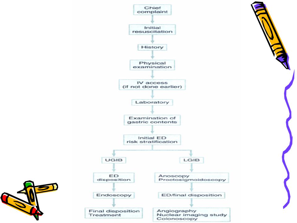
After initial resuscitation of the patient, it is important to identify whether the hemorrhage is proximal or distal to the ligament of Treitz (i.e., UGIB or LGIB) If the patient's vomitus can be inspected for blood or has been reported by the patient as bloody or “coffee grounds” or if melenic stool is present, an upper GI bleed should be the first consideration
 If the patient s vomitus can be inspected for blood or has been reported by the patient as bloody or coffee grounds or if melenic stool is present, an upper GI bleed should be the first consideration.")
73
Placement of a nasogastric tube is generally not necessary and rarely yields information that is independently useful for either diagnosis or risk stratifications

74
Aspiration of bloody contents diagnoses UGIB (or bleeding from nasal or oral passageways), but it does not differentiate if the bleeding is ongoing or has already stopped There is a 10% incidence of failure to aspirate blood in established UGIB

75
False-negative results may occur if the bleeding is intermittent or has already stopped and the stomach cleared or if the bleeding is in the duodenum, and edema or spasm of the pylorus has prevented reflux of blood into the stomach.

76
The presence of bile in an otherwise clear aspirate excludes the possibility of active bleeding above the ligament of Treitz, but is rarely seen and should not be used to exclude UGIB in a patient with documented melena False-positive results may occur from nasal bleeding

77
Gastric contents should not be tested for occult blood because visual inspection of the vomitus or aspirate is insufficient to diagnose subtle bleeding, and testing is unreliable

78
In patients who have hematochezia, an upper GI origin for the bleeding is often associated with signs and symptoms of shock because rapid transit time of large quantities of blood is producing the hematochezia

79
Because up to 11% of patients with hematochezia have UGIB, a nasogastric tube is indicated in most cases of LGIB If the gastric aspirate does not appear bloody, the nasogastric tube should be removed, LGIB should be considered, and anoscopy/proctosigmoidoscopy performed

80
Gastric tubes are safe in most patients, but pharyngeal and esophageal perforation, cardiac arrest, ethmoid sinus fracture with brain trauma, and bronchial intubation have been reported The old approach of placing a nasogastric tube in all patients with suspected UGIB predated endoscopy and has no place in modern emergency medicine

81
No evidence exists that gastric tube placement aggravates hemorrhage from varices or Mallory-Weiss tears

82
Gastric lavage may be necessary to prepare a patient for endoscopy
Before gastric lavage, patients with evidence of a possible perforated viscus (e.g., severe pain, peritoneal signs) should undergo radiologic assessment looking for free air. Lavage should not be performed in the presence of pneumoperitoneum
 should undergo radiologic assessment looking for free air. Lavage should not be performed in the presence of pneumoperitoneum.")
83
Gastric lavage does not reduce blood loss in patients with UGIB, and iced lavage is not recommended
Gastric lavage, in preparation for endoscopy, is best performed with a large-bore Ewald tube, passed orally while the patient is in the left lateral decubitus position with the bed in Trendelenburg position

84
Additional holes may be cut in the distal portion of the Ewald tube to improve aspiration of blood and clots. Clots that cannot be aspirated continue to cause pink return and give the false impression of continued bleeding The irrigant need not be sterile; regular tap water may be used

85
The irrigant should be delivered and removed by gravity in volumes of 200 to 300 mL until the return is clear Little irrigant is absorbed by the patient Gastric rupture has been reported as a rare complication of gastric lavage

86
Anoscopy/Proctosigmoidoscopy
Patients with mild rectal bleeding who do not have obviously bleeding hemorrhoids should have anoscopy/proctosigmoidoscopy performed If bleeding internal hemorrhoids are discovered, and the patient does not have portal hypertension, the patient may be discharged with appropriate treatment and follow-up evaluation for hemorrhoids

87
If hemorrhoids are not detected, it is important to determine if the stool above the rectum contains blood

88
The absence of blood above the rectum in a patient who is actively bleeding indicates that the source of bleeding is in the rectum The presence of blood above the anoscope or sigmoidoscope does not invariably indicate a proximal source of bleeding because retrograde passage of blood into the more proximal colon commonly occurs

89
Endoscopy Endoscopy is the most accurate diagnostic tool available for the evaluation of UGIB. It identifies a lesion in 78% to 95% of patients with UGIB if it is performed within 12 to 24 hours of the hemorrhage Accurate identification of the bleeding site allows for risk stratification with respect to predicting rebleeding and mortality

90
Endoscopy-based triage significantly reduces hospitalization rates and costs of treating upper GI bleeding Significant advances in endoscopic hemostasis also make it of therapeutic value in select patients (e.g., banding or sclerosing of varices). Colonoscopy is an effective tool for diagnosis and selected treatment of LGIB
. Colonoscopy is an effective tool for diagnosis and selected treatment of LGIB.")
91
Angiography Angiography can detect the location of UGIB in two thirds of patients studied Since the advent of endoscopy, however, the use of angiography has decreased significantly, and today angiography is used in only 1% of patients with UGIB Angiography is used more commonly in patients with LGIB and usually in consultation with a general surgeon

92
Angiography rarely diagnoses the cause of bleeding, it does identify the site of bleeding in approximately 40% of patients who have LGIB and 65% of patients who eventually require surgical intervention

93
Angiography ideally is performed during active bleeding; this may be apparent by persistently unstable vital signs or continued transfusion requirements to establish or maintain an optimal hemoglobin or hematocrit level Arterial embolization can be used in selected cases of LGIB

94
Gastric Acid Secretion Inhibition
All patients with documented peptic ulcer disease should be treated with a proton-pump inhibitor (e.g., omeprazole) There is no benefit to initiating this therapy or administering H2 antihistamines in the emergency department for patients with UGIB
 There is no benefit to initiating this therapy or administering H2 antihistamines in the emergency department for patients with UGIB.")
95
When the diagnosis of peptic ulcer has been confirmed by endoscopy, it is appropriate to start a proton-pump inhibitor Medical therapy is an adjunct, not a substitute for endoscopic evaluation, as appropriate

96
Octreotide (Somatostatin Analogues)
Patients with documented esophageal varices should be treated with an intravenous infusion of octreotide at pg/hr for a minimum of 24 hours while being observed in the intensive care unit (ICU). It is a useful addition to endoscopic sclerotherapy and decreases rebleeding occurrences
. It is a useful addition to endoscopic sclerotherapy and decreases rebleeding occurrences.")
97
Vasopressin Intravenous vasopressin has been used in the treatment of patients with UGIB, most commonly in patients with variceal hemorrhage Controlled studies have not shown a positive effect of vasopressin on overall mortality

98
These results, combined with a relatively high rate of serious complications (9% major and 3% fatal), suggest that use of vasopressin should be limited The recommended dose of vasopressin is 20 U IV over 20 minutes, then unit/min Consultation with a gastroenterologist is advisable

99
Sengstaken-Blakemore Tube
The Sengstaken-Blakemore tube stops hemorrhage in approximately 80% of patients bleeding from esophageal varices The Linton tube is superior to the Sengstaken-Blakemore tube in patients with bleeding gastric varices

100
In general, these tubes should not be used without endoscopic documentation of the source of bleeding because complications are common and significant (14% major, 3% fatal)
")
101
Indication : an exsanguinating patient with probable variceal bleeding in whom endoscopy is not immediately available and vasopressin has not slowed the hemorrhage Consultation with a surgeon or gastroenterologist is advisable

102
Surgery Surgery is indicated for all hemodynamically unstable patients with active bleeding who do not respond to appropriate intravascular volume replacement, correction of any coagulopathy, and endoscopic intervention (if available)
")
103
The mortality for patients undergoing emergency operations for GI bleeding is approximately 23%
Generally, surgery is indicated whenever the risk of ineffective medical therapy and continued hemorrhage outweighs that of surgical morbidity and mortality

104
Emergency surgery should be considered when blood replacement exceeds 5 U within the first 4 to 6 hours or when 2 U of blood is needed every 4 hours after replacing initial losses to maintain normal cardiac output

105
DISPOSITION

106
Risk Stratification Risk stratification involves combining historical, clinical, and laboratory data to determine the risk of death and rebleeding in patients presenting to an emergency department with GI bleeding

107
Patients can be sorted into four categories:
Very low risk Low risk Moderate risk high risk

108
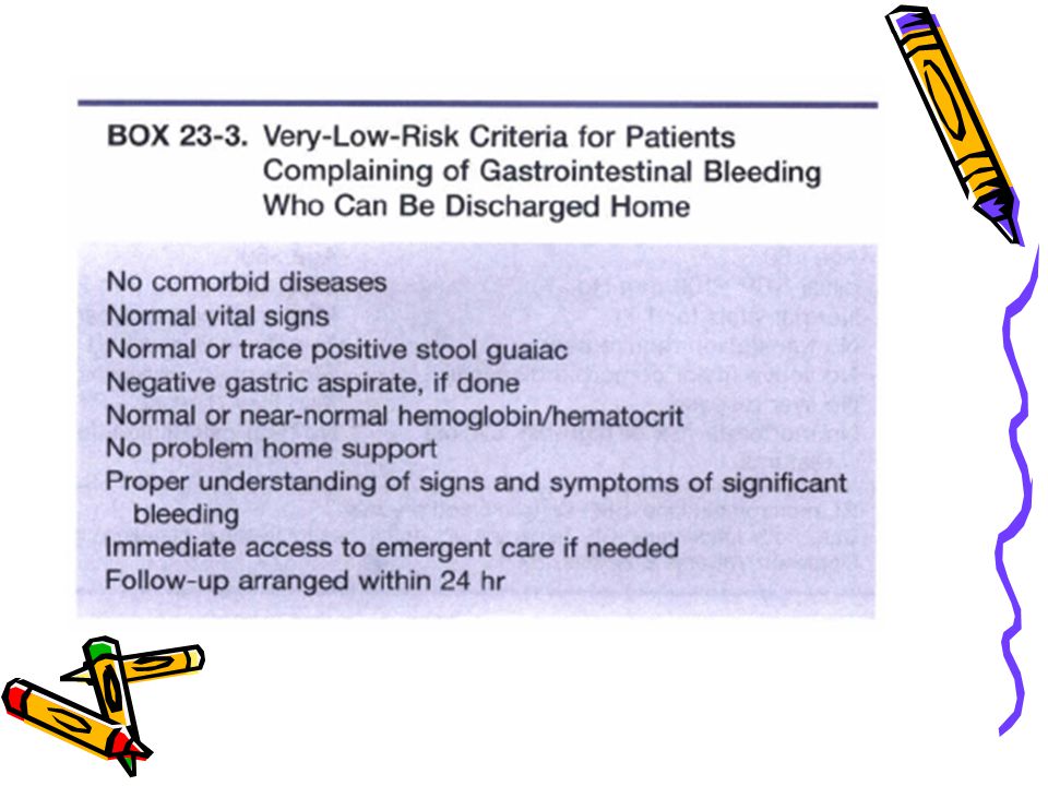
Very low risk : patients present to the emergency department with a chief complaint of vomiting blood or passing blood from their rectum but with little or no objective evidence of significant GI bleeding These patients can be sent home without further diagnostic tests

110
Before discharge, patients should be educated about the signs and symptoms of significant GI bleeding and when to return to the emergency department or call their primary care physician They should be given specific education about the possible or actual cause of the bleeding and specific treatment for the cause of the bleeding

111
They should be educated about the side effects of any medications
Patents should be given specific follow-up evaluation within 24 to 36 hours They should be instructed to avoid aspirin, nonsteroidal anti-inflammatory drugs, and alcohol

112
Nearly all patients with significant GI bleeding were admitted to the hospital
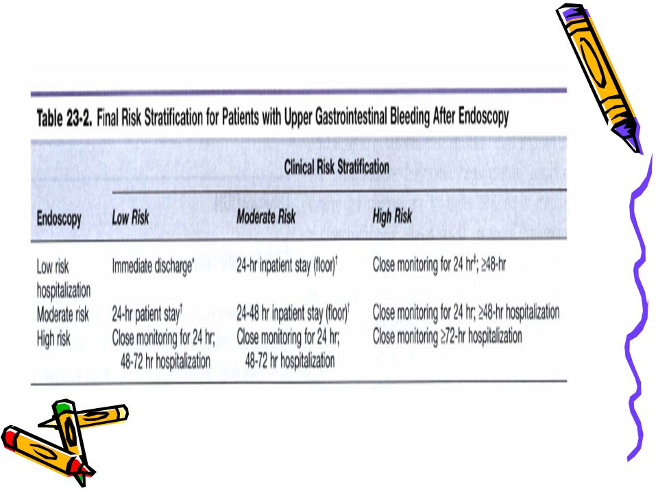
Combining clinical and endoscopic criteria provides an accurate estimation of the risk of rebleeding and mortality in patients with UGIB

113
These combined criteria have been used to identify patients with UGIB who are at low risk and can be discharged home and patients at moderate or high risk who need to be admitted to an appropriate care site in the hospital

114
Risk stratification for patients with LGIB is not as well studied, and so nearly all patients with significant LGIB are admitted. Risk stratification can be used for patients with LGIB, however, to decide an appropriate inpatient care site

117
Patients with clinical evidence of GI bleeding should undergo endoscopy as soon as it is available for final risk stratification, inpatient triage, and determination of appropriate treatment

118
If endoscopy is not immediately available, patients with low clinical risk may be admitted to an emergency department observation unit or short-stay hospital bed until endoscopy can be performed Patients with moderate clinical risk criteria may be admitted to an inpatient floor, intermediate care unit, or ICU depending on the individual patient and the capabilities of the institution

119
Patients with high clinical risk should be admitted to a closely monitored step-down unit or an ICU
The timing of endoscopy depends on availability, the acuity of the patient, the need for emergent therapy, the need to determine final care site, and the need to minimize length of stay

120
Patients with LGIB that is not clearly due to hemorrhoids, fissure, or proctitis should be admitted to an inpatient bed Patients with low risk may be admitted to the floor and set up for a nuclear medicine imaging study (e.g., red blood cell–labeled study) or colonoscopy
 or colonoscopy.")
121
Patients with high-risk criteria should be admitted to a step-down unit or ICU and be considered for angiography to identify the site of LGIB Patients with moderate-risk criteria need to be individualized for the most appropriate inpatient care site (floor, intermediate care bed, or ICU), and the best diagnostic studies (nuclear imaging or angiography)
, and the best diagnostic studies (nuclear imaging or angiography)")
122
Consultation with a surgeon should be obtained if it appears that more than 2 U of blood will be required after the initial emergency department resuscitation or if there is reasonable suspicion that operative intervention may be needed. This is especially true of patients older than 65 years of age

123
In general, the older the patient, the more aggressive the surgical management ought to be
Patients with a history of varices, persistent postural changes in heart rate, or significant bright red blood per rectum are more likely to require surgery than patients without these findings

124
Patients who have abdominal aortic grafts who enter the emergency department with GI bleeding should receive prompt vascular surgical consultation in the emergency department for the possibility of aortoenteric fistula

Similar presentations


 682-3793; (p) 413-3222.>")


 bleeding refers to any bleeding that starts in the gastrointestinal tract. Bleeding may come from.>")


