Download presentation
Presentation is loading. Please wait.

1
DIAGNOSTIC RADIOLOGY & BODY SCANS CHAPTERS 20 & 21

2
X-RAYS Electromagnetic radiation of short wavelength. Penetrate most substances, including tissue. Also called ionizing radiation. Cause fluorescence (emission of light) on photographic plates. Harmful, in a dose-dependent fashion.
 on photographic plates. Harmful, in a dose-dependent fashion..")
3
X-RAYS AS A DIAGNOSTIC TOOL 3 PRIMARY WAYS 1) Radiography. 2) Fluoroscopy. 3) Tomography.
 Radiography. 2) Fluoroscopy. 3) Tomography.")
4
RADIOGRAPHY Radiograph = an X-Ray image (picture), or what we call “the X-Ray.” “Like negatives of photographs.” X-Rays that penetrate the tissues and reach the film turn the film black. As such: AIR = BLACK (ISH) FAT = DARK GRAY WATER = LIGHTER GRAY BONE = WHITE (ISH)
 FAT = DARK GRAY WATER = LIGHTER GRAY BONE = WHITE (ISH).")
5
FLUOROSCOPY See text. Not recommended A real-time X-Ray Typically used with dye studies such as barium swallow, angiography, etc. Prolongs radiation exposure.

6
TOMOGRAPHY A tomogram is a radiograph that generates cross-sectional images at different tissue planes. CT = computed tomography, or CAT scan = computed axial tomography – uses computers, produces digital images. (More later)
.")
7
DIGITAL RADIOGRAPH A digital image without film. Stored in a computer database. Can be accessed by anyone with access to the database, can be emailed, etc. No need to chase down the hard copy films.

8
RADIATION SAFETY Exposure is cumulative. RISKS 1) Genetic damage, cancer. 2) Sterility. 3) Alterations in the composition of individual cells. 4) Bone marrow production.
 Sterility. 3) Alterations in the composition of individual cells. 4) Bone marrow production..")
9
RADIATION SAFETY Occupational exposure to ionizing radiation is associated with: 1) Leukemia. 2) Skin cancer. See text re: The “rem,” the “rad”, and the “gray.” X-Ray exposure is measured in millirads.
 Skin cancer. See text re: The rem, the rad , and the gray. X-Ray exposure is measured in millirads..")
10
RADIATION SAFETY 3 ways to protect against ionizing radiation: 1) Time - as short as possible. 2) Distance - as far away as possible. 3) Shielding.
 Distance - as far away as possible. 3) Shielding..")
11
RADIATION SAFETY PREGNANCY – potential for teratogenesis etc. is highest during organogenesis, which occurs during the 1 st 12 weeks. Need to ask about pregnancy. In a perfect world, X-Rays would be done between menses and ovulation. Shielding the pelvis advised when uncertain.

12
BODY SCANS CT MRI DEXA PET SPECT

13
CT / CAT Computed tomography, computerized axial tomography. Uses X-Rays in a 180° fashion. Radiation exposure is “small.” Image generated based on amount of radiation absorbed. Can depict all types of tissues except nerves.

14
CT / CAT ADVANTAGES 1) Excellent detail. 2) Quick results. DISADVANTAGES 1) Cost. 2) Time of exposure to radiation.
 Cost. 2) Time of exposure to radiation..")
15
CT / CAT 1 st CT developed in 1972 to evaluate brain abnormalities. Now in widespread use for other organ systems. USES: evaluation of neoplasms / masses, hematomas, abscesses, foreign body localization, trauma. Quicker than MRI.

16
MRI Magnetic resonance imaging. See text for details of the physics involved. In short, uses radio waves and a magnetic field that detects changes in absorption of energy by hydrogen ions. As such, no radiation exposure.

17
MRI ADVANTAGES 1) No radiation exposure. Can be used during pregnancy. 2) Better detail than CT. 3) Imaging modality of choice for the CNS. DISADVANTAGES 1) Cost – 1/3 more than CT. 2) Takes longer, results not available as fast as CT.
 Better detail than CT. 3) Imaging modality of choice for the CNS. DISADVANTAGES 1) Cost – 1/3 more than CT. 2) Takes longer, results not available as fast as CT..")
18
→

19
CIRRHOSIS

20
STONE RIGHT URETER ←

21
MRI Absolute Contraindications Brain Aneurysm Clip Implanted neural stimulator Implanted cardiac pacemaker or defibrillator Cochlear implant Ocular foreign body (e.g. metal shavings) Other implanted medical devices: (e.g. Swan Ganz catheter) Insulin pump Metal shrapnel or bullet.
 Other implanted medical devices: (e.g. Swan Ganz catheter) Insulin pump Metal shrapnel or bullet..")
22
MRI Relative Contraindications 1) Penile and non metallic valve prosthesis 2) Pregnancy: We try to avoid scanning in the first trimester since we are not sure if there are any adverse effects to MRI. (Pregnant women never receive the contrast agent gadolinium) 3) Claustrophobic or anxious patients can not tolerate the MRI scanner. 4) Obese patients may not fit into the small opening of the scanner. In addition the maximum weight that the MRI table can sustain is 350 lbs.
 3) Claustrophobic or anxious patients can not tolerate the MRI scanner. 4) Obese patients may not fit into the small opening of the scanner. In addition the maximum weight that the MRI table can sustain is 350 lbs..")
23
DEXA Dual Energy X-Ray Absorptiometry. The modality of choice for measuring bone mineral density (BMD) in evaluating osteoporosis. Low radiation exposure, quick.
 in evaluating osteoporosis. Low radiation exposure, quick..")
24
Dexa T Scores NormalOsteopaeniaOsteoporosis A normal bone density is when the T score measures greater than –1.0 (greater than minus 1.0) If the bone mineral density is measured as a T score between –1.0 and –2.5 (minus 1.0 and minus 2.5) this indicates the presence of osteopaenia. Osteopaenia is not osteoporosis. It does however indicate that there is a reduction in bone mineral density, which is not as severe as osteoporosis. This condition represents an earlier phase of bone mineral density loss in the skeleton. If the bone mineral density is measured as a T score of –2.5 or less (minus 2.5 or less) this indicates the presence of osteoporosis at the site of measurement.
 this indicates the presence of osteoporosis at the site of measurement..")
25
PET SCAN Positron emission tomography. The positron is the antiparticle or the antimatter counterpart of the electron. The positron has an electric charge of +1, a spin of 1/2, and the same mass as an electron. (Wikipedia)antiparticleantimatter electron electric chargespin Measures function rather than structure.
antiparticleantimatter electron electric chargespin Measures function rather than structure..")
26
PET SCAN Patient is injected with a metabolically active biochemical substance, such as glucose, water, ammonia, which has been tagged with a radioactive isotope that emits a positron. Binding of these substances with electrons found in the tissue being studied causes emission of gamma rays, which are converted into color-coded images. Degree of gamma ray production reflects cellular utilization / metabolism of the tagged substance.

27
PET SCAN

28
APPLICATIONS OF PET SCANS High dollar machine, high dollar test. Costs 1/3 more than MRI. Availability typically limited to research institutions, and used more as a research tool than a diagnostic one. Findings of altered metabolic function can direct innovations in treatment.

29
APPLICATIONS OF PET SCANS Neuroimaging - dementia, stroke, epilepsy, Parkinson’s disease. Cardiac – to assess myocardial viability. Psychiatric – schizophrenia, mood disorders, substance abuse. See http://en.wikipedia.org/wiki/Positron_emi ssion_tomography

30
SPECT Single Photon Emission Computed Tomography Uses radiopharmaceuticals labeled with a positron-emitting isotope such as Technitium – 99 (Tc-99m). Detects gamma rays emitted by the natural radioactive decay of the isotope. Degree of gamma ray production reflects more the degree of perfusion of the organ being studied than its function.

31
APPLICATIONS OF SPECT SCANS Neuroimaging- dementia, neoplasms, infection, epilepsy. Cardiac- ischemic heart disease. Others- thyroid, bone, white cells. See: http://en.wikipedia.org/wiki/SPECT

32
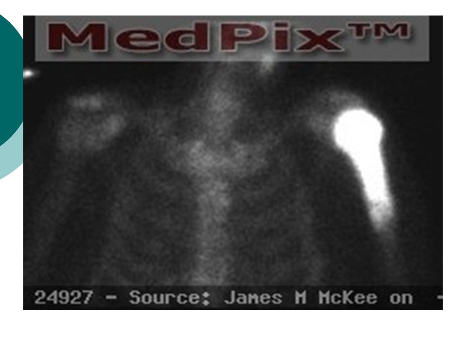
63 YOM W/ PROSTATE CANCER

33
Tc-99 Scan

35
FUNCTIONAL MRI Measures signal changes in the brain that are due to changes in neural activity. Increased neural activity → need for increased oxygen → increased oxygenated hemoglobin as relative to deoxygenated hemoglobin. Because deoxygenated hemoglobin attenuates the MR signal, the vascular response leads to a signal increase that is related to the neural activity.

36
FUNCTIONAL MRI A fMRI scan showing regions of activation in orange, including the primary visual cortex.

37
IMAGES Images compliments of the Department of Radiology at the Uniformed Services University of the Health Sciences (USUHS), Bethesda, Md. See: www.rad.usuhs.edu

38
THE CHEST X-RAY Can be used to assess a wide variety of pathologic conditions of the chest: heart, lungs, mediastinum, bone, esophagus, trachea, diaphragm. Such as: infection, tumor, lymphoma, foreign bodies, rib fractures, cardiac enlargement, presence of free air (pneumothorax, pneumomediastinum), fluid accumulation (pulmonary edema, pleural effusion), changes such as sarcoid, amyloid.
, fluid accumulation (pulmonary edema, pleural effusion), changes such as sarcoid, amyloid..")
39
THE CHEST X-RAY Usually take an AP (antero-posterior), or PA (postero-anterior), and a lateral view. Can also take an oblique view. If known, the part you’re interested in should be closest to the film. To assess free air or fluid, patients position can be manipulated to allow gravity to affect layering of the fluid or air.

40
22 YEAR OLD SMOKER W/ COUGH, WHEEZING ←

41
←

42
← CYSTIC TERATOMA

43
46 YOF W/ CHEST PAIN AFTER A ROUND OF GOLF ← ←

44
22 YOM W/ NIGHT SWEATS, WT LOSS ← →

45
ABDOMINAL X-RAYS The “flat plate,” or scout film, done as the sole diagnostic film or prior to a contrast study. Manipulation of position of the patient can assess presence of free air (under the diaphragm), fluid, or the presence of “air-fluid levels” as seen in bowel obstruction. KUB- kidneys, ureter, bladder. Term used interchangeably w/ flat plate.
, fluid, or the presence of air-fluid levels as seen in bowel obstruction. KUB- kidneys, ureter, bladder. Term used interchangeably w/ flat plate..")
46
AIR-FLUID LEVELS IN BOWEL OBSTRUCTION

47
43 YOF W/ ABDOMINAL PAIN

48
32 YOF W/ G. E. REFLUX

49
32 YOF W/ REFLUX

51
32 YOF W/ REFLUX

52
SKELETAL X-RAYS Most useful in assessing fractures, but also good for: joint dislocation / subluxation, changes in bone architecture (erosions, thickening, density), presence of abnormal calcifications / deposits (osteophytes, tophi), etc
, presence of abnormal calcifications / deposits (osteophytes, tophi), etc")
53
18 YOM W/ RIGHT THIGH PAIN

55
OSTEOSARCOMA

56
53 YOM W/ SWELLING, LEFT MIDDLE FINGER

57
TOPHACEOUS GOUT →

58
18 YOM FELT SOMETHING “POP” WHEN THROWING A SPLIT-FINGER FASTBALL

59
UNICAMERAL BONE CYST W/ PATHOLOGIC FRACTURE

60
MAMMOGRAPHY Radiographic images of the breast, primarily for early detection of breast cancer, prior to the appearance of a palpable mass. Also useful for evaluation of palpable masses: benign neoplasms, fibrocystic breast disease, etc. Used as an aid in placement of the biopsy needle.

61
SCREENING MAMMOGRAPHY For early detection of breast cancer. Guidelines as to who, when, and how often vary by organization, and depend on the patients risk status. American Cancer Society: baseline between 35 and 40, every 1-2 years between 40-50, and yearly after 50, along with monthly self-breast examination and annual physical exam.

62
SCREENING MAMMOGRAPHY ↓

63
CONTRAST STUDIES Involves the introduction of a radiopaque substance (barium, dye, etc) into an organ, vessel, duct, etc Allows for the identification of the anatomy of the structure being studied, its shape, contour, size, etc. “Filling defect” – describes an area where dye should be but isn’t.

64
CONTRAST STUDIES Barium swallow, upper GI, small bowel series. Barium enema (B.E.) Oral cholecystograms (OCG). Cholangiograms. Intravenous pyelogram (IVP). Angiograms- arteriograms, venograms. Lymphangiograms. Hysterosalpingograms (HSG). Myelograms. Arthrograms.
 Oral cholecystograms (OCG). Cholangiograms. Intravenous pyelogram (IVP). Angiograms- arteriograms, venograms. Lymphangiograms. Hysterosalpingograms (HSG). Myelograms. Arthrograms..")
65
→ ← “APPLE CORE” LESION OF ADVANCED COLON CANCER AIR-CONTRAST B.E.

66
ACHALASIA

67
2 DAY OLD INFANT W/ INCREASING ABDOMINAL DISTENTION

69
16 YOF W/ RLQ PAIN ←

70
↓

71
HSG BICORNUATE UTERUS

72
30 YOF W/ NEW ONSET SEIZURES

73
ANGIOGRAM MENINGIOMA

74
ANGIOGRAM 19 YOF W/ HYPERTENSION

75
RENAL ARTERY STENOSIS

Similar presentations


 CT scanning or (CAT scanning) is using X-rays to create a 3D image of the inside of an object. CT stands for computed tomography.>")


