Download presentation
Presentation is loading. Please wait.

1
Definition- a specialized technique for producing radiographs showing only a section or a slice of a patient Each tomograph shows the tissues within the focal through sharply defined and in focus

2
Fig.14.1

3
Maxilla and mandible are captured on one film Tube head and film move around patient’s head in a coordinated motion.

4
Beam of radiation-narrow slit Film requires intensifying screens Film placement is extraoral Object to film distance varies Exposure time varies from 8 to 22 seconds Image quality is fair to moderate but significantly less details than periapical

5
Screens have fluorescent crystals that “glow” or produce visible light when hit by an x-ray Convert x-ray into visible light and expose the film Reduce the patient dose significantly Film has higher contrast than intraoral radiograph

6
Intensifying screens have two types of crystals-calcium tungstate and rare earth Intensifying screens have to be matched to the type of the film that responds to blue or green light

7
Real or actual shadows- structures in or close to the focal through Ghost or artefactual shadows- structures on the opposite side or a long way from the focal through. The 8 o upward angulation of the X- ray beam means that these ghost shadows appear at a higher level than the structures that have caused them

8

10
An X-ray tubehead, A cassette and cassette carriage assembly Patient-positioning apparatus including light beam markers

12
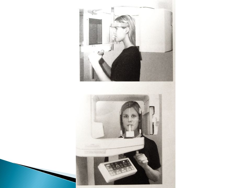
Patients should be asked to remove any earrings, jewellery, hair pins, spectacles, dentures or orthodontic appliances. The procedure and equipment movements should be explained, to reassure patients. A protective lead apron should be used only for pregnant patients

13
Mid-saggital plane should be perpendicular to the floor Frankfurth plane should be parallel to the floor The patient should bite into notch Tell patient to put tongue in roof of mouth and to keep still during the exposure

14
Confirm the exposure settings Move outside the room or behind the barrier Watch patient during entire exposure

15
Switch on the lateral ceph machine by pressing the C button Unscrew the knock on the pan machine turning counterclock wise and plucking it out Adjust the program( no 1) at the window of the pan Press R button on the panel of the machine
 at the window of the pan Press R button on the panel of the machine")
16
A large area is imaged allowing comparison on symmetrical structures The image is easy for patients to understand and is therefore a useful teaching aid Positioning is relatively easy The radiation dose is one-third of the dose for the full-mouth survey

17
Structures or abnormalities not in the focal through may not be evident Soft tissues or air shadows can overlie the required hard tissues structures Ghost shadows can overlie the structures in the focal through

18
The tomographic movement together with the distance between the focal through and film produce distortion and magnification of the final image ( approx. 1.3) The use of intensifying screens results in some loss of resolution Not suitable for children under 5 years or on some disabled patients
 The use of intensifying screens results in some loss of resolution Not suitable for children under 5 years or on some disabled patients.")
Similar presentations


 penetrates tissue to form a.>")