Download presentation
Presentation is loading. Please wait.

1
Ophthalmic Life and Death A Case Presentation
Dr Phillip Hayes Central Coast day Hospital 2012

2
Clinical history 34 year old woman IDDM
Presented initially to GP June 2007 Nearly constant Headache 6 months around L temporal Presumed migraine Only partial response to NSAID Referred Neurology outpatients

3
Clinical History Seen at Neurology OP July 2007
Continuous unilateral headache ? Hemicrania Admitted to Gosford Hosp for further investigation MRI / MRV to exclude cortical vein thrombosis

4
What is a good way to screen for Thrombosis Intracranial Venous sinuses causing headache.

5
What is a good way to screen for Thrombosis Intracranial Venous sinuses causing headache?
Ophthalmoscopy to exclude Increased ICP by excluding Papilloedema

6
Background History Type 1 Diabetes Mellitus Endometriosis Smoker
Diagnosed at aged 22 Actrapid / Protaphane Poorly controlled No documented complications Endometriosis Microgynon (OCP) Smoker Prior history of Migraines
 Smoker. Prior history of Migraines.")
7
Clinical History Admitted Neurology 19/7/07
? Migraine, ? Venous thrombosis OP CT scan Brain – no acute intracranial pathology Treated with paracetamol, NSAID, amitriptyline with significant improvement Anxious for discharge MRI arranged as outpatient with follow-up Subsequently did not attend this appointment

8
Clinical History Re-admitted via ED 24/09/07 (2 months later)
Severe L temporal headaches over 2/52 Associated with gradual loss of vision L eye Extremely unwell Vomiting Missed several insulin doses

9
Examination Vomiting, distressed, afebrile,
L temporal tenderness & hyperaesthesia Impression of L proptosis L RAPD noted Acuity R 6/6 and L 6/36 No ophthalmoplegia Painful on prolonged L lateral gaze No nystagmus or diplopia

10
Examination L RAPD noted Vomiting, distressed, afebrile,
L temporal tenderness & hyperaesthesia Impression of L proptosis L RAPD noted Acuity R 6/6 and L 6/36 No ophthalmoplegia Painful on prolonged L lateral gaze No nystagmus or diplopia

12
RAPD An objective sign of vision loss
Localises pathology to between the Retina and the Optic Chiasm It compares the quantitative neural signal between each eye Media opacity doesn’t produce RAPD Can occasionally occur with normal VA

13
Examination Vomiting, distressed, afebrile,
L temporal tenderness & hyperaesthesia Impression of L proptosis L RAPD noted Acuity 6/6 R and 6/36 L No ophthalmoplegia Painful on prolonged L lateral gaze No nystagmus or diplopia

14
Summary Anxious, young, sick, poorly controlled Diabetic
Severe Left Headache Left eye: reduce vision with RAPD Do we do LP, MRI, look at the retina or call an Ophthalmologist?

15
Further Examination Ophthalmology review
Confirmed RAPD Possible Left Proptosis No ophthalmoplegia Normal optic discs Narrow arterioles, no diabetic retinopathy No retinal pathology Remainder of Cranial Nerves and PN exam normal

16
What next? What is needed for diagnosis.

17
What next? What is needed for diagnosis.
Urgent Neuro imaging and basic blood work. Should we start treatment??

18
Initial Management Commenced on Insulin dextrose infusion
Opiate analgesia to no effect Several IV anti-emetics

19
Investigations pH 7.41 (Normal), trace ketonuria
Biochemistry – unremarkable WCC 12.8 CRP 37, ESR 25 TFT normal, CK 22 HbA1C 16%

21
t

23
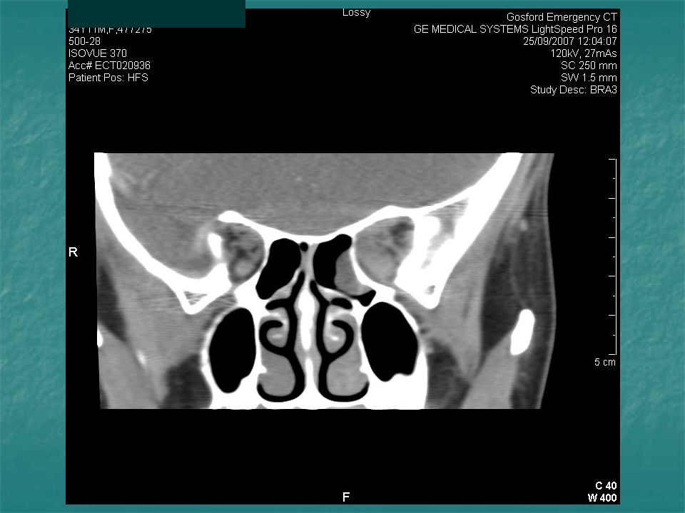
Imaging CT Brain & orbits MRI Diffuse swelling of muscles of L orbit
Inflammatory changes at orbital apex Probable compression of optic nerve MRI Inflammation of extra-ocular muscles and soft tissue of L orbital apex

25
Orbital Inflammation Graves 50%
Thyroid Eye Disease (Throid Orbitopathy) Infection 45% Orbital Cellulitis (Pseudotumour) Idiopathic Non Specific Orbital inflammation Systemic
 Infection. 45% Orbital Cellulitis. (Pseudotumour) Idiopathic. Non Specific Orbital inflammation. Systemic.")
26
Idiopathic Orbital inflammation (pseudotumour)
Pain + proptosis, injection, chemosis, ophthalmoplegia Individual muscles, trochlea, or lacrimal gland Makes up 5% of orbital conditions Tendons usually involved( vs thyroid eye disese) Idiopathic or associated condition with systemic
 Idiopathic or associated condition with systemic.")
27
Idiopathic Orbital inflammation (pseudotumour)
")
28
Idiopathic Orbital inflammation (pseudotumour)
May need biopsy to exclude other pathology but findings usually non-specific Trial of steroids with rapid resolution is supportive Other immunosuppressive therapies

29
Idiopathic Orbital inflammation (pseudotumour)
A Subgroup of these diverse group of conditions is localised to the orbital apex Termed “ The Orbital Apex Syndrome”

30
The orbital apex

31
Orbital Apex Syndrome Headache Peri-orbital / facial pain
Minimal Proptosis Reduced visual acuity RAPD Diplopia, Field defect Ophthalmoplegia Optic atrophy (not seen early)
")
32
Orbital Apex Syndrome Orbital apex Syndrome involving dysfunction of
Entry portal for all nerves & vessels to eye & origin of all extraocular muscles except inferior oblique Syndrome involving dysfunction of Optic nerve III, IV, VI, V1 Radiological evidence of inflammation in posterior orbit

33
Orbital Apex Syndrome This is the most likely diagnosis in this diabetic woman. However, it is not really a diagnosis but a description of a clinical syndrome.

34
Orbital Apex Syndrome Inflammatory Infectious Neoplastic
Sarcoid, SLE, Wegner’s, Churg-straus, GCA Tolosa-hunt syndrome Idiopathic Orbital inflammation Dysthyroid eye disease Infectious Fungi – Mucormycosis, Aspergillosis Strep, staph, actinomyces, anaerobes, TB Neoplastic Lymphoma, nasopharyngeal ca Iatrogenic / Trauma Vascular

36
Orbital Apex Syndrome Infection
Something that responds to Systemic Steriods Something that can kill you rapidly like Murcomycosis Something else like a tumour

37
Mucormycosis Zygomycetes Broad, irregularly branched, with few septa
Rhinocerebral mucormycosis Pulmonary, GI, Renal, Cutaneous, CNS Broad, irregularly branched, with few septa Thrive in acidic high glucose media Predisposed hosts Diabetes Immunosuppressed IVDU Iron overload

38
Mucormycosis Zygomycetes fungus

39
Mucormycosis Zygomycetes Broad, irregularly branched, with few septa
Rhinocerebral mucormycosis Pulmonary, GI, Renal, Cutaneous, CNS Broad, irregularly branched, with few septa Thrive in acidic high glucose media Predisposed hosts Diabetes Immunosuppressed IVDU Iron overload

40
Mucormycosis Inhalation of spores to paranasal sinuses of susceptible host Infarction and necrosis with vascular invasion Usually aggressive - very fast pace Typically in a diabetic with DKA (70%)
")
41
Mucormycosis

42
Mucormycosis Enter the orbit via ethmoid or maxillary sinus
Often febrile Orbital cellulitis picture

43
Mucormycosis

44
Orbital Mucormycosis Vision loss Headache Neurological symptoms
Infection progresses to the orbital apex, cavernous sinus and brain

45
Mucormycosis

46
Mucormycosis

47
Mucormycosis Diagnosis Treatment
Sinus inspection and biopsy of sinus or orbit along with washings Direct microscopy or histopath or culture Imaging to identify degree of adjacent tissue involvement Treatment Aggressive surgical debridement (orbital exenteration) Amphotericin IV Optimize metabolic factors
 Amphotericin IV. Optimize metabolic factors.")
48
Mucormycosis Prognosis
Overall mortality of 25-50% in rhinocerebral mucormycosis Delayed diagnosis and advanced or extensive disease lead to increased mortality Pulmonary involvement - 80% mortality

50
Further Progress After urgent CT and MRI she was reviewed by Ophthalmologist. Dx “orbital apex syndrome” with need to exclude mucormycosis in view of Diabetic status. ENT review same night Sinuses grossly normal, no evidence mucor in naso-pharynx Transferred to Westmead Hospital for orbital and sinus biopsy

51
Westmead Biopsy of L posterior ethmoidal sinus & L orbital soft tissue
Commenced steroids and anti-fungals

52
Westmead Biopsy of L posterior ethmoidal sinus & L orbital soft tissue
Commenced steroids and anti-fungals Biopsy Non-specific inflammation, No granulomata No fungal hyphae. No positive culture

53
Westmead Biopsy of L posterior ethmoidal sinus & L orbital soft tissue
Commenced steroids and anti-fungals Biopsy Non-specific inflammation, No granulomata No fungal hyphae. No positive culture Anti-fungals ceased Discharged on tapering regime high dose steriods Acuity 6/6 L eye F/U with ophthalmology & endocrine

54
Non Specific Orbital Apex Syndrome
Diabetes was not related to her vision loss. She responded well to Systemic steriods. The optic neuropathy was probably compressive but may have been peri-neuritic. Apart from her symptoms and radiology there was not much clinical information except subjective and objective vision loss.

55
Non Specific Orbital Apex Syndrome
Diabetes was not related to her vision loss. She responded well to Systemic steriods. The optic neuropathy was probably compressive but may have been peri-neuritic. Apart from her symptoms and radiology there was not much clinical information except subjective and objective vision loss.

56
Which of the folllowing can cause a RAPD?
Dense unilateral Cataract Dense Vitreous Haemorrhage Temporal Retinal Detachment All the above

57
Which of the folllowing can cause a RAPD?
Dense unilateral Cataract Dense Vitreous Haemorrhage Temporal Retinal Detachment All the above

58
In orbital apex syndrome a common finding is
Vision loss Proptosis Ptosis Lid retraction

59
In orbital apex syndrome a common finding is
Vision loss Proptosis Ptosis Lid retraction

60
The most common Orbital inflammation is
Orbital Cellulitis Orbital Pseudotumour Tolosa Hunt Syndrome Thyroid Orbitopathy

61
The most common Orbital inflammation is
Orbital Cellulitis Orbital Pseudotumour Tolosa Hunt Syndrome Thyroid Orbitopathy

62
A useful test to screen for raised intracranial pressure is
Consensual pupillary light reflex MRI/ MRV Fundus Fluorescein Angiogram Bimicroscopy of optic nerve head

63
A useful test to screen for raised intracranial pressure is
Consensual pupillary light reflex MRI/ MRV Fundus Fluorescein Angiogram Bimicroscopy of optic nerve head

64
A diabetic patient that is unwell with eye pain and swelling and sudden vision loss could have
Mucormycosis Dense Vitreous haemorrage Hypogylcaemic crisis All of the above

65
A diabetic patient that is unwell with eye pain and swelling and sudden vision loss could have
Mucormycosis Dense Vitreous haemorrage Hypogylcaemic crisis All of the above

66
Thank you

67
Tolosa-Hunt syndrome Idiopathic inflammatory granulomatous process of cavernous sinus Unilateral orbital pain Opthalmoplegia (III,IV,VI,) Rare 1 per million per year Inflammation may extend beyond CS Optic disc oedema or pallor reported but loss of visual acuity rare
 Rare 1 per million per year. Inflammation may extend beyond CS. Optic disc oedema or pallor reported but loss of visual acuity rare.")
68
Tolosa-Hunt syndrome Steroid responsive(usually in 72 hours for pain) but permanent deficits can occur and relapse common Low index of suspicion for misdiagnosis even if response to steroids Lymphoma, vasculitis, and some infections will respond Diagnosis of exclusion

Similar presentations


 Intracranial (15-20%) Bony (5-10%) Radiography – Computed tomography (CT) best for.>")


