Download presentation
Presentation is loading. Please wait.

1
Non-Infective Inflammatory disease Dr. Mohammad Shehadeh
Orbit 4 Non-Infective Inflammatory disease Dr. Mohammad Shehadeh

2
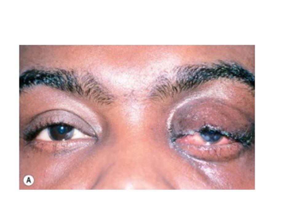
Idiopathic orbital inflammatory disease
previously referred to as orbital pseudotumour is an uncommon disorder characterized by non-neoplastic, non-infective, space-occupying orbital lesions The inflammatory process may involve any or all of the orbital soft tissues, resulting in, for example, myositis, dacryoadenitis, optic perineuritis or scleritis Unilateral disease is the rule in adults, although in children bilateral involvement may occur

3
Diagnosis Presentation is with acute periorbital redness, swelling and pain Signs Congestive proptosis and ophthalmoplegia may occur. Optic nerve dysfunction, particularly if the inflammation involves the posterior orbit.

4
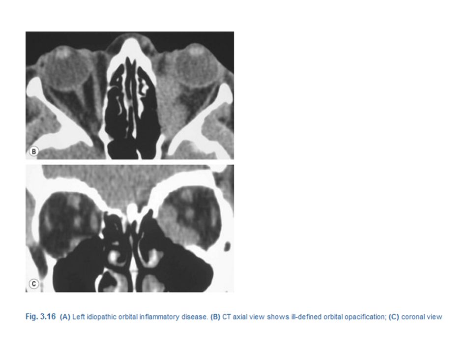
CT shows ill-defined orbital opacification and loss of definition of contents
Course. This follows one of the following patterns: Spontaneous remission after a few weeks without sequelae. Intermittent episodes of activity with eventual remission. Severe prolonged inflammation eventually leading to progressive fibrosis of orbital tissues, resulting in a ‘frozen orbit’ characterized by ophthalmoplegia, which may be associated with ptosis and visual impairment caused by optic nerve involvement.

7
Treatment Observation, for relatively mild disease, in anticipation of spontaneous remission. Biopsy is generally required in persistent cases to confirm the diagnosis and rule out neoplasia. NSAIDs are often effective and may precede steroid therapy. Systemic steroids should be administered only after the diagnosis has been confirmed, as they may mask other pathology such as infection and Wegener granulomatosis. Oral prednisolone, initially 60–80 mg/day, is later tapered and discontinued, depending on clinical response, although it may need to be reintroduced in the event of recurrence. Radiotherapy may be considered if there has been no improvement after 2 weeks of adequate steroid therapy. Even a low dose treatment (i.e. 10 Gy) may produce remission. Antimetabolites such as methotrexate or mycophenolate mofetil may be necessary in the context of resistance to both steroids and radiotherapy. Systemic infliximab, a tumour necrosis factor inhibitor, may be effective in recurrent or recalcitrant cases that have failed to respond to conventional therapy.
 may produce remission. Antimetabolites such as methotrexate or mycophenolate mofetil may be necessary in the context of resistance to both steroids and radiotherapy. Systemic infliximab, a tumour necrosis factor inhibitor, may be effective in recurrent or recalcitrant cases that have failed to respond to conventional therapy.")
8
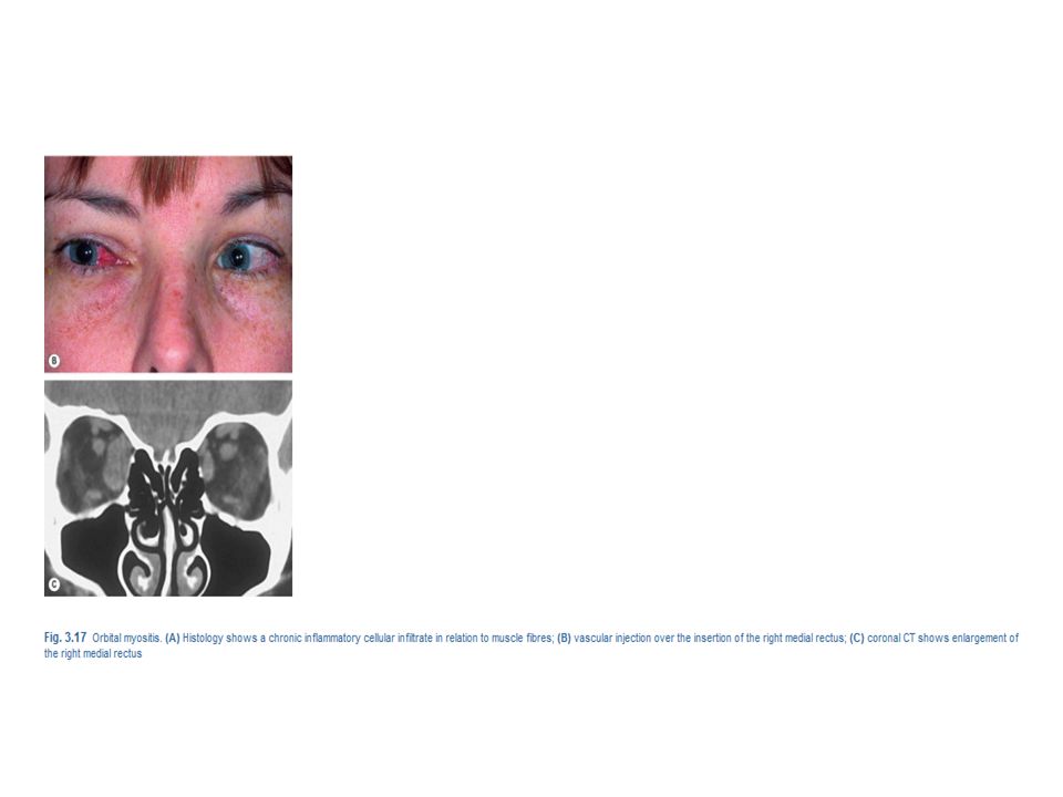
Orbital myositis Orbital myositis is an idiopathic, non-specific inflammation of one or more extraocular muscles and is considered a subtype of IOID Presentation is usually in early adult life with acute pain, exacerbated by eye movement, and diplopia. Signs Lid oedema, ptosis and chemosis. Pain and diplopia associated with eye movements. Vascular injection over the involved muscle . In chronic cases the affected muscle may become fibrosed, with permanent restrictive myopathy.

9
CT shows enlargement of the affected muscles (Fig. 3
CT shows enlargement of the affected muscles (Fig. 3.17C), with or without involvement of the tendons of insertion Course Acute non-recurrent involvement which resolves spontaneously within 6 weeks. Chronic disease characterized by either a single episode persisting for longer than 2 months (often for years) or recurrent attacks.
, with or without involvement of the tendons of insertion. Course. Acute non-recurrent involvement which resolves spontaneously within 6 weeks. Chronic disease characterized by either a single episode persisting for longer than 2 months (often for years) or recurrent attacks.")
10
Treatment is aimed at relieving discomfort and dysfunction, shortening the course and preventing recurrences. NSAIDs may be adequate in mild disease. Systemic steroids are generally required and usually produce dramatic improvement, although recurrences occur in 50% of cases. Radiotherapy is also effective, particularly in limiting recurrence.

12
Acute dacryoadenitis Lacrimal gland involvement occurs in about 25% of patients with IOID. More commonly, dacryoadenitis occurs in isolation, resolves spontaneously and does not require treatment. Occasionally it may be caused by mumps, mononucleosis, and rarely bacteria.

13
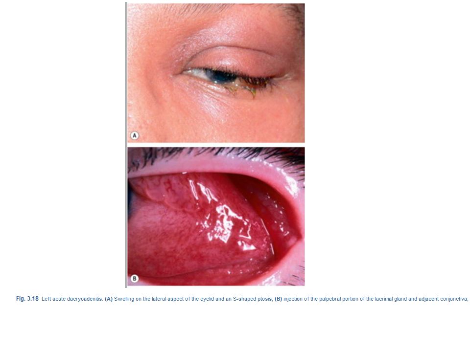
Presentation is with acute discomfort in the region of the lacrimal gland.
Signs Swelling of the lateral aspect of the eyelid giving rise to a characteristic S-shaped ptosis and slight downward and inward dystopia (Fig. 3.18A) Tenderness over the lacrimal gland fossa. Injection of the palpebral portion of the lacrimal gland and adjacent conjunctiva Lacrimal secretion may be reduced.
 Tenderness over the lacrimal gland fossa. Injection of the palpebral portion of the lacrimal gland and adjacent conjunctiva Lacrimal secretion may be reduced.")
14
CT shows enlargement of the gland and involvement of adjacent tissues

16
Orbital Varices Varices consist of weakened segments of the orbital venous system of variable length and complexity. Intrinsic to the circulation, they enlarge with increased venous pressure, their distensability varying with the residual thickness and strength of their walls

17
Signs Intermittent non-pulsatile proptosis not associated with a bruit. As the orbital veins are devoid of valves, rapidly reversible proptosis may be precipitated or accentuated by increasing venous pressure through coughing, straining, Valsalva manoeuvre, assuming the dependent position or external compression of the jugular veins.

18
Complications acute haemorrhage and thrombosis. Patients with long-standing lesions may develop atrophy of surrounding fat and enophthalmos associated with a deepened superior sulcus in the resting position Treatment by surgical excision is technically difficult and often incomplete because the lesions are friable and bleed easily. Indications include recurrent thrombosis, pain, severe proptosis and optic nerve compression.

Similar presentations


 Intracranial (15-20%) Bony (5-10%) Radiography – Computed tomography (CT) best for.>")


 Nicola Davis.>")


