Download presentation
Presentation is loading. Please wait.

1
Fungal Diseases of Paranasal Sinuses

2
Non invasive fungal rhinosinusitis.
Classification Non invasive fungal rhinosinusitis. Allergic fungal rhinosinusitis Mycetoma Invasive fungal rhinosinusitis Acute invasive Chronic invasive Granulomatous

3
Non-invasive fungal rhinosinusitis

4
Allergic fungal rhinosinusitis
Most common form FRS. Characterized by dark thick, inspissated mucus filling PNS. On M/E necrotic and degraanulating eosinophils, charcot leyden crystals & few fungal hyphae Most common organisms isolated are bipolaris and curvularia fungi

6
Clinical Manifestations
Classically unilateral disease Slow progressive nasal congestion PND Nasal obstruction Anosmia Thick mucinous debris in nasal discharge

7
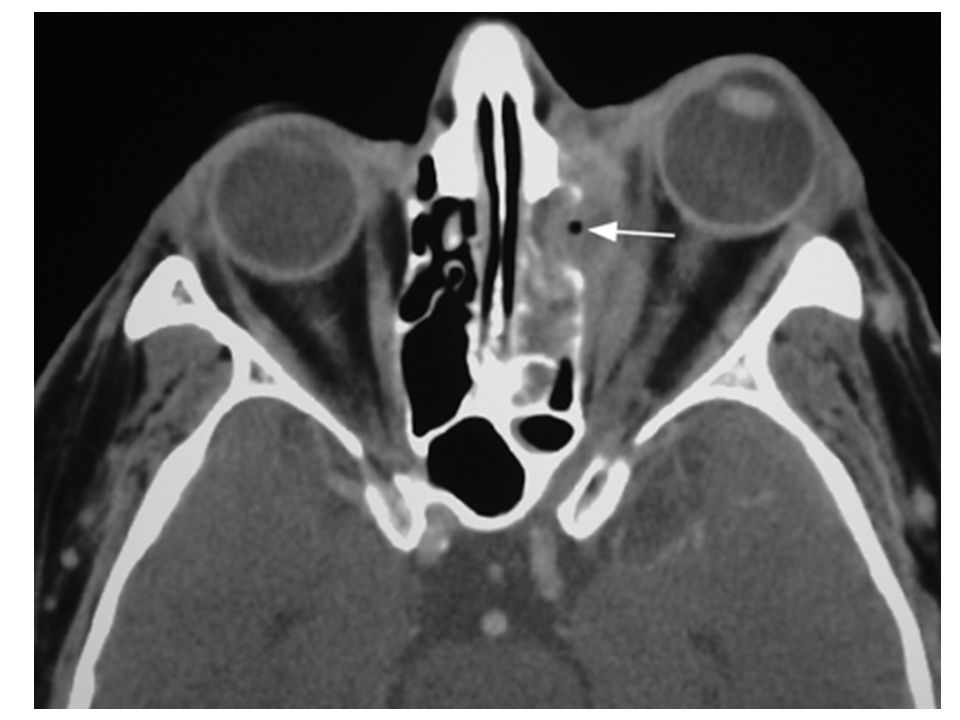
Patients are typically young, atopic and immunocompetent.
The patients may have unilateral or bilateral nasal polyposis and thick yellow to green colored nasal discharge. CT Scan demonstrates opacification of multiple sinuses, mucoceles and bone remodelling.

8
The hyperdensity is due to one or a combination of the following:
Inspissated secretions, fungus or blood

9
Treatment AFRS is treated through a combination of medical and surgical methods. Surgery is employed to open and evacuate PNS Immunotherapy Leukotrine inhibitors Systemic steroids Macrolides Anti-fungals

10
Mycetoma Classically described in immunocompetent persons
It is a non-invasive fungal infection. The most frequently isolated organism is Aspergillosis fumigatus.

11
The patients present with old symptoms of:
Nasal obstruction Unilateral purulent nasal discharge Cacosmia Seldom proptosis. In most patients only one sinus (maxillary sinus) is implicated The mycetoma grows in the sinus exerting mass effect
 is implicated. The mycetoma grows in the sinus exerting mass effect.")
12
Radiological findings include partial or complete opacification of the sinus, thickening of bony walls and sclerosis or bone destruction. CT scan of the sinus reveals opacification of the involved sinus with flocculent calcifications. The diagnostics points on histopathology are collection of dense, matted fungal hyphae that are lying separate from mucosa.

13
Mycetoma

14
Treatment The “typical peanut butter” appearance of the mycetoma is seen when opening involved area. Opening the sinus and removal of all the debris is mainstay of treatment. Medical therapy is generally not needed.

16
Invasive fungal rhinosinusitis

17
A life threatening disorder
Fungi invade sinus mucosa, bone, adjacent structures such as eyes or brain. Several organisms have been shown to be causative organisms for the different forms. Most often the causative organisms are Asmycota phylum, Aspergillus species and Mucorales. Different species may coexist.

18
Common immunodeficiency-associated risk factors include:
Patients at risk are immunocompromised with the exception of chronic granulomatous form. Common immunodeficiency-associated risk factors include: Diabetes, AIDS, Hematologic malignancies with leukopenia, leukopenia for other reasons Immunomodulation for organ transplant.

19
Patients who are immunocompromised and present with signs of rhinosinusitis should be examined with nasal endoscopy. Sloughing, crusting, necrosis or hypovascular areas should raise the suspicion for fungal sinusitis.

20
Patients may present with
Epistaxis Infection of facial soft tissues Peri-orbital oedema Proptosis Decreased vision Mental status change Seizures

21
Mortality depends on several factors:
Form of disease Extent of involvement Use of combination surgical and medical therapy Patient immune factors

22
Acute Invasive Fungal Rhinosinusitis
Acute form is present for less than four weeks. It progresses rapidly, can manifest within hours. It may prove fatal in 50-80% patients. Therefore it should be considered an emergency. Tissue involvement can spread rapidly from sinuses to adjacent tissues. Most commonly caused by Aspergillus fumigatus

25
Treatment Treatment is both medical and surgical. Surgical debridement
This may involve radical excision of tissues including removal of orbital contents, overlying soft tissues of face and some involved intracranial tissues.

26
Therapy against underlying immunocompromise.
Medical Treatment Two forms Anti-fungal therapy Therapy against underlying immunocompromise. Aspergillus fusiform: Voriconazole Candid: Fluconazole For neutropenic patients: Amphotericin B No agent identified: Amphotericin B

27
Chronic Invasive Fungal Rhinosinusitis
It is a slowly progressive disease. It is seen both in immuno-competent and immuno-compromised patients. The disorder is usually caused by aspergillus. The condition begins as a fungal ball and then becomes invasive perhaps as a result of immuno-suppression. It has a low degree of invasion.

28
The patients present with previous symptoms of Nasal obstruction
Unilateral facial discomfort An enlarging mass or silent proptosis. CT findings show a hyper dense mass with associated erosion of sinus walls.

30
Treatment Initiate medical treatment with systemic antifungals once invasion is diagnosed. Amphotericin B (2 g/d) is recommended. This can be replaced by ketoconazole or itraconazole once the disease is under control. Surgical treatment is mandatory and can be approached endoscopically in some patients. Consider an external approach when adequate debridement cannot be achieved endoscopically.

Similar presentations