Download presentation
Presentation is loading. Please wait.

1
Structure of the thoracic wall
Dr. Hassan S Shaibah

2
Formed by: Thoracic part of vertebral column (posteriorly). Sternum & costal cartilages (anteriorly). Ribs & intercostal spaces (Laterally). Superiorly: suprapleural membrane. Inferiorly: Diaphragm.
. Superiorly: suprapleural membrane. Inferiorly: Diaphragm.")
3
Sternum Lies in the middle line. Consists of: manubrium sterni,
body of the sternum xiphoid process. Manubrium articulates with: the clavicle first rib upper part of the second costal cartilage. Manubrium lies opposite T3 & T4

4
3 4 Middle Post Superior A 9 8

5
2.Body of the sternum: Articulates with the manubrium (manubriosternal joint). On each side it articulates with 2nd to 7th costal cartilages. The sternum possesses red hematopoietic marrow throughout life, it is a common site for marrow biopsy.

6
3. The xiphoid process: It is a thin plate of cartilage that becomes ossified at its proximal end during adult life. Not attched to ribs or costal cartilages Forms Xiphisternal Joint Lies at level of T9

7
The sternal angle (angle of Louis):
Formed by articulation of manubrium with body of sternum. It lies at level of 2nd costal cartilage, point from which all costal cartilages and ribs are counted. It lies opposite the intervertebral disc between T4 & T5.

8
Ribs There are 12 pairs of ribs, all of which are attached posteriorly to the thoracic vertebrae. The ribs are divided into three categories: True ribs: The upper 7 pairs are attached anteriorly to the sternum by their costal cartilages. False ribs: The 8th, 9th, and 10th pairs of ribs are attached anteriorly to each other and to the 7th rib by means of their costal cartilages and small synovial joints. Floating ribs: The 11th and 12th pairs have no anterior attachment.

9
A. Typical Rib Has an inferior border costal groove, which is related to the intercostal vessels and nerve. The anterior end of each rib is attached to the corresponding costal cartilage.

10
A rib has a head, neck, tubercle, shaft, and angle.
The head has 2 facets for articulation with same numerically corresponding vertebral body and that of the vertebra immediately above.

11
The tubercle has a facet for articulation with the transverse process of the numerically corresponding vertebra.

12
B. Atypical Rib: 1. The first rib:
It is related nerves of the brachial plexus and subclavian artery and vein. Scalenus anterior muscle is attached to its upper surface and inner border. Anterior to the scalenus anterior, the subclavian vein crosses the rib. Posterior to the muscle attachment, the subclavian artery & brachial plexus cross the rib and lie in contact with the bone.

13
Cervical Rib It arises from the transverse process of C7
It may have a free anterior end, OR may be connected to the first rib by a fibrous band, or may articulate with the first rib. It can cause pressure on the brachial plexus in some patients, producing pain down the medial side of the forearm and hand and wasting of the small muscles of the hand. It can also exert pressure on the overlying subclavian artery and interfere with the circulation of the upper limb. The Thoracic Outlet Syndrome

19
Joints of the Chest Wall
A. Joints of the Sternum: The manubriosternal joint between the manubrium and the body of the sternum. A small amount of angular movement is possible during respiration. The xiphisternal joint between the xiphoid process (cartilage) and the body of the sternum. The xiphoid process usually fuses with the body of the sternum during middle age.
 and the body of the sternum. The xiphoid process usually fuses with the body of the sternum during middle age.")
20
1. Joints of the Heads of the Ribs:
B: Joints of the Ribs: 1. Joints of the Heads of the Ribs: The 1st rib & 3 lowest ribs have a single joint their corresponding vertebral body. The 2nd to 9th ribs, head articulates with the corresponding vertebral body and that of the vertebra above it.

21
2. Joints of the Tubercles of the Ribs
B: Joints of the Ribs: 2. Joints of the Tubercles of the Ribs articulates with transverse process of the corresponding vertebra. (This joint is absent on the 11th and 12th ribs.)
")
22
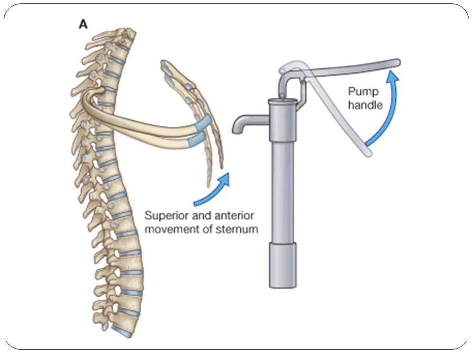
Movements of the Ribs and Costal Cartilages
1st ribs & their costal cartilages are fixed to manubrium & are immobile. The raising and lowering of the ribs during respiration are accompanied by movements in both joints of the head & tubercle, permitting the neck of each rib to rotate around its own axis.

25
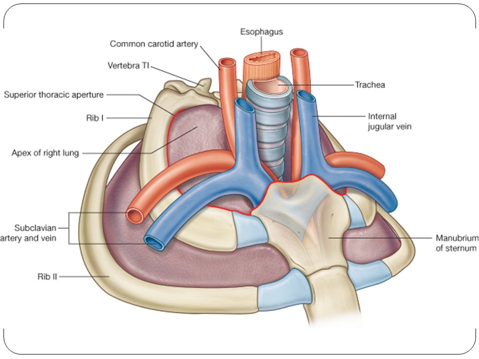
Openings of the Thorax Posteriorly: T1
Thoracic outlet: The chest cavity communicates with root of the neck through an opening called the thoracic outlet. It is called an outlet because important vessels and nerves emerge from the thorax. The opening is bounded: Posteriorly: T1 Laterally: 1ST ribs and their costal cartilages. Anteriorly: manubrium Contains the esophagus and trachea and many vessels and nerves. Because of the obliquity of the opening, The apices of the lung and pleurae project upward into the neck.

26
2. The thoracic cavity communicates with the abdomen through a large opening.
It is bounded: Posteriorly by T12 Laterally by costal margin. c. Anteriorly by the xiphisternal joint. This opening is closed by the diaphragm.

27
Intercostal Spaces * They are spaces between the ribs.
Contain three muscles of respiration: The external intercostal The internal intercostal The innermost intercostal muscle. The intercostal n. and blood vessels run between the intermediate and deepest layers of muscles. They are arranged in the following order from above downward: intercostal vein, intercostal artery, and intercostal nerve (i.e., VAN).
.")
29
I. Intercostal Muscles 1. The external intercostal muscle:
most superficial layer. Its fibers are directed downward and forward from the inferior border of the rib above to the superior border of the rib below. The muscle extends forward to the costal cartilage where it is replaced by an aponeurosis, the anterior (external) intercostal membrane.
 intercostal membrane.")
30
It forms the intermediate layer.
2. The internal intercostal muscle: It forms the intermediate layer. Its fibers are directed downward and backward from the subcostal groove of the rib above to the upper border of the rib below. The muscle extends backward from the sternum in front to the angles of the ribs behind, where the muscle is replaced by an aponeurosis, the posterior (internal) intercostal membrane.
 intercostal membrane.")
31
3. The innermost intercostal muscle
It forms the deepest layer. It is an incomplete muscle layer and crosses more than one intercostal space within the ribs. It is related internally to fascia (endothoracic fascia) and parietal pleura and externally to the intercostal nerves and vessels. It is divided into three portions which are separate from one another.
 and parietal pleura and externally to the intercostal nerves and vessels. It is divided into three portions which are separate from one another.")
32
Action the intercostal muscles can raise the 2nd to the 12th ribs toward the first rib, as in inspiration. 1st to the 11th ribs will be lowered by the contraction of the intercostal muscles, as in expiration. Nerve Supply The intercostal muscles are supplied by the corresponding intercostal nerves.

33
Important : a. The intercostal nerves and blood vessels (the neurovascular bundle) run between the middle and innermost layers of muscles. b. They are arranged in the following order from above downward: intercostal vein, intercostal artery, and intercostal nerve (i.e., VAN).
.")
Similar presentations


oolong weight loss tea