Download presentation
Presentation is loading. Please wait.

1
Renal Diseases Renal cysts and Tumors

2
CYSTIC DISEASES OF THE KIDNEY
Fluid filled spaces within the kidney May involve cortex or medulla or both May be unilateral or bilateral May be unilocular or multilocular May be congenital or acquired May be sporadic or genetically determined Clinical significance may be trivial or grave

3
CLASSIFICATIONS OF RENAL CYSTIC DISEASES
Polycystic kidney diseases: 1. Autosomal recessive (ARPKD) classic infantile polycystic disease with congenital hepatic fibrosis 2. Autosomal dominant (ADPKD) Simple renal cysts Acquired renal cystic disease
 classic infantile polycystic disease. with congenital hepatic fibrosis. 2. Autosomal dominant (ADPKD) Simple renal cysts. Acquired renal cystic disease.")
4
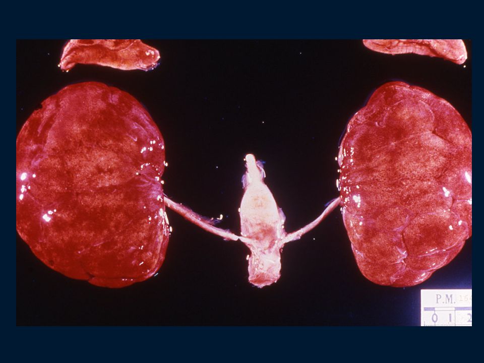
Autosomal Recessive Polycystic Kidney Disease ( ARPKD )
Rare, 1: live births Abnormal gene located on chromosome 6p21 May be still born or neonatal death due to pulmonary insufficiency Progressive renal failure Low specific gravity, mild proteinuria, urine concentrating defect, anemia, hypertension CCF Bilaterally symmetrical enlargement of kidneys Smooth surface with innumerable 1-2 mm cysts Diffuse fusiform dilatation of the collecting ducts

5
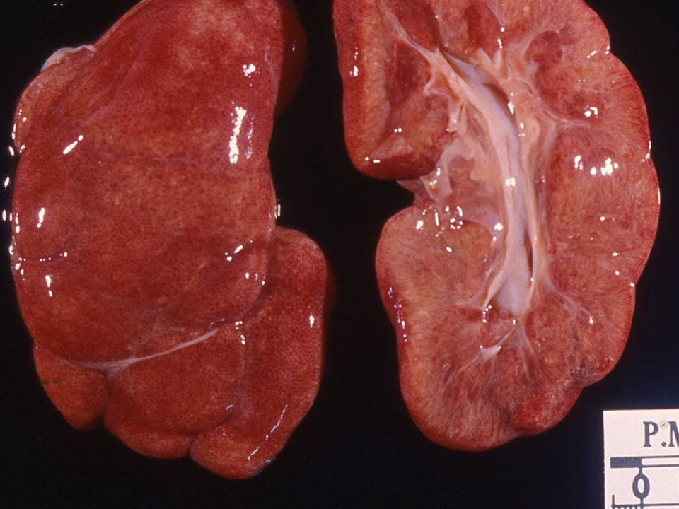
ARPKD ( Cont. ) Enlarged but normally shaped pelvi-calyceal system
Normal reniform shape complete with fetal lobation & normal sized (undilated) ureter Normal glomeruli and tubules Normal interstitium and no dysplasia Congenital hepatic fibrosis is almost always present Normal numbers of nephrons, no interstitial fibrosis and no dysplasia
 ureter. Normal glomeruli and tubules. Normal interstitium and no dysplasia. Congenital hepatic fibrosis is almost always present. Normal numbers of nephrons, no interstitial fibrosis and no dysplasia.")
9
Autosomal Dominant Polycystic Kidney Disease (ADPKD )
World wide, 1:1000 in general population Accounting for 6% of patients in dialysis and transplant program Abnormal gene on short arm of chromosome 16 i.e. 16p13.3(PKD-1). 4q12-22(PKD-2), 3rd locus found Present in 3rd-5th decade Dull loin pain, flank masses. azotemia, positive family history, hypertension, hematuria, colic etc. Prograssive renal failure anemia & GFR, Ur & Cr
. 4q12-22(PKD-2), 3rd locus found. Present in 3rd-5th decade. Dull loin pain, flank masses. azotemia, positive family history, hypertension, hematuria, colic etc. Prograssive renal failure anemia & GFR, Ur & Cr.")
10
ADPKD (CONT.) Associated cysts in liver of biliary type, in pancreas (10%), spleen,thyroid & seminal vesicle Vascular anomalies including Berry aneurysms(33%), aortic aneurysm, aortic root dilatation and mitral valve prolapse Diverticular disease of the colon (80%)
, aortic aneurysm, aortic root dilatation and mitral valve prolapse. Diverticular disease of the colon (80%)")
11
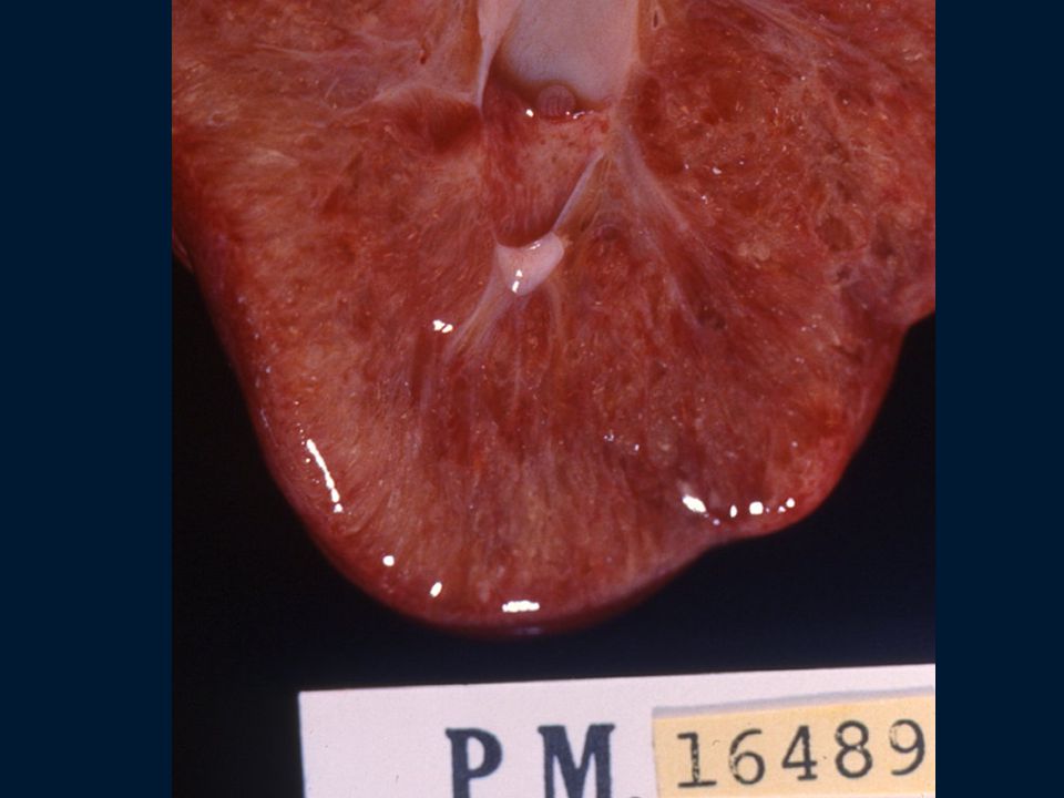
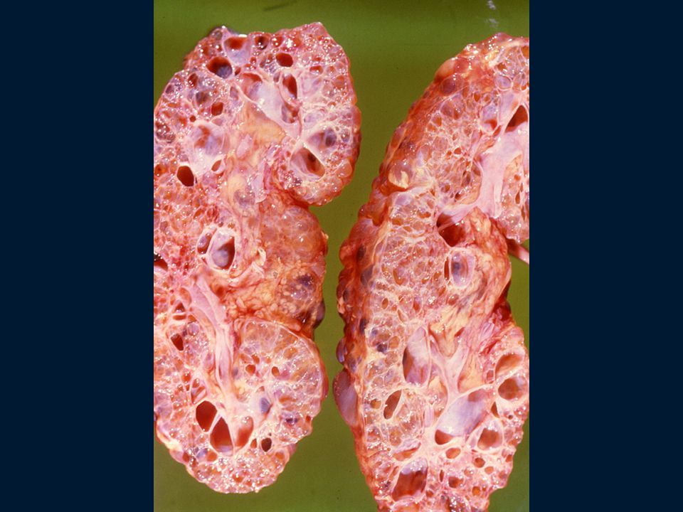
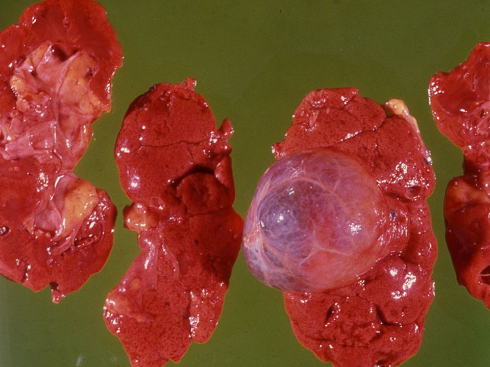
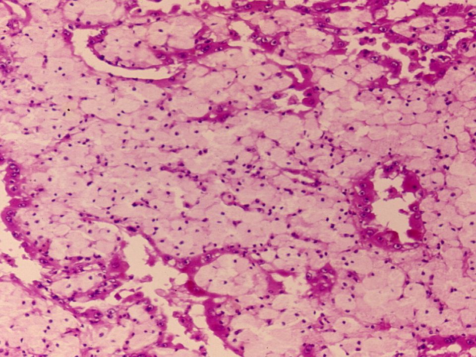
ADPKD- Pathological Features
Bilaterally enlarged kidneys (up to 4000 gms) Diffuse cystic (1-2% cystic nephrons) change with uninvolved intervening parenchyma Varying sized, numerous to innumerable generally spherical unilocular cysts, distributed in cortex and medulla obscuring normal reniform shape and corticomedullary junction, containing yellowish to turbid to brown to black colored fluid Distorted pelvi-calyceal system Cysts arising from any part of nephron or collecting duct
 Diffuse cystic (1-2% cystic nephrons) change with uninvolved intervening parenchyma. Varying sized, numerous to innumerable generally spherical unilocular cysts, distributed in cortex and medulla obscuring normal reniform shape and corticomedullary junction, containing yellowish to turbid to brown to black colored fluid. Distorted pelvi-calyceal system. Cysts arising from any part of nephron or collecting duct.")
12
ADPKD- PATHOLOGICAL FEATURES
Cysts lined by single layer of cuboidal epithelium Neoplastic change - uncommon

15
Simple Renal Cysts Extremely common as age advances
Incompletely understood pathogenesis Commonly associated with scarred kidneys Asymptomatic with normal renal function May be solitary/multiple/unilateral/bilateral Generally unilocular, round to oval of varying sizes

16
SIMPLE RENAL CYSTS (Cont.)
Arises as dilated tubules or collecting ducts Thin, translucent fibrous wall containing clear or amber colored serous fluid

19
ACQUIRED RENAL CYSTIC DISEASE
Secondary to end-stage renal disease as well as prolonged dialysis ( 9 months -7 yrs.) Kidneys are generally small but may be normal or even enlarged Common in cortex Seems to be arising in either proximal or distal tubules Neoplastic change are common
 Kidneys are generally small but may be normal or even enlarged. Common in cortex. Seems to be arising in either proximal or distal tubules. Neoplastic change are common.")
21
RENAL TUMOURS In infants and children :
Nephroblastoma ( Wilms’ tumour ) In adults : Renal cell carcinoma Renal cell adenoma Renal oncocytoma
 In adults : Renal cell carcinoma. Renal cell adenoma. Renal oncocytoma.")
22
NEPHROBLASTOMA ( Wilms’ tumour )
Embryonal tumour arising from nephrogenic blastemal cells can differentiate in to several cell lines - blastemal, epithelial and stromal many replicate developing kidneys Common in young children / uncommon in neonates and infants 90% in < 6yrs. old ( mean: 3yrs. in boys and 3.5yrs. in girls )
")
23
NEPHROBLASTOMA Etiology and Pathogenesis
Generally unknown World wide i.e. … No environmental factors Variable incidence in racial groups : blacks> whites> Orientals Familial tendencies: 1% autosomal dominant with variable penetrance & expressivity Genetic predispositions WT-1 gene (11p13); WT-2 gene (11p15.5)
; WT-2 gene (11p15.5)")
24
NEPHROBLASTOMA Clinical Features
Most common genitourinary cancer Age: 1-3yrs., 98% in <10yrs Abdominal mass, pain, & hematuria Usually unicentric, may be multicentric (7%) or bilateral (5%) Imaging technique to reveal smaller lesions No specific tumor markers identified
 or bilateral (5%) Imaging technique to reveal smaller lesions. No specific tumor markers identified.")
25
NEPHROBLASTOMA Pathologic findings (gross)
Usually solitary, sharply (well) defined masses with pseudocapsule Variable size & weight ( gms. with a mean of 550 gms.) Uniform, pale gray to tan, divided by prominent fibrous septa in to lobules May be cystic, hemorrhagic or necrotic No specific location
 defined masses with pseudocapsule. Variable size & weight ( gms. with a mean of 550 gms.) Uniform, pale gray to tan, divided by prominent fibrous septa in to lobules. May be cystic, hemorrhagic or necrotic. No specific location.")
27
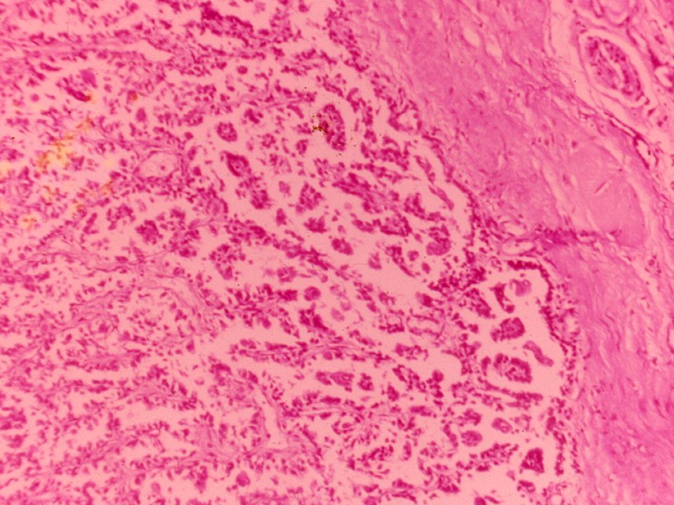
NEPHROBLASTOMA Microscopic findings
Generally triphasic pattern : blastemal, epithelial and stromal cell type may contains heterologous elements “favorable” or “unfavorable” histology on the bases of nuclear anaplasia i.e marked nuclear enlargement (3x) abnormal mitoses i.e increased DNA
 abnormal mitoses i.e increased DNA.")
28
NEPHROBLASTOMA - Spread
Local Regional i.e. . . lymphatic Distant : lungs liver

29
NEPHROBLASTOMA prognosis and treatment
Depends upon : stage, age and histology Surgery with chemotherapy for : stage I & II with favorable histology surgery with chemotherapy and radiotherapy for higher stages and unfavorable histology

30
RENAL CELL CARCINOMA Hypernephroma / Grawitz’s tumour
3% of all adult malignancies world wide, no racial predispositions M:F = 1.6:1, 6th decade (mean 55 yrs) seems to be arising from mature renal tubules tobacco - smoked / chewed
 seems to be arising from mature renal tubules. tobacco - smoked / chewed.")
31
RENAL CELL CARCINOMA ( Cont. )
Chromosomal abnormalities : 3p13 - clear or granular cell type trisomy 17/ tri-or tetrasomy 7 - papillary type Rare familial association Associated with acquired cysts and / or in patients on chronic hemodialysis

32
RENAL CELL CARCINOMA Clinical Features & Diagnosis
classic triad : hematuria, flank pain and abdominal mass may be clinically occult, 30% presents with metastatic lesion Polycythemia due to erythropoietin constitutional symptoms imaging techniques - useful

38
RENAL CELL CARCINOMA mode of dissemination
Direct extension Vascular Lymphatic Lungs, lymph nodes, liver, bone brain, skin etc.

39
RENAL CELL CARCINOMA prognosis
Influenced by multiple factors : tumour size infiltrative margins histological type tumour stage - most important Can be expressed in terms of histological types

40
RENAL CELL ADENOMA Incidental findings at autopsy (22%)
Well demarcated, unencapsulated Pale yellow-gray, discrete cortical mass Up to 2 cms. in maximum dimension

Similar presentations


>")


![Case Report # [] Submitted by:Kandra Vogt, MSIV Faculty reviewer:Sandra A. A. Oldham, M.D. Date accepted:31 August 2007 Radiological Category:Principal.](/18/5671912/big_thumb.jpg "Case Report # [] Submitted by:Kandra Vogt, MSIV Faculty reviewer:Sandra A. A. Oldham, M.D. Date accepted:31 August 2007 Radiological Category:Principal.>")


