Download presentation
Presentation is loading. Please wait.

1
The Integumentary System
The body’s largest organ is the skin ~ 9-11 pounds (7% total body wt) An average adult male covers 20 square feet An average adult female covers 17 square feet Skin is constantly flaking away and replaced completely every 4 weeks On average, every person sheds 40 pounds of skin in a lifetime Everything you see when you look at someone is dead!
 An average adult male covers 20 square feet. An average adult female covers 17 square feet. Skin is constantly flaking away and replaced completely every 4 weeks. On average, every person sheds 40 pounds of skin in a lifetime. Everything you see when you look at someone is dead!")
2
The Integumentary System
Would you be enticed by an advertisement for a coat that is waterproof, stretchable, washable, and permanent-press, that automatically repairs small cuts, rips and burns, and that is guaranteed to last a lifetime with reasonable care?

3
Integumentary System Integument means “covering” Structures
Skin (cutaneous membrane) Cut- or cutis- = the true skin; -ous = possessing, full of Covers 15 to 20 square feet Average weight is 9 pounds (7% of body weight) Composed of two layers Epidermis (epithelial) and dermis (fibrous CT) Skin derivatives Sweat glands, oil glands, hairs and nails
 Cut- or cutis- = the true skin; -ous = possessing, full of. Covers 15 to 20 square feet. Average weight is 9 pounds (7% of body weight) Composed of two layers. Epidermis (epithelial) and dermis (fibrous CT) Skin derivatives. Sweat glands, oil glands, hairs and nails.")
4
Skin (Integument) Functions
Mechanical (physical) protection (barrier) Hardness of keratinized cells (intact epidermis) Chemical protection (barrier) Melanin produced by melanocytes provides shield to prevent UV damage “Acid mantle” Acidic secretions that inhibits bacteria Desiccation Prevention of drying out (keratin waterproofs)
 protection (barrier) Hardness of keratinized cells (intact epidermis) Chemical protection (barrier) Melanin produced by melanocytes provides shield to prevent UV damage. Acid mantle Acidic secretions that inhibits bacteria. Desiccation. Prevention of drying out (keratin waterproofs)")
5
Skin (Integument) Functions
Biological protection (barrier) Langerhans’ cells in epidermis and macrophages (phagocytes) in dermis ingest foreign substances and pathogens
 Langerhans’ cells in epidermis and macrophages (phagocytes) in dermis ingest foreign substances and pathogens.")
6
Skin (Integument) Functions
Temperature Regulation Normal conditions sweat glands produce ~ 500 mL of sweat a day If body temperature rises, nervous system stimulates blood vessels to dilate and sweat glands to secrete sweat Heat loss Activate sweat glands produce sweat which evaporate and cool the body Allow blood to flush in skin capillary beds and radiate from surface of the skin Heat retention Not allowing blood to flush into skin capillary beds

7
Skin Functions Cutaneous sensation
Cutaneous sensory receptors (nervous system) Exteroceptors (respond to external stimuli) Detect pain, light pressure, deep pressure, and temperature (hot and cold) Meissner’s corpuscles (dermal papillae) Light pressure and touch Pacinian corpuscles (deep dermis) Deep pressure Free nerve endings Painful stimuli like chemicals, temperature change, or pain
 Exteroceptors (respond to external stimuli) Detect pain, light pressure, deep pressure, and temperature (hot and cold) Meissner’s corpuscles (dermal papillae) Light pressure and touch. Pacinian corpuscles (deep dermis) Deep pressure. Free nerve endings. Painful stimuli like chemicals, temperature change, or pain.")
8
Skin (Integument) Functions
Metabolic functions (chemical reactions) Modified cholesterol molecules in skin are converted to vitamin D by sunlight Without vitamin D calcium cannot be absorbed in the intestines Blood reservoir Dermal vascular supply is extensive and can hold large volumes of blood (about 5% of entire blood volume)
 Modified cholesterol molecules in skin are converted to vitamin D by sunlight. Without vitamin D calcium cannot be absorbed in the intestines. Blood reservoir. Dermal vascular supply is extensive and can hold large volumes of blood (about 5% of entire blood volume)")
9
Skin (Integument) Functions
Excretion Nitrogen-containing wastes, water and salts are eliminated in sweat

10
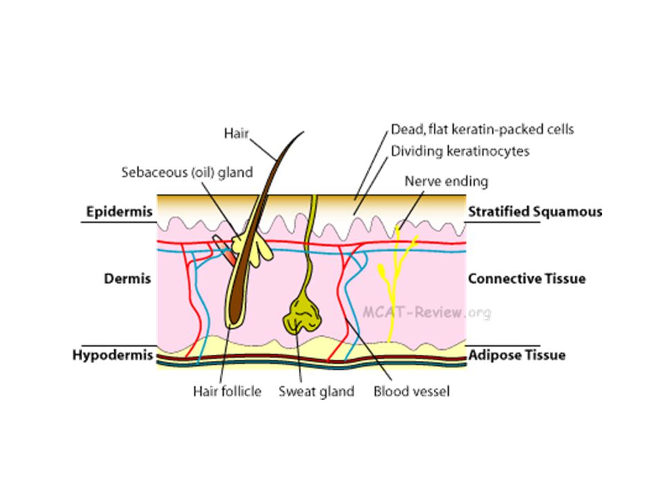
Skin Structure

11
Skin Structure Epidermis (outer layer) (Epi - = upon, on)
Keratinized stratified squamous epithelium Avascular (without blood vessels) Oxygen and nutrients diffuse from dermis Dermis (inner layer) Dense fibrous CT with collagen and elastic fibers Much thicker Contains blood vessels, nerve fibers, etc. Hypodermis (underlying layer or subcutaneous) Adipose tissue Anchors skin to the underlying structures Shock absorber and insulator to reduce heat loss
 Oxygen and nutrients diffuse from dermis. Dermis (inner layer) Dense fibrous CT with collagen and elastic fibers. Much thicker. Contains blood vessels, nerve fibers, etc. Hypodermis (underlying layer or subcutaneous) Adipose tissue. Anchors skin to the underlying structures. Shock absorber and insulator to reduce heat loss.")
13
Cells of the Epidermis Keratinocytes (most) Melanocytes
Langerhans’ (dendritic) cells Tactile (Merkel) cells
 cells. Tactile (Merkel) cells.")
14
Cells of the Epidermis Keratinocytes Primary cell type of epidermis
Produces keratin Protein that gives epidermis its protective function Arise in deepest layer (stratum basale) New cells push old cells upward and by the time they reach the surface they are dead cells filled with keratin Millions of cells rub off each day Completely new epidermis every days Persistent friction causes a thickening of the epidermis called a callus
 New cells push old cells upward and by the time they reach the surface they are dead cells filled with keratin. Millions of cells rub off each day. Completely new epidermis every days. Persistent friction causes a thickening of the epidermis called a callus.")
15
Cells of the Epidermis Melanocytes Spider-shaped epithelial cells
Produce the pigment melanin Found in the deepest layer of epidermis (stratum basale) keratinization In keratinocytes the melanin accumulate on ‘sunny side’ or superficial side of nucleus and forms a shield that protects it from UV radiation – natural sunscreen
 keratinization. In keratinocytes the melanin accumulate on ‘sunny side’ or superficial side of nucleus and forms a shield that protects it from UV radiation – natural sunscreen.")
16
Cells of the Epidermis Langerhans’ (dendritic) cells
White blood cells that arise from bone marrow and migrate to epidermis Ingest foreign substances Key activators of immune system Tactile (Merkel) cells Shaped like a spiky hemisphere Rarely found and associated with sensory nerve ending Sensory receptor for touch
 cells. Shaped like a spiky hemisphere. Rarely found and associated with sensory nerve ending. Sensory receptor for touch.")
17
Layers of Epidermis Variation of thickness depends on location in body
Four or five structurally different layers called strata Thick skin – palms, fingertips, soles of feet consists of all five layers Thin skin – rest of the body the stratum lucidum is not present Deep to superficial Stratum basale Stratum spinosum Stratum granulosum Stratum lucidum (thick skin only) Stratum corneum
 Stratum corneum.")
18
Layers of Epidermis Stratum basale
Deepest layer – closest to dermis Cells rest on underlying dermis Basal cells 90% are keratinocytes 10% are melanocytes and other types Stratum spinosum (prickly cell layer) Several cell layers thick of flattened cells with bundles of keratin Langerhans’ (dendritic) cells are most abundant in this layer
 Several cell layers thick of flattened cells with bundles of keratin. Langerhans’ (dendritic) cells are most abundant in this layer.")
19
Layers of Epidermis Stratum granulosum Stratum lucidum (clear layer)
Very thin region (3-5 layers) Last layer that can obtain nutrients from diffusion Process of keratinization begins here and cells die Cells flatten and ‘toughen’ up Stratum lucidum (clear layer) 2-3 rows of clear, flat, dead cells Translucent layer that looks like a band Visible only in thick skin
 Last layer that can obtain nutrients from diffusion. Process of keratinization begins here and cells die. Cells flatten and ‘toughen’ up. Stratum lucidum (clear layer) 2-3 rows of clear, flat, dead cells. Translucent layer that looks like a band. Visible only in thick skin.")
20
Layers of Epidermis Stratum corneum Outermost layer
20 to 30 cells thick of dead cells High levels of keratin Protection against abrasion and penetration, water repellant (prevents water loss) Average person sheds 8 lb of dead skin cells in a year
 Average person sheds 8 lb of dead skin cells in a year.")
21
Epidermis Check-up While walking barefoot in the barn, Jeremy stepped on a rusty nail that penetrated the depth of the epidermis on the sole of his foot. Name the layers the nail pierced from the superficial skin surface to the junction with the dermis. What is the stratum basale’s function? Given that epithelia are avascular, what layer would be expected to have the best-nourished cells? Stratum basale stratum spinosum stratum granulosum stratum lucidum stratum corneum To produce new keratinocytes and keratin Stratum basale

22
Dermis (Derm = skin) Second major skin region It is your “hide”
Strong, flexible connective tissue layer Cells Fibroblasts, macrophages, mast cells and white blood cells Matrix Embedded with collagen, elastic and reticular fibers Rich in nerve fibers, blood vessels, and lymphatic vessels Major portions of hair follicles, sweat and oil glands reside in dermis

23
Dermis (Derma = skin) Two major layers of dermis Papillary layer
Reticular layer

24
Dermis (Derma = skin) Papillary layer
Papilla = any nipple-like projection Thin superficial layer Areolar connective tissue Superior surface is called the dermal papillae Projections that protrude into epidermis Some contain capillary loops, free nerve endings, and Meissner’s corpuscles Where we get our fingerprint

25
Dermis – your ‘hide’ Reticular layer Deepest layer
Dense connective tissue 80% of the thickness of the dermis Highly vascular - lots of blood vessels Matrix contains thick bundles of collagen fibers (strength and resiliency) and elastic fibers (stretch-recoil) Wrinkling of the skin is caused by loss of the elasticity of the skin Many appendages are located here: sweat glands, oil glands and nerve receptors
 and elastic fibers (stretch-recoil) Wrinkling of the skin is caused by loss of the elasticity of the skin. Many appendages are located here: sweat glands, oil glands and nerve receptors.")
26
Skin Structure Figure 4.4

27
Dermis Check Up What layer of the dermis is responsible for producing finger-print patterns? What cell component of the hypodermis makes it a good shock absorber? You have just gotten a paper cut. It is very painful, but it doesn’t bleed. Has the cut penetrated into the dermis or just the epidermis? What component of the layer was affected? Papillary layer – dermal papillae Adipocytes Epidermis – tactile cells

28
Normal Skin Color Skin Color Variations Skin Appendages

29
Normal Skin Color Three pigments contribute to skin color
Melanin (only one made in skin) Carotene Hemoglobin
 Carotene. Hemoglobin.")
30
Normal Skin Color Melanin (Melan - = black)
Pigment found in eyes, skin and hair Function is to protect cells from UV radiation – a natural sunscreen Range in color from yellow to tan to reddish-brown to black Produced by melanocytes and phagocytized by keratinocytes

31
Normal Skin Color Melanin
Exposure to sun = increase in melanin production Darker-skinned people tend to live near the equator and those with lighter skin live near the poles Freckles and moles are local accumulation of melanin Albinism (albin- = white) No melanin produced; eyes, skin and hair are white
 No melanin produced; eyes, skin and hair are white.")
32
Normal Skin Color Carotene
Yellow-orange pigment found in certain plant products such as carrots Found predominantly in the stratum corneum Can be changed by altering diet and it is not dangerous to health Carotene can be converted to Vitamin A in the liver Essential for normal vision and protection of epithelial tissues

33
Normal Skin Color Hemoglobin
Oxygen carrying pigment found in red blood cells and gives them their red color Produces a pinkish hue of fair skin Caucasian skin contains small amounts of melanin, so the color can show through easier Oxygen content determines the extent of red coloring When capillaries are open the blood is rushed to the surface causing redness

34
Skin Color Variations Cyanosis (Blue color disorder)
Hemoglobin is poorly oxygenated The skin and blood appear blue Occurs during heart failure and respiratory disorders In darker skinned individuals, the skin does not appear cyanotic because of so much melanin Apparent in mucous membranes and nail beds Redness (Erythema) Capillaries dilate and open up to bring blood to the surface Caused by irritation or injury Other causes are embarrassment, fever, hypertension or allergy
 Capillaries dilate and open up to bring blood to the surface. Caused by irritation or injury. Other causes are embarrassment, fever, hypertension or allergy.")
35
Skin Color Variations Pallor or blanching (Paleness or white)
Blood is either shunted or directed away from surface Fear, anger, anemia or low blood pressure. Jaundice (yellow cast) Caused by the buildup of yellow bile pigments from the liver called bilirubin They accumulate in the blood and are deposited in body tissues like the skin or whites of eyes
 Caused by the buildup of yellow bile pigments from the liver called bilirubin. They accumulate in the blood and are deposited in body tissues like the skin or whites of eyes.")
36
Skin Color Variations Hematomas (Bruises)
Collection of blood, usually clotted, in a tissue or organ, usually caused by a break in a blood vessel Black and blue bruises Hema- = blood and -oma = tumor

38
Appendages of the Skin Derivatives of the epidermis called skin appendages Nails, sweat glands, sebaceous (oil) glands and hair Each appendage plays a unique role in maintaining homeostasis

39
Sudoriferous (sweat) glands
Distributed over the entire skin except nipples, lips, and some genitalia Most numerous gland ~2.5 million/person Two types Eccrine and apocrine Simple, coiled tubular glands Sweat is secreted into and through the gland’s duct system to the surface of the skin Sudor = sweat

40
Eccrine glands Found almost everywhere and most numerous
Abundant on palms, soles and forehead Secretory part lies coiled in dermis Duct extends to open in a pore Eccrine gland secretion (sweat) Sweat 99% water with some salts, vitamin C, antibodies and traces of wastes Composition of sweat depends on genetics and diet Acidic with pH between 4-6
 Sweat. 99% water with some salts, vitamin C, antibodies and traces of wastes. Composition of sweat depends on genetics and diet. Acidic with pH between 4-6.")
41
Apocrine Glands Found in axillary and genital areas (about 2000 of them) Found deeper in dermis and larger than eccrine glands Ducts empty into hair follicles Apocrine gland sweat composition Same as eccrine sweat plus fatty substances and proteins Odorless and appears milky or white Bacteria eats organic molecules in the sweat and causes body odor Begin functioning at puberty

42
Functions of Sweat Controlled by the nervous system
Can be heat-induced or emotionally induced (cold sweat) Heat-induced begins on forehead and spreads inferiorly Emotionally induced begins on the palms, soles and then spreads Excretes waste products Acidic nature inhibits bacteria growth
 Heat-induced begins on forehead and spreads inferiorly. Emotionally induced begins on the palms, soles and then spreads. Excretes waste products. Acidic nature inhibits bacteria growth.")
43
Sudoriferous glands Ceruminous glands Modified apocrine glands
Found in lining of external ear canal Produce cerumen (ear wax) Functions to deter insects and block entry of foreign materials. Mammary glands Specialized sweat glands Secretes milk
 Functions to deter insects and block entry of foreign materials. Mammary glands. Specialized sweat glands. Secretes milk.")
44
Sebaceous (Oil) Glands
Found all over the body except on palms and soles of feet Secrete an oily substance called sebum Softens and lubricates hair and skin Bactericidal (bacteria-killing) action Develop and secrete into hair follicles Stimulated by hormones and activated at puberty
 action. Develop and secrete into hair follicles. Stimulated by hormones and activated at puberty.")
45
Sebaceous (Oil) Glands
Blackheads Appears on surface of skin when a sebaceous gland duct is blocked and the sebum accumulates Acne An active inflammation of the sebaceous glands accompanied by ‘pimples’ (pustules) Usually caused by bacterial infection
 Usually caused by bacterial infection.")
46
Plotting of Sweat Glands

47
Hair Millions of hairs are distributed all over the body except on the palms, soles, lips, nipples, and parts of external genitalia Flexible strands produced by hair follicles Consist largely of dead, keratinized cells Melanocytes provide pigment for hair color Hair Functions Not really used for warmth anymore Sense insects on the skin before they bite us Hair on scalp guards the head from physical trauma, heat loss and sunlight Eyelashes shield the eyes Nose hairs filter large particles from the air

48
Hair Regions of a hair Hair Shaft Projects from the skin
Extends halfway down the embedded portion Hair Root Remainder of hair deep in the follicle

49
Hair Anatomy Hair has 3 concentric layers of keratinized cells
Medulla - central core Consists of large cells and open spaces Cortex – surrounds medulla Several layers of flattened cells Cuticle - outside of cortex Single layer of cells that overlap each other Looks like shingles on a roof Most heavily keratinized and provides strength “Split ends” occurs when the cuticle wears away

50
Hair Follicle Epithelial tissue folds down from the epidermis into the dermis Deep end of the follicle forms a hair bulb Hair follicle is the wall around the hair Two sheaths Outer connective tissue root sheath derived from the dermis Inner epithelial root sheath derived from the epidermis Figure 4.7a

51
Hair Follicle Hair papilla Hair matrix (growth zone)
Dermal connective tissue protrudes into the hair bulb Contains capillaries that supplies the nutrients to the hair Hair matrix (growth zone) Actively dividing area of the hair bulb Produces the hair Figure 4.7a
 Actively dividing area of the hair bulb. Produces the hair. Figure 4.7a.")
52
Hair Follicle Arrector pili
Each hair follicle has a smooth muscle attached to it Hair follicles approach surface of the skin at a slight angle Muscle contraction pulls the hair follicle to an upright position Causes a dimple in the skin – goosebumps Response to fear or cold temperatures

53
Hair Growth Hair growth is influenced by many factors
Nutrition and hormones Hirsutism (excessive hairiness) Results from an adrenal gland or ovarian tumor that secretes an abnormally high amount of androgens Androgens control the growth of hair and are produced by ovaries and adrenal glands These tumors are usually removed as soon as possible
 Results from an adrenal gland or ovarian tumor that secretes an abnormally high amount of androgens. Androgens control the growth of hair and are produced by ovaries and adrenal glands. These tumors are usually removed as soon as possible.")
54
Hirsuitism

55
Hair Thinning and Baldness
A hair follicle only has a limited number of cycles in it Hair grows fastest from the teen years to the 40s and then it slows Alopecia (scientific term for baldness) When the hairs are not replaced as they are shed leads to hair thinning or baldness due to age True Baldness Male pattern baldness that is genetically determined, sex-influenced condition Hair thinning can occur because of high fever, surgery, emotional trauma, certain drugs, and protein-deficient diets If the cause is removed or corrected, the hair will grow back Hair loss due to severe burns or follicular damage is permanent
 When the hairs are not replaced as they are shed leads to hair thinning or baldness due to age. True Baldness. Male pattern baldness that is genetically determined, sex-influenced condition. Hair thinning can occur because of high fever, surgery, emotional trauma, certain drugs, and protein-deficient diets. If the cause is removed or corrected, the hair will grow back. Hair loss due to severe burns or follicular damage is permanent.")
57
Skin Homeostatic Imbalances
A disease or condition as a result of a disturbance in homeostasis Skin Homeostatic Imbalances Cancer Burns Bacterial, viral or yeast infections

58
Skin Cancer Cancer Two types of tumors
When cells fail to follow normal controls of cell division and multiply excessively it results in a tumor Two types of tumors Benign (most tumors like warts) Does not spread and grows slowly Malignant (mal = ‘bad’) Grow relentlessly and may become killers Metastasis Cells tend to break away from the mass and travel via blood or lymph to other body organs where they can grow into new masses
 Does not spread and grows slowly. Malignant (mal = ‘bad’) Grow relentlessly and may become killers. Metastasis. Cells tend to break away from the mass and travel via blood or lymph to other body organs where they can grow into new masses.")
59
Skin Cancer Types 1 in 5 Americans will develop skin cancer at some point Huge risk factor is too much UV radiation which damages DNA bases Three types of skin cancer Basal cell carcinoma Squamous cell carcinoma Melanoma

60
Skin Cancer Types Basal cell carcinoma
Least malignant but most common skin cancer (80%) Stratum basal cells proliferate and invade the dermis and hypodermis Occur most often on sun-exposed areas of face Appear as shiny, dome-shaped nodules that develop a central ulcer with a pearly, beaded edge – sore that doesn’t heal
 Stratum basal cells proliferate and invade the dermis and hypodermis. Occur most often on sun-exposed areas of face. Appear as shiny, dome-shaped nodules that develop a central ulcer with a pearly, beaded edge – sore that doesn’t heal.")
61
Skin Cancer Types Basal cell carcinoma

62
Skin Cancer Types Squamous cell carcinoma
Second most common skin cancer Affects men more than women Arises from keratinocytes of the stratum spinosum Lesion appears as a scaly reddened papule that arises usually on head and hands Grows and metastasizes to lymph nodes quickly Early removal allows a good chance of cure

63
Skin Cancer Types Basal Cell Carcinoma Squamous Cell Carcinoma
Cell type Stratum basale (basal layer) keratinocytes Stratum spinosum keratinocytes Rank them by how common they are to get 1st 2nd Seriousness and metastasis Least malignant Grows and metastasizes quickly Appearance Shiny, dome-shaped nodules with a central ulcer and a pearly edge Scaly, reddened papule
 keratinocytes. Stratum spinosum keratinocytes. Rank them by how common they are to get. 1st. 2nd. Seriousness and metastasis. Least malignant. Grows and metastasizes quickly. Appearance. Shiny, dome-shaped nodules with a central ulcer and a pearly edge. Scaly, reddened papule.")
64
Skin Cancer Types

65
Skin Cancer Types Malignant melanoma Most deadly of skin cancers
Highly metastatic and resistant to chemotherapy Rare (2-3% of all skin cancers) Cancer of melanocytes and can begin wherever there is pigment Appears as a spreading brown to black patch Early detection is key to survival Detection uses ABCD rule
 Cancer of melanocytes and can begin wherever there is pigment. Appears as a spreading brown to black patch. Early detection is key to survival. Detection uses ABCD rule.")
66
ABCD Rule A = Asymmetry Two sides of pigmented mole do not match
B = Border irregularity Borders of mole are not smooth C = Color Different colors in pigmented area D = Diameter Spot is larger then 6 mm in diameter (pencil eraser) Skin Cancer Video
 Skin Cancer Video.")
67
Skin Homeostatic Imbalances
Burns Tissue damage inflicted by intense heat (fire or materials), electricity, radiation (sunlight), or certain chemicals Denature cell proteins and cause cell death Immediate threat to life is loss of body fluids containing proteins and electrolytes Results in dehydration and electrolyte imbalance After initial crises, infection and sepsis becomes the main threat
, electricity, radiation (sunlight), or certain chemicals. Denature cell proteins and cause cell death. Immediate threat to life is loss of body fluids containing proteins and electrolytes. Results in dehydration and electrolyte imbalance. After initial crises, infection and sepsis becomes the main threat.")
68
Extent of Burn - Rules of Nines
Volume of fluid lost can be estimated by computing the percentage of body surface burned Tool to approximate the extent of burns using the rule of nines Body is divided into 11 areas Each area represents about 9% of total body area Head and neck = 9 % Upper limbs = 18% Trunk = 36% Lower limbs = 36% Genitals =1%

69
Severity of Burns Burns are classified according to their severity (depth) First-degree burns (epidermis) Skin is red and swollen Second degree burns (epidermis and upper dermis) Skin is red with blisters Third-degree burns (entire skin layer) High risk for fluid loss Burn is gray-white, cherry red or black Nerve endings are destroyed so it is not painful at first Skin regeneration may occur but will take too long Usually skin grafting is required
 Skin is red with blisters. Third-degree burns (entire skin layer) High risk for fluid loss. Burn is gray-white, cherry red or black. Nerve endings are destroyed so it is not painful at first. Skin regeneration may occur but will take too long. Usually skin grafting is required.")
70
Critical Burns Burns are considered critical if:
Over 25% of body has second degree burns Over 10% of the body has third degree burns There are third degree burns of the face, hands, or feet Most third degree burns will never heal and may require a skin graft Skin Grafting Removal and transplanting of healthy skin from one area to another

71
Infections & Allergies
Psoriasis A chronic condition characterized by reddened epidermal lesions covered with dry, silvery scales. Athlete’s Foot An itchy, red, peeling condition of the skin between the toes resulting from a fungal infection. Cold Sores Small fluid filled blistered that itch and sting, caused by a herpes simplex infection.

72
Infections & Allergies
Contact Dermatitis Itching, redness, and swelling of the skin, progressing to blistering. Caused by the exposure of skin to chemicals that provoke allergic responses. (it is = swelling) Impetigo Pink, water-filled, raised lesions that develop (mouth and nose) a yellow crust and eventually rupture . Caused by staphylococcus infection.
 Impetigo. Pink, water-filled, raised lesions that develop (mouth and nose) a yellow crust and eventually rupture . Caused by staphylococcus infection.")
Similar presentations


 glands Sebaceous glands.>")


 Largest organ of the body (15% of body weight) Skin thickness variable, normally 1-2 mm Protection –chemical barrier (waterproof)>")


