Download presentation
Presentation is loading. Please wait.

1
Increased water in brain parenchyma.
Edema Increased water in brain parenchyma.

2
Types of Herniations Subfalcine herniation (cingulate gyrus)
Transtentorial Herniation (uncal gyral) Tonsillar Herniation
 Tonsillar Herniation.")
3
Mass Tumor Blood clot Abscess Local edema S T

4
Transtentorial Herniation
[involve Uncal gyral]

5
Transtentorial Herniation
[Uncus]

6
Cerebral Herniation Complication of Intracranial Hypertension
Tonsillar herniation Cerebellar tonsils herniate into the foramen magnum. Causes "coning" of the cerebellar tonsils Produces cardiorespiratory arrest Coma and Death

7
It can occur as an complication of Lumber Puncture

8
Complications of Tonsillar Herniation & Increased ICP
Hemorrhagic lesion of the mid Brain and Pons : Secondary Brain stem or Duret hemorrhage Linear hemorrhage

9
Duret hemorrhage: pathogenesis
Kinking of the penetrating median and paramedian pontine arteries that branch off the basilar artery.

10
Duret hemorrhage : causes
Tonsillar Herniation Intracranial Neoplasm Intracranial hemorrhage (basal ganglia )
")
11
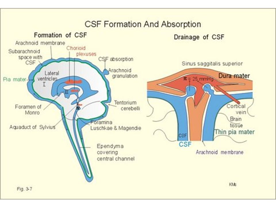
Hydrocephalus Accumulation of excessive CSF within the ventricular system & enlargement

12
CT scan of Hydrocephalous

13
Overproduction of CSF (choroid plexus tumor)
")
14
Communicating Hydrocephalous.
Meningitis. Subarachnoid hemorrhage Obstruction in subarachnoid space

15
Non communicating Hydrocephalous (Obstructive)
Medulloblastoma, Ependymoma No communication between ventricles and subarachonoid space.

16
Congenital hydrocephalous (present at birth)
Aqueductal stenosis (narrowing) is the most frequent cause. Blockage of fourth ventricle outlet (Dandy Walker Syndrome) – due to congenital malformation
 is the most frequent cause. Blockage of fourth ventricle outlet (Dandy Walker Syndrome) – due to congenital malformation.")
17
The gross shows hydrocephalus of the frontal horns of the lateral ventricles.

18
Hydrocephalous before the fusion of the cranial Sutures [Head circumference increase]
![Hydrocephalous before the fusion of the cranial Sutures [Head circumference increase]](http://slideplayer.com/slide/3451305/12/images/18/Hydrocephalous+before+the+fusion+of+the+cranial+Sutures+%5BHead+circumference+increase%5D.jpg "Hydrocephalous before the fusion of the cranial Sutures [Head circumference increase]")
19
Hydrocephalous after the fusion of the Sutures, produce Ventricular expansion and Increased Intracranial Pressure

20
Anencephaly Anencephaly showing absence of the brain and opening of the spinal canal. 20 20

21
Spina bifida Types of spina bifida: A: Spina bifida occulta B: Meningocele C: Meningomyelocele 21

22
Syringomyelia Note the collapsed cystic cavity (syrinx) in the center of the cervical spinal cord 22

23
Brain contusion The contrecoup injury involves the frontal and temporal lobes (left arrows) The coup lesion (site of impact) involves the cerebellum (right arrow). 23
 involves the cerebellum (right arrow). 23.")
24
Epidural and Subdural Hematoma
A, Schematic of epidural hematoma and subdural hematoma. B, Epidural hematoma. Note the blood is located on top of the dura (arrow). C, Subdural hematoma. The reflected dura shows the outer membrane of an organized venous clot covering the convexity of the brain. 24 24
. C, Subdural hematoma. The reflected dura shows the outer membrane of an organized venous clot covering the convexity of the brain")
25
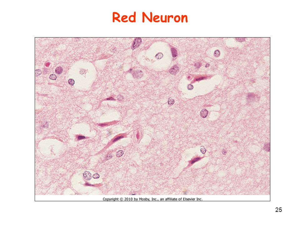
Red Neuron Red neurons. Note the brightly eosinophilic staining cells with the pyknotic nuclei within spaces representing apoptotic neurons. 25 25
26
Border zone infarct: Watershed infarct : Follows a Hypotensive episode.
Lesion lies at the boundary between the anterior and middle cerebral artery territories.

27
Atherosclerotic (thrombotic) stroke
27

28
Amaurosis fugax Cholesterol embolus to retinal artery. Note the yellow embolus trapped at the bifurcation of the retinal artery (arrow). This produces a sudden, painless loss of vision ("curtain coming down") followed in a variable period of time by restoration of vision ("curtain coming up") as the embolus dislodges. This is called amaurosis fugax. 28 28
. This produces a sudden, painless loss of vision ( curtain coming down ) followed in a variable period of time by restoration of vision ( curtain coming up ) as the embolus dislodges. This is called amaurosis fugax")
29
Embolic Stroke Embolic stroke showing a wedge-shaped hemorrhagic infarction (arrow) along the periphery of the cerebral cortex in the distribution of the middle cerebral artery. It is hemorrhagic because blood flow was reestablished when the embolus dislodged and converted a pale infarct to a hemorrhagic infarct. 29 29
 along the periphery of the cerebral cortex in the distribution of the middle cerebral artery. It is hemorrhagic because blood flow was reestablished when the embolus dislodged and converted a pale infarct to a hemorrhagic infarct")
30
Intracerebral Hemorrhage
Intracerebral hemorrhage, showing a large blood clot within the basal ganglia area of the brain. 30 30

31
Berry Aneurysms 31

32
Subarachnoid Hemorrhage
Subarachnoid hemorrhage. Note the presence of blood covering the surface of the brain. 32 32

33
Lacunar Infarcts The arrows show multiple small cystic spaces (liquefactive necrosis) that are most prominent in the basal ganglia. Sections under these lesions showed hyaline arteriolosclerosis. The arrows show multiple small cystic spaces (liquefactive necrosis) that are most prominent in the basal ganglia. 33 33
 that are most prominent in the basal ganglia. Sections under these lesions showed hyaline arteriolosclerosis. The arrows show multiple small cystic spaces (liquefactive necrosis) that are most prominent in the basal ganglia")
34
Lacunar infarcts Cause : Chronic hypertension Site: The pons.

35
Pyogenic Meningitis

36
Microscopically, a neutrophilic exudate is seen involving the meninges

37
Bacterial infections. A, Pyogenic meningitis
Bacterial infections. A, Pyogenic meningitis. A thick layer of suppurative exudate covers the brain stem and cerebellum, and thickens the leptomeninges. B, Cerebral abscesses in the frontal white matter (arrows). (A, From Golden JA, Louis DN: Images in clinical medicine: acute bacterial meningitis.
. (A, From Golden JA, Louis DN: Images in clinical medicine: acute bacterial meningitis.")
38
Mechanism for the conversion of PrPc through protein-protein interactions. The initiating molecules of PrPsc may arise through inoculation (as in directly transmitted cases) or through an extremely low-rate spontaneous conformational change. The effect of the mutations in PrPc is to increase the rate of the conformational change once PrPsc is able to recruit and convert other molecules of PrPc into the abnormal form of the protein. Although the model is drawn with no other proteins involved, it is possible that other proteins play critical roles in the conversion of Prpc to PrPsc.
 or through an extremely low-rate spontaneous conformational change. The effect of the mutations in PrPc is to increase the rate of the conformational change once PrPsc is able to recruit and convert other molecules of PrPc into the abnormal form of the protein. Although the model is drawn with no other proteins involved, it is possible that other proteins play critical roles in the conversion of Prpc to PrPsc..")
39
Spongiform encephalopathy of gray matter : brain lesion in CJD

40
Complications: sequel
Edema can lead to herniation and death. Communicating hydrocephalous.

41
Meningeal Syphilis 1 of 2 Neurosyphilis is a tertiary stage of syphilis – only in 10% with untreated syphilis May involve spinal Meninges: produce thickening. Produce meningeal fibrosis and secondary Hydrocephalous.

42
Neutrophils in the abscess.

43
Abscess in the brain in a patient who had septicemia.

44
Atrophy There is marked atrophy seen superiorly and laterally.

45
The cortical atrophy leads to compensatory dilation of the cerebral ventricles [hydrocephalus ex vacuo ]
![The cortical atrophy leads to compensatory dilation of the cerebral ventricles [hydrocephalus ex vacuo ]](http://slideplayer.com/slide/3451305/12/images/45/The+cortical+atrophy+leads+to+compensatory+dilation+of+the+cerebral+ventricles+%5Bhydrocephalus+ex+vacuo+%5D.jpg "The cortical atrophy leads to compensatory dilation of the cerebral ventricles [hydrocephalus ex vacuo ]")
46
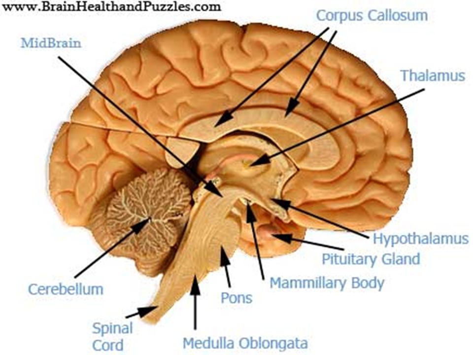
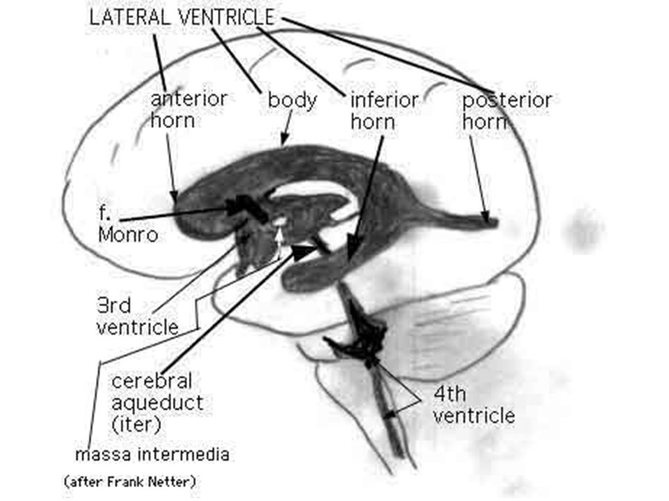
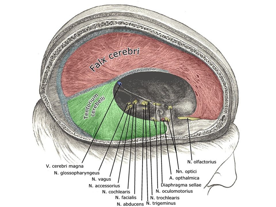
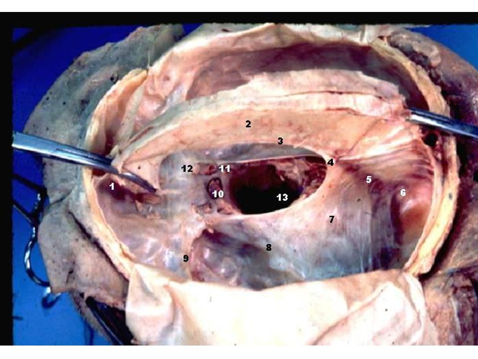
For anatomy, not for exam
Self Study For anatomy, not for exam

Similar presentations


>")


