Download presentation
Presentation is loading. Please wait.

1
Fluid and Electrolytes & Renal Disorders

2
Topics for the Day Fluids and Electrolytes: review of normal physiology * Fluid imbalances * Electrolyte Disturbances * Beginning acid-base imbalance * Renal Disorders Fluid Types *

4
Electrolytes Solutes that form ions (electrical charge)
Cation (+) Anion (-) Major body electrolytes: Na+, K+, Ca++, Mg++ Cl-, HCO3-, HPO4--, SO4-
 Anion (-) Major body electrolytes: Na+, K+, Ca++, Mg++ Cl-, HCO3-, HPO4--, SO4-")
5
Fluid & Electrolytes Fluid: Water
Electrolytes: ions dissolved in water Sodium, potassium, bicarbonate, etc. Also used medically for non ions (glucose) Osmolarity – osmols/kg solvent Osmolality – osmols/liter solution In clinical practice are used interchangeably
 Osmolarity – osmols/kg solvent. Osmolality – osmols/liter solution. In clinical practice are used interchangeably.")
6
Electrolyte Distribution
Major ICF ions K+ HPO4-- Major ECF ions NA+ CL-, HCO3- Intravascular (IVF) vs Interstitial (ISF) Similar electrolytes, but IVF has proteins
 vs Interstitial (ISF) Similar electrolytes, but IVF has proteins.")
7
Mechanisms Controlling Fluid and Electrolyte Movement
Diffusion Selective Permeability Facilitated diffusion Active transport Osmosis 2*Na + BUN + Glucose/18 Hydrostatic pressure Oncotic pressure

11
Cells are selectively permeable

12
Sodium is the largest Determinant of Osmolality
Na+: 135 – 145 mEq/L Ca+: 8.5 – 10.5 mEq/L K+: 3.5 – 5 mEq/L Osmolality~ 2*(Na+) = 2*( mEq/L) Normal (Isotonic) 280 – 300 Low (hypotonic) < 280 High (hypertonic) > 300
 = 2*( mEq/L) Normal (Isotonic) 280 – 300. Low (hypotonic) < 280. High (hypertonic) > 300.")
13
Fluid Exchange Between Capillary and Tissue: Sum of Pressures
Fig. 17-8

14
Fluid Shifts Plasma to interstitial fluid shift results in edema
Elevation of hydrostatic pressure Decrease in plasma oncotic pressure Elevation of interstitial oncotic pressure

15
Fluid Movement between ECF and ICF
Water deficit (increased ECF) Associated with symptoms that result from cell shrinkage as water is pulled into vascular system Water excess (decreased ECF) Develops from gain or retention of excess water
 Associated with symptoms that result from cell shrinkage as water is pulled into vascular system. Water excess (decreased ECF) Develops from gain or retention of excess water.")
16
Fluid Spacing First spacing: Normal distribution of fluid in ICF and ECF Second spacing: Abnormal accumulation of interstitial fluid (edema) Third spacing: Fluid accumulation in part of body where it is not easily exchanged with ECF (e.g. ascites)
")
17
Regulation of Water Balance
Hypothalamic regulation Pituitary regulation Adrenal cortical regulation Renal regulation Cardiac regulation Gastrointestinal regulation Insensible water loss

18
F&E Balance Renin Epinephrine Angiotensin I Atria (ANP)
Ventricles (BNP) Endothelium (CNP) Angiotensin II Aldosterone
 Endothelium (CNP) Angiotensin II. Aldosterone.")
19
Fluid Status Indicators
Physical exam Mucous membranes Turgor Blood Hematocrit Plasma BUN Urine Output (volume) Specific Gravity* < 1.003: less conc > 1.030: more conc Electrolytes
 Specific Gravity* < 1.003: less conc. > 1.030: more conc. Electrolytes.")
20
F&E Balance Fluids Electrolytes (Sodium!!!) Normal Contracted Expanded
Isotonic Hypertonic Hypotonic

22
Extracellular Fluid Deficit
Causes Inadequate intake, diuresis, excess sweating, burns, diarrhea, vomiting, hemorrhage Treatment Stop underlying disorder Replace fluids appropriately Treat complications

23
D5W Hypotonic ½ NS ½ NS (0.45%) Crystalloids Isotonic NS (0.9%) Lactated Ringer Hypertonic Plasmalyte IV Fluids 3% Saline Albumin D5W in ½ NS Dextran Colloids D10W FFP PRBCs

24
Volume Deficit Isotonic Deficit Hypertonic Deficit Hypotonic Deficit
Electrolyte drinks Isotonic saline (0.9%) injection Hypertonic Deficit Drinking Water Hypotonic saline (0.45%) injection, D5W Hypotonic Deficit Isotonic Saline Hypertonic saline (3%)
 injection. Hypertonic Deficit. Drinking Water. Hypotonic saline (0.45%) injection, D5W. Hypotonic Deficit. Isotonic Saline. Hypertonic saline (3%)")
25
Extracellular Fluid Excess
Causes The Three failures: heart, liver, kidney Treatment Remove fluid --> ???? Treat underlying disorder

26
Electrolyte Normal Values (memorize!!!!!)
Sodium 135 – 145 Potassium 3.5 – 5 Chloride 106 – 106 Calcium 9 – 11 BUN 10 – 20 Creatinine 0.7 – 1.2 CO2 (really bicarb) 22 – 26 Magnesium: 1.5 – 2.5
 22 – 26. Magnesium: 1.5 – 2.5.")
27
Electrolyte Disorders: Signs & Symptoms (most common*)
Hypokalemia Bradycardia ECG changes CNS changes Fatigue Hyperkalemia Ventricular fibrillation Weakness Potassium (K) Hyponatremia CNS deterioration Hypernatremia Thirst Increased interstitial fluid Sodium (Na) Deficit Excess Electrolyte
 Hyponatremia. CNS deterioration. Hypernatremia. Thirst. Increased interstitial fluid. Sodium (Na) Deficit. Excess. Electrolyte.")
28
Electrolyte Disorders Signs and Symptoms
Hypomagnesemia Hyperactive DTRs CNS changes Hypermagnesemia Loss of deep tendon reflexes (DTRs) Depression of CNS Depression of neuromuscular function Magnesium (Mg) Hypocalcemia Tetany Chvostek’s, Trousseau’s signs Muscle twitching ECG changes Hypercalcemia Thirst CNS deterioration Increased interstitial fluid Calcium (Ca) Deficit Excess Electrolyte
 Depression of CNS. Depression of neuromuscular function. Magnesium (Mg) Hypocalcemia. Tetany. Chvostek’s, Trousseau’s signs. Muscle twitching. ECG changes. Hypercalcemia. Thirst. CNS deterioration. Increased interstitial fluid. Calcium (Ca) Deficit. Excess. Electrolyte.")
29
Hypernatremia Manifestations Impaired LOC Produced by clinical states
Thirst, lethargy, agitation, seizures, and coma Impaired LOC Produced by clinical states Central or nephrogenic diabetes insipidus Reduce levels gradually to avoid cerebral edema

30
Hypernatremia Treatment
Treat underlying cause If oral fluids cannot be ingested, IV solution of 5% dextrose in water or hypotonic saline Diuretics if necessary

31
Hyponatremia Results from loss of sodium-containing fluids
Sweat, diarrhea, emesis, etc. Or from water excess Inefficient kidneys Drowning, excessive intake Manifestations Confusion, nausea, vomiting, seizures, and coma

32
Treatment Oral NaCl If caused by water excess
Fluid restriction is needed If Severe symptoms (seizures) Give small amount of IV hypertonic saline solution (3% NaCl) If Abnormal fluid loss Fluid replacement with sodium- containing solution
 Give small amount of IV hypertonic saline solution (3% NaCl) If Abnormal fluid loss. Fluid replacement with sodium- containing solution.")
33
Hyperkalemia High serum potassium caused by
Massive intake Impaired renal excretion Shift from ICF to ECF (acidosis) Drugs Common in massive cell destruction Burn, crush injury, or tumor lysis False High: hemolysis of sample
 Drugs. Common in massive cell destruction. Burn, crush injury, or tumor lysis. False High: hemolysis of sample.")
34
Hyperkalemia Manifestations Weak or paralyzed skeletal muscles
Ventricular fibrillation or cardiac standstill Abdominal cramping or diarrhea

36
Treatment Emergency: Calcium Gluconate IV Stop K intake
Force K from ECF to ICF IV insulin Sodium bicarbonate Increase elimination of K (diuretics, dialysis, Kayexalate)
")
37
Hypokalemia Low serum potassium caused by
Abnormal losses of K+ via the kidneys or gastrointestinal tract Magnesium deficiency Metabolic alkalosis

38
Hypokalemia Manifestations Most serious are cardiac
Skeletal muscle weakness Weakness of respiratory muscles Decreased gastrointestinal motility

39
Hypokalemia KCl supplements orally or IV
Should not exceed 10 to 20 mEq/hr To prevent hyperkalemia and cardiac arrest No Pee no Kay!!!!!!!!!!!!!!!!!!!!!!!!!

40
Calcium Obtained from ingested foods
More than 99% combined with phosphorus and concentrated in skeletal system Inverse relationship with phosphorus Otherwise…

41
Calcium Bones are readily available store
Blocks sodium transport and stabilizes cell membrane Ionized form is biologically active Bound to albumin in blood Bound to phosphate in bone/teeth Calcified deposits

42
Calcium Functions Transmission of nerve impulses
Myocardial contractions Blood clotting Formation of teeth and bone Muscle contractions

43
Calcium Balance controlled by Bone used as reservoir
Parathyroid hormone Calcitonin Vitamin D/Intake Bone used as reservoir

44
Hypercalcemia High serum calcium levels caused by
Hyperparathyroidism (two thirds of cases) Malignancy (parathyroid tumor) Vitamin D overdose Prolonged immobilization
 Malignancy (parathyroid tumor) Vitamin D overdose. Prolonged immobilization.")
45
Hypercalcemia Manifestations Decreased memory Confusion Disorientation
Fatigue Constipation

46
Treatment Excretion of Ca with loop diuretic
Hydration with isotonic saline infusion Synthetic calcitonin Mobilization

47
Hypocalcemia Low serum Ca levels caused by Decreased production of PTH
Acute pancreatitis Multiple blood transfusions Alkalosis Decreased intake

48
Hypocalcemia Manifestations Weakness/Tetany
Positive Trousseau’s or Chvostek’s sign Laryngeal stridor Dysphagia Tingling around the mouth or in the extremities

49
Treatment Treat cause Oral or IV calcium supplements
Not IM to avoid local reactions Treat pain and anxiety to prevent hyperventilation-induced respiratory alkalosis

50
Phosphate Primary anion in ICF
Essential to function of muscle, red blood cells, and nervous system Deposited with calcium for bone and tooth structure

51
Phosphate Involved in acid–base buffering system, ATP production, and cellular uptake of glucose Maintenance requires adequate renal functioning Essential to muscle, RBCs, and nervous system function

52
Hyperphosphatemia High serum PO43 caused by Manifestations
Acute or chronic renal failure Chemotherapy Excessive ingestion of phosphate or vitamin D Manifestations Calcified deposition: joints, arteries, skin, kidneys, and corneas Neuromuscular irritability and tetany

53
Hyperphosphatemia Management Identify and treat underlying cause
Restrict foods and fluids containing PO43 Adequate hydration and correction of hypocalcemic conditions

54
Hypophosphatemia Low serum PO43 caused by
Malnourishment/malabsorption Alcohol withdrawal Use of phosphate-binding antacids During parenteral nutrition with inadequate replacement

55
Hypophosphatemia Manifestations CNS depression Confusion
Muscle weakness and pain Dysrhythmias Cardiomyopathy

56
Hypophosphatemia Management Oral supplementation
Ingestion of foods high in PO43 IV administration of sodium or potassium phosphate

57
Magnesium 50% to 60% contained in bone
Coenzyme in metabolism of protein and carbohydrates Factors that regulate calcium balance appear to influence magnesium balance

58
Magnesium Acts directly on myoneural junction
Important for normal cardiac function

59
Hypermagnesemia High serum Mg caused by
Increased intake or ingestion of products containing magnesium when renal insufficiency or failure is present

60
Hypermagnesemia Manifestations Lethargy or drowsiness Nausea/vomiting
Impaired reflexes*** Respiratory and cardiac arrest

61
Hypermagnesemia Management Prevention Emergency treatment
IV CaCl or calcium gluconate Fluids to promote urinary excretion

62
Hypomagnesemia Low serum Mg caused by Prolonged fasting or starvation
Chronic alcoholism Fluid loss from gastrointestinal tract Prolonged parenteral nutrition without supplementation Diuretics

63
Hypomagnesemia Manifestations Confusion
Hyperactive deep tendon reflexes Tremors Seizures Cardiac dysrhythmias

64
Hypomagnesemia Management Oral supplements (MgO, MgSO4)
Increase dietary intake Parenteral IV or IM magnesium when severe

65
Elemenary Acid-Base balance
Buffer systems Carbonic Acid Bicarbonate Metabolic: bicarb low → metabolic acidosis high → metabolic alkalosis Respiratory: carbon dioxide

66
Metabolic Panel and acid-base
“CO2” on a BMP means bicarb!!!!!! normal 22 – 26 <22 = ? >26 =?

67
Metabolic Acidosis Manifestat
Acidosis causes HYPERKALEMIA!!! Neuro: Drowsiness, Confusion, H/A, coma CV: ↓BP, dysrhythmia (K+), dilation GI: NVD, abd pain Resp: increased resp (comp)
, dilation. GI: NVD, abd pain. Resp: increased resp (comp)")
68
Metabolic Alkalosis Manifestat
Alkalosis causes HYPOKALEMIA!!! Neuro: Dizziness, Irritability, Nervous, Confusion CV: ↑HR, dysrhythmia (K+) GI: NV, anorexia Neuromuscular: Tetany, tremor, paresthesia, seizures Resp: decreased resp (comp)
 GI: NV, anorexia. Neuromuscular: Tetany, tremor, paresthesia, seizures. Resp: decreased resp (comp)")
69
MEMORIZE Arterial pH, PaCO2, HCO3-!!!!!!!

70
Interpretation of ABGs
Diagnosis in six steps Evaluate pH Analyze PaCO2 Analyze HCO3- Determine if Balanced or Unbalanced Determine if CO2 or HCO3- matches the alteration Decide if the body is attempting to compensate

71
Interpretation of ABG pH over balance PaCO2 = “respiratory” balance
HC03- = “metabolic” balance If all three normal = balanced Match direction. e.g., if pH and PaCO2 are both acidotic, then primary respiratory acidosis If other is opposite, then partial compensation; if pH normal, then fully compensated.

72
Interpretation of ABGs
pH 7.36 PaCO2 67 mm Hg PaO2 47 mm Hg HCO3 37 mEq/L What is this?

73
Interpretation of ABGs
pH 7.18 PaCO2 38 mm Hg PaO2 70 mm Hg HCO3- 15 mEq/L What is this?

74
Interpretation of ABGs
pH 7.60 PaCO2 30 mm Hg PaO2 60 mm Hg HCO3- 22 mEq/L What is this?

75
Interpretation of ABGs
pH 7.58 PaCO2 35 mm Hg PaO2 75 mm Hg HCO3- 50 mEq/L What is this?

76
Interpretation of ABGs
pH 7.28 PaCO2 28 mm Hg PaO2 70 mm Hg HCO3- 18 mEq/L What is this ?

77
Putting it all together
Always pay attention to Patient history Vital signs Symptoms and physical exam findings Lab Values Always ask: What is causing this abnormal finding? What can be done to fix it?

78
D5W Hypotonic ½ NS ½ NS (0.45%) Crystalloids Isotonic NS (0.9%) Lactated Ringer Hypertonic Plasmalyte Fluids 3% Saline Albumin D5W in ½ NS Dextran Colloids D10W FFP PRBCs

79
IV Fluids Purposes Maintenance Replacement
When oral intake is not adequate Replacement When losses have occurred

80
D5W (Dextrose = Glucose)
Hypotonic Provides 170 cal/L Free water Moves into ICF Increases renal solute excretion Used to replace water losses and treat hyponatremia Does not provide electrolytes

81
Normal Saline (NS) Isotonic No calories More NaCl than ECF
30% stays in IVF 70% moves out of IV space

82
Normal Saline (NS) Expands IV volume Does not change ICF volume
Preferred fluid for immediate response Risk for fluid overload higher Does not change ICF volume Blood products Compatible with most medications

83
Lactated Ringer’s Isotonic More similar to plasma than NS Expands ECF
Has less NaCl Has K, Ca, PO43, lactate (metabolized to HCO3) CONTRAINDICATED in lactic acidosis Expands ECF
 CONTRAINDICATED in lactic acidosis. Expands ECF.")
84
D5 ½ NS Hypertonic Common maintenance fluid
KCl added for maintenance or replacement

85
D10W Hypertonic Max concentration of dextrose that can be administered in peripheral IV Provides 340 kcal/L Free water Limit of dextrose concentration may be infused peripherally

86
Plasma Expanders Stay in vascular space and increase osmotic pressure
Colloids (protein solutions) Packed RBCs Albumin Plasma Dextran
 Packed RBCs. Albumin. Plasma. Dextran.")
Similar presentations
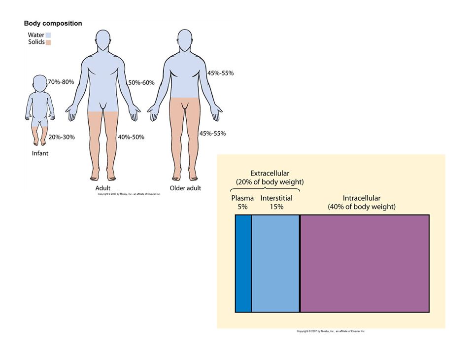


 of TBW is intracellular (ICF) 2/3 (65%) of TBW is intracellular (ICF) 1/3 extracellular.>")


 Integumentary System (chapters 44- 46)>")


