Download presentation
Presentation is loading. Please wait.

1
Neurological Pathophysiology

2
Edema in the CNS Increase in tissue mass that results from the excess movement of body fluid from the vascular compartment or its abnormal retention in the tissue. Why is this a special problem in the brain and spinal cord? Enclosed space Lack of lymphatics Lack of anastomoses in venous drainage

3
Vasogenic edema Occurs when the blood-brain barrier is upset
Inflammation due to infection Toxic agents that damage capillary endothelium Abnormal capillaries associated with malignant neoplasm Leakage of proteins fluid into interstitium → swelling Plasma filtrate accumulation alters ionic balance and impairs function

4
Cytotoxic edema Intracellular phenomenon Hypoxia
Cardiac arrest Near drowning Strangulation Focal edema due to blockage of an end artery Toxic substances that: Impair sodium/potassium pump Impair production of ATP

5
In practice, swelling often caused by both
Treatment is different If swelling is due to cytotoxicity, can give I.V. bolus of a hypertonic solution such as mannitol to draw water into the vasculature and out of the brain If the cause is vasogenic would this help? No! would draw fluid into interstitial space and increase swelling!!

6
Increased intracranial pressure (IICP)
Normal intracranial pressure is 5-15 mm Hg May be due to: Tumor growth Edema Excess cerebrospinal fluid Hemorrhage

7
Contents of cranium Tissue of the Central Nervous System
Cerebrospinal Fluid (CSF) Blood An increase in any one of these increases intracranial pressure. Clinical hallmarks of IICP: Headache Vomiting Papilledema – swelling of the optic discs
 Blood. An increase in any one of these increases intracranial pressure. Clinical hallmarks of IICP: Headache. Vomiting. Papilledema – swelling of the optic discs.")
8
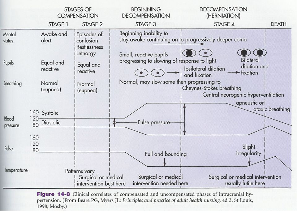
Since the brain is encased in the cranium, the only way pressure can be relieved is by decreasing cranial contents. Most readily displaced is CSF If ICP still high, cerebral blood volume is altered: Stage 1 – vasoconstriction and external compression of the venous system Compensating, so few symptoms

9
If ICP continues to increase, may exceed brain’s ability to adjust.
Stage 2: IICP (gradually rising) causes a decrease of oxygenation of neural tissue Systemic vasoconstriction occurs to increase blood pressure to get blood to brain Clinical manifestations transient: episodes of confusion, restlessness, drowsiness, and slight pupillary and breathing changes
 causes a decrease of oxygenation of neural tissue. Systemic vasoconstriction occurs to increase blood pressure to get blood to brain. Clinical manifestations transient: episodes of confusion, restlessness, drowsiness, and slight pupillary and breathing changes.")
10
When ICP begins to = arterial pressure, there is a lack of compensation- beginning decompensation Stage 3 Hypoxia and hypercapnia → cytotoxic edema Decreasing levels of arousal Widened pulse pressure May begin Cheynes-Stokes respirations Bradycardia – due to increased pressure in carotid arteries Pupils small and sluggish Surgical or medical intervention needed

11
When all compensatory mechanisms have been exhausted:
Stage 4: Dramatic rise in ICP in a short time Autoregulation is lost, and get vasodilation, further increasing intracranial volume ↓ cerebral perfusion = severe hypoxia and acidosis Brain contents shift (herniate) from area of high pressure to areas of lower pressure ↓ blood flow
 from area of high pressure to areas of lower pressure ↓ blood flow.")
12
Small hemorrhages develop
Ipsilateral pupil dilation and fixation, progressing to bilateral fixed and dilated pupils When mean systolic arterial pressure equal ICP, cerebral blood flow ceases

13
Treatment Remove the cause of the IICP
Mechanical hyperventilation to medicated and comatose patient Reduce blood pressure through diuretics, which slows production of CSF and decreases blood-brain volume Drugs, us. Barbiturates to slow brain metabolism and ↓ effects of hypoxia Emergency craniotomy to relieve pressure

15
Brain Trauma Highest risk: Male: female = 3:1 15 to 30 years of age
Infants 6 mo. to two years Young school age children Elderly persons Male: female = 3:1

16
Most likely causes of head injury:
Transportation accidents Falls Sports related events Violence

17
Two major categories of head trauma:
closed (blunt) trauma open (penetrating) trauma
 trauma. open (penetrating) trauma.")
18
Open (penetrating) trauma
Break in dura results in exposure of brain tissues to environment. Results in focal (localized) injury May be due to skull fracture or wound – intracerebral hematoma Traumatic pneumocephalus - injury to a nasal sinus that allows air into brain or ventricles - cerebrospinal rhinorrhea
 injury. May be due to skull fracture or wound – intracerebral hematoma. Traumatic pneumocephalus - injury to a nasal sinus that allows air into brain or ventricles - cerebrospinal rhinorrhea.")
19
Blunt Head Trauma More common than open trauma.
Involves head hitting hard surface or rapidly moving object strikes head Dura is intact – no brain tissue exposed May cause focal or diffuse axonal injury (DAI)
")
20
Serious injury decrease due to :
Seat belt use Improved management

21
Mild cerebral concussion
% of all head injuries Not severe Diffuse axonal injury – no visible signs on brain May see transient dizziness, paralysis, unconsciousness, unequal pupils and shock. Reactive period: vomiting, Temp o, rapid pulse, headache, and cerebral irritation lasting hours.

22
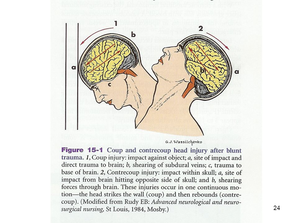
Contusions (bruise) : impacts which lead to hemorrhage and possibly hematoma
Coup (strike) – head strikes against object shearing forces cause small tears in blood vessels (subdural vessels) edema severity = force smaller area = greater force
 – head strikes against object. shearing forces cause small tears in blood vessels (subdural vessels) edema. severity = force. smaller area = greater force.")
23
Contrecoup (rebound) – brain hits opposite side of skull
shearing forces and damage opposite to site of impact

25
Extradural (Epidural) Hematomas
1-2% of major head injuries Most common in year olds Often caused by temporal skull fracture or injury Artery is often the source of bleeding Get herniation (shift) of temporal lobe of brain through tentorial notch
 of temporal lobe of brain through tentorial notch.")
27
Subdural hematomas 10 - 20 % of persons with traumatic brain injury
Develop rapidly (within hours) Typically on top of skull Often due to tearing of veins or dural sinuses Acts as an expanding mass → IICP→ herniation of brain
 Typically on top of skull. Often due to tearing of veins or dural sinuses. Acts as an expanding mass → IICP→ herniation of brain.")
28
Intracerebral hematomas
2-3% of head injuries Single or multiple Usually frontal and temporal lobes May occur in deep white matter Small blood vessels injured by shearing forces Acts as expanding mass, compresses tissue, and causes edema May appear days after head injury

29
Clinical manifestations of contusions
Loss of consciousness, loss of reflexes Transient cessation of breathing Brief bradycardia Decreased blood pressure As hematoma enlarges: headache, vomiting, drowsiness, confusion, seizure, hemiparesis

30
Treatment Contusions: Control intracranial pressure
Drugs can relieve fluid pressures; may alter Na+ conc. in brain fluids Manage symptoms hematomas: Surgical ligation

31
Cerebrovascular Disease
Most frequent of all neurological problems Due to blood vessel pathology: Lesions on walls of vessels leading to brain Occlusions of vessel lumen by thrombus or embolus Vessel rupture Alterations of blood quality

32
CV disease leads to two types of brain abnormalities :
Ischemia (with or without infarct) Hemorrhage
 Hemorrhage.")
33
Cerebrovascular Accident (Stroke)
Clinical expression of cerebrovascular disease: a sudden, nonconvulsive focal neurological deficit Incidence: third leading cause of death in U.S. – half a million people a year – one third will die from it

34
Incidence Highest risk > 65 years of age
But about 1/3 (28%) are < 65 years old Tends to run in families More often seen in females More often seen in Blacks, perhaps due to increased incidence of hypertension
 are < 65 years old. Tends to run in families. More often seen in females. More often seen in Blacks, perhaps due to increased incidence of hypertension.")
35
Three types : Global hypoperfusion – shock
Ischemia – thrombotic and embolic Hemorrhagic

36
Risk Factors Arterial hypertension Heart disease
Myocardial infarction or endocarditis Atrial fibrillation Elevated plasma cholesterol Diabetes mellitus Oral contraceptives Smoking Polycythemia and thrombocythemia

37
Occlusive strokes Occurs with blockage of blood vessel by a thrombus or embolus May be temporary or permanent Thrombotic stroke: 3 clinical types: TIAs Stroke-in-evolution Completed stroke

38
Transient Ischemic Attacks
Last for only a few minutes, always less than 24 hours All neurological deficits resolve Symptom of developing thrombosis

39
Causes: Thrombus formation Vasospasm Other: Atherosclerosis Arteritis
Hypertension Vasospasm Other: Hypotension Anemia Polycythemia

40
Symptoms depend on location
Ophthalmic branch of internal carotid artery – amaurosis fugax – fleeting blindness Anterior or middle cerebral arteries – contralateral monoparesis, hemiparesis, localized, tingling numbness in one arm, loss of right or left visual field or aphasia

41
Treatment Without Tx 80% have a recurrence in symptoms, and 1/3 go on to have a full stroke within 5 years Give anticoagulants prophylactically , usually ½ to 1 aspirin / day

42
Stroke-in-evolution Can have abrupt onset, but develop in a step-by-step fashion over minutes to hours, occasionally, from days to weeks Characteristic of thrombotic stroke or slow hemorrhage

43
Thrombotic CVA Involves permanent damage to brain due to ischemia, hypoxia and necrosis of neurons Most common form of CVA Causes: Atherosclerosis assoc. with hypertension Diabetes mellitus, and vascular disease Trauma

44
May take years to develop, often asymptomatic until major narrowing of arterial lumen
Anything that lowers systemic B.P. will exacerbate symptoms (60 % during sleep) Area affected depends on artery and presence of anastomoses Area affected initially is greater than damage due to edema Infarcted tissue undergoes liquifaction necrosis
 Area affected depends on artery and presence of anastomoses. Area affected initially is greater than damage due to edema. Infarcted tissue undergoes liquifaction necrosis.")
45
Embolic stroke Second most common CVA
Fragments that break from a thrombus outside the brain, or occasionally air, fat, clumps of bacteria, or tumors

46
Common causes Atrial fibrillation Myocardial infarction Endocarditis
Rheumatic heart disease and other defects

47
Impact is the same for thrombotic stroke
Rapid onset of symptoms Often have a second stroke

48
Hemorrhagic Stroke Third most common, but most lethal
Bleeding into cerebrum or subarachnoid space

49
Causes: Ruptured aneurysms Vascular malformations Hypertension
Bleeding into tumors Bleeding disorders Head trauma

50
Often a history of physical or emotional exertion immediately prior to event
Causes infarction by interrupting blood flow to region downstream from hemorrhage Further damage by hematoma or IICP Onset less rapid than embolic CVA, evolving over an hour or two

51
Usually chronic hypertension, and B.P. may continue to rise
About half report severe headache In about 70 % hematoma expands, destroying vital brain centers, shifts of brain tissue, and death

52
Degenerative Disorders
Progressive neurodisorders Long-lasting Permanent effects Many present as syndromes No cure, but much research

53
Alzheimer Disease (Dementia of Alzheimer Type)
")
54
Dementia is a loss of ordered neural function
Discrimination and attending to stimuli Storing new memories and retrieving old Planning and delay of gratification Abstraction and problem solving Judgement and reasoning Orientation in time and space Language processing Appropriate use of objects Planning and execution of voluntary movements

55
Course : slow progression (5years or more)
At first affects only short term memory, but gradually extends to long term Many experience restlessness Many patients retain insight, which leads to anxiety and depression Personality may be lost Ultimately, mute and paralyzed Death comes from infection

56
Onset may be as young as 50, and incidence increases with age:
6 % of people over 65 years have AD Almost half over 85 have AD Diagnosis is by ruling out all other causes –specific diagnosis only by biopsy or autopsy

57
Pathology restricted to cerebral cortex, hippocampus, amygdala, and another basal nucleus called nucleus of Meynert Nucleus of Meynert produces Acetylcholine – loss results in impaired neural function

58
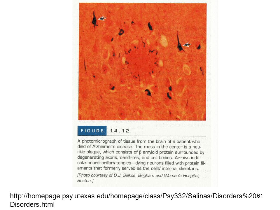
Pathology Pyramidal cells die; loss of white matter
Gyri shrink and and ventricles and sulci expand – walnut like appearance Neurofibrillary tangles Neuritic (senile) plaques : filaments, microglia, astrocytes around core of amyloid Amyloid angiopathy
 plaques : filaments, microglia, astrocytes around core of amyloid. Amyloid angiopathy.")
60
www.infoaging.org/ d-alz-8-r-tangles.html

61
http://homepage. psy. utexas
62
About 10 % of cases are familial, usually early onset
Gene on chromosome 21 – carries gene for amyloid protein Almost all people with Down syndrome who live beyond 45 years develop AD Also linked to mutations on chromosomes 14 and 19 Prions have been isolated

63
Treatment So far resistant May revolve around amyloid protein
See high levels of aluminum – chelating agents temporarily arrest or reverse some symptoms THA(tetrahydroaminoacridine) used experimentally – liver toxicity Arthritics have lower incidence of AD Therapy centers on problems of failing cognitive skills
 used experimentally – liver toxicity. Arthritics have lower incidence of AD. Therapy centers on problems of failing cognitive skills.")
64
Parkinson Disease – movement disorder
Described over 180 years ago by James Parkinson Combination of slowed, reduced movements and restless tremoring Paralysis agitans Slowly degenerative CNS disorder affecting 80,000 adults in North America

65
Parkinson Disease Degenerative disease of the basal ganglia involving the failure of dopamine-secreting neurons (substantia nigra) Can be primary or secondary Secondary caused by trauma, infection, neoplasm, atherosclerosis, toxins and drug intoxication

67
Primary Parkinson Disease
Begins after the age of 40, with peak age of onset between 58 – 62 Course of years – slowly progressive More prevalent in males (slightly to 2X)
")
68
Substantia nigra – two nuclei in midbrain
Outflow pathway from basal nuclei to cortex via thalamus Damage impairs flow of motor programs Expressed as difficulty initiating movements, general lack and slowing of movement (bradykinesia)
")
69
Most disabling symptoms are muscle rigidity and bradykinesia
Loss of feedback loop impairs flow of programs and expresses as resting tremor Most disabling symptoms are muscle rigidity and bradykinesia Muscle strength is more or less normal Poor balance Face becomes immobile and inexpressive Autonomic function is decreased: orthostatic hypotension, excess sweating, constipation, etc.

70
Some patients suffer dementia similar to Alzheimer Disease
Not fatal, but shortens life expectancy

71
Treatment Active exercise and good nutrition
Strategies to overcome bradykinesia Only when symptoms are severe are drugs given – levodopa Side effects: cardiac arrhythmias, gastrointestinal hemorrhage, psychiatric problems, unpredictable involuntary movement disorders

72
Possible therapies Foreign or autograft of tissue still experimental
Lesions in the subthalamic nucleus, thalamus or internal segment of globus pallidus promising Stem cell research?

73
Multiple Sclerosis Focal, chronic, progressive, usually exacerbating and remitting demyelination of CNS tracts. Lesions can occur in a wide variety of locations and give rise to complex symptoms Areas of demyelination are called plaques, and can occur anywhere oligodendrocytes provide myelin sheath

74
Onset Onset is between 20 and 40 years, rarely before 15 or after 50
Females: Males 2:1

75
Clinical presentation depends on site of lesion
Involvement of optic nerve produces monocular visual disturbances – first symptom in ¼ of patients Half of people with optic neuritis are diagnosed with MS

76
Common symptoms: Double vision
Tingling in the back and anterior thigh upon neck flexion – Lhermitte’s sign Symptoms worsen when patient becomes heated – Uhthoff’s sign

77
Diagnosis CSF obtained by lumbar puncture shows slight increase in protein; on electrophoresis shows specific banding pattern – antibodies within CSF suggest immune reaction Changes in velocities of visual and auditory pathways Plaques visible on MRI

78
Cause Myelin undergoes breakdown and phagocytic destruction – antibodies may play a role Decreased signal conduction due to edema and demyelination that exposes potassium channels that short-circuit signal (edema resolves and have partial remyelination)
")
79
Epidemiology hints at interaction between a viral illness in the teen years and a genetic predisposition Growing up in northern temperate climates increases rise Non Asian heritage increases risk

80
Course Pattern of exacerbation and remission
Stresses can trigger exacerbation: infection, medication, stress, fatigue Course is unstable and unpredictable 10 % undergo severe, rapid progressive deterioration Some have died with 7 months of first symptom due to acute brain inflammation and infection

81
A significant number never severely incapacitated
Some experience only a single episode Death is usually attributable to complications of MS (infections due to decreased function)
")
82
Symptoms Increased urinary frequency
Lesions in frontal or temporal lobes can cause emotional outbursts Depression and euphoria can be problems Unpredictable progression taxes ability to cope Occasionally, plaques can cause paraplegia or quadriplegia

83
Therapies Only corticosteroids and ACTH appear to have effects – reduce the duration of exacrerbation, but have no impact on long term outcome Interferon β Maintaining a healthy lifestyle and outlook

84
Acute encephalopathies:
Reye’s Syndrome First recognized in 1963 Characterized by encephalopathy and fatty changes in several organs, esp. liver Incidence has declined in past 20 years due to awareness of ingestion of aspirin during illness and development of Reye’s syndrome.

85
Reye’s syndrome Typically develops in a healthy child of 6 mo. to 15 years recovering from varicella, influenza B, upper respiratory tract infection, or gastroenteritis. Stage I: vomiting, lethargy, drowsiness Stage II: disorientation, delirium, aggressiveness and combativeness, central neurological hyperventilation, shallow breathing, hyperactive reflexes, stupor

86
Reye’s syndrome Stage III: Insensitivity to pain, coma, hyperventilation, rigidity Stage IV: deepening coma, loss of ocular reflexes; large, fixed pupils; divergent eye movements Stage V: seizures, loss of deep tendon reflex, flaccidity, respiratory arrest Mortality is 10 or more Formerly 40 to as high as 80% In a few cases death is due to liver failure

87
Reye’s syndrome Cause: Treatment: May have a genetic predisposition
May be due in part to exhaustion of glycogen stores and use of fatty acids -Mitochondrial injury Treatment: Aggressive intensive care Treatment for brain edema and IICP Fluids I.V. and control of blood electrolytes Prevent hyperthermia

88
Seizure disorders Seizure is and abnormal discharge of electrical activity within the brain. It is a rapidly evolving disturbance of brain function that may produce impaired consciousness, abnormalities of sensation or mental function or convulsive movements. Convulsions are episodes of widespread and intense motor activity

89
Epilepsy A recurrent disorder of cerebral function marked by sudden, brief attacks of altered consciousness, motor activity or sensory phenomenon. Convulsive seizures are the most common form Some, but not all, recurrent seizures are due to epilepsy

90
Epilepsy Second most common neurological disorder
Incidence increases with age, with 30% initially occurring before 4 years and % before 20 years. Causes: brain tumor, scar tissue, neurological disease, great majority of cases are idiopathic.

91
Signs and symptoms vary:
petit mal – almost imperceptible alterations in consciousness grand mal – generalized tonic-clonic seizures – dramatic loss of consciousness, falling, generalized tonic-clonic convulsions of all extremities, incontinence, and amnesia for the event.

92
Some attacks are proceeded by a prodrome – a set of symptoms that warn of a seizure
As the seizure begins, the patient may experience an aura – mental, sensory or motor phenomena Others have no warning

93
Phases of a grand mal seizure
Tonic phase ( seconds) – muscle contraction Epileptic cry – respiration stops Clonic phase – (1/2 -2 minutes) muscle spasms; respiration is ineffective; autonomic nervous system active Terminal phase (about 5 minutes) –limp and quiet, EEG flat lines
 – muscle contraction. Epileptic cry – respiration stops. Clonic phase – (1/2 -2 minutes) muscle spasms; respiration is ineffective; autonomic nervous system active. Terminal phase (about 5 minutes) –limp and quiet, EEG flat lines.")
94
Seizure activity lasts more than 30 minutes
5-8 % are at risk of status epilepticus – a series of GTCS without regaining consciousness – medical emergency Seizure activity lasts more than 30 minutes Acidosis Elevated pCO2 Hypoglycemia Fall in blood pressure Can lead to severe brain damage or death

95
Epileptogenic focus Group of brain neurons susceptible to activation
Plasma membranes may be more permeable to ion movement Firing of these neurons may be greater in frequency and amplitude Electrical activity can spread to other hemisphere and then to the spinal cord

96
Treatment Treat any underlying metabolic disorders, or tumors
Most cases can be controlled through routine use of antiepileptic medications – usually only one drug to minimize side effects Surgical intervention if drugs ineffective Supportive therapy – patients learn to cope effectively with stress, eat well, and get sufficient rest and avoid triggers.

97
Eliciting stimuli: Hypoglycemia Fatigue Emotional or physical stress
Fever Hyperventilation Environmental stimuli

98
Patients are normal between attacks
Can participate in sports, drive a car (if no seizures for 6 mo – 1 year) Should not drink alcohol
 Should not drink alcohol.")
Similar presentations


 (MND) are a group of neurological disorders that selectively affect motor neurons.>")


 Stroke - Overview Third leading cause of death in industrialized countries. Total cost of strokes in the U.S. is roughly.>")


