Download presentation
Presentation is loading. Please wait.

1
Muscles and Movement IB Biology

2
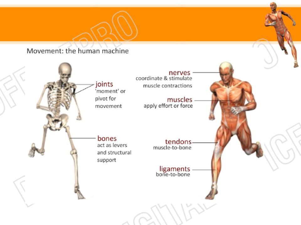
11.2.1 State the role of bones, ligaments, muscles, tendons and nerves in human movement.

4
Joints Also called articulations or arthrosis Two or more bones contact each other Provide mobility Determine the type or range of motion of a particular area of the body

5
Bones Are organs Provide a framework to support the body
Protection of soft tissue and organs Act as levers for body movement Form blood cells in the bone marrow Allow storage of minerals – calcium and phosphorus Bone to bone connection by ligaments Strengthen joint Provide stability

6
Muscles and Tendons Tendons – cords of dense connective tissue
Connect muscles to bone Muscles – provide force for movement by shortening the length of their fibers (cells) Muscles only move by contracting fibers Most muscles occur as antagonistic pairs
 Muscles only move by contracting fibers. Most muscles occur as antagonistic pairs.")
7
Nerves Sensory nerve endings housed in ligaments
Allow for monitoring of the position of the joints Help prevent over-extension of the joint and it parts

8
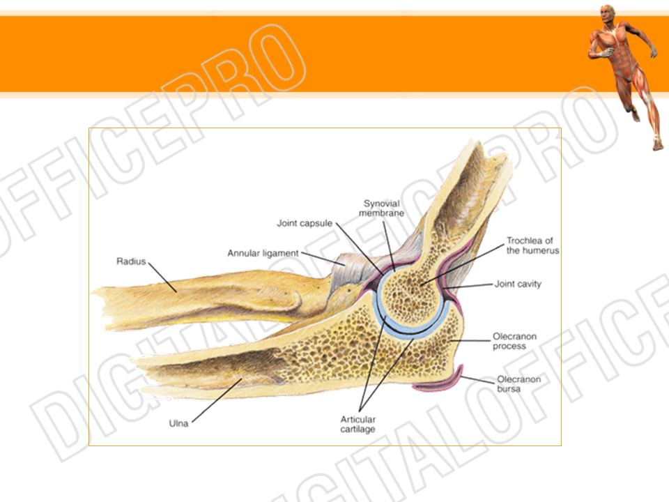
11.2.2 Label a diagram of the human elbow joint, including cartilage, synovial fluid, joint capsule, named bones and antagonistic muscles (biceps and triceps).
.")
9
Hinge joints Works like a door

11
11.2.3 Outline the functions of the structures in the human elbow joint named in

12
Joint Part Function Cartilage Reduces friction & absorbs compression Synovial fluid Lubricates to reduce friction & provides nutrients to the cells of the cartilage Joint capsule Surrounds the joint, encloses the synovial cavity, & unites the connecting bones Tendons Attach muscle to bone Ligaments Connect bone to bone Biceps muscle Contracts to bring about flexions of the arm (reduce an angle) Triceps muscle Contracts to cause extension of the arm (increase an angle) Humerus Acts as a lever that allows anchorage of the muscles of the elbow Radius Acts as a lever for the biceps muscle Ulna Acts as a lever for the triceps muscle
 Triceps muscle. Contracts to cause extension of the arm (increase an angle) Humerus. Acts as a lever that allows anchorage of the muscles of the elbow. Radius. Acts as a lever for the biceps muscle. Ulna. Acts as a lever for the triceps muscle.")
13
Compare the movements of the hip joint and the knee joint

14
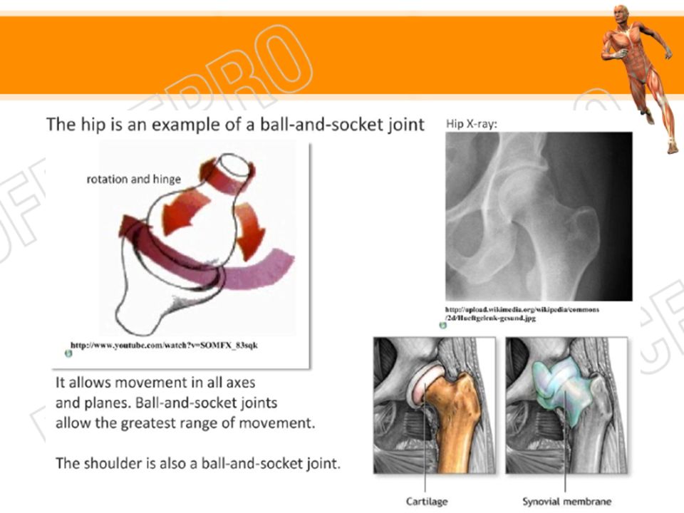
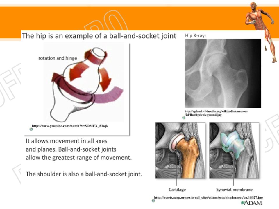
Ball and Socket Joints The hip is ball and socket joint
Permits movement in several directions, including rotational movement Head of femur (ball) fits into acetabulum (depression) of the hip
 fits into acetabulum (depression) of the hip.")
17
Hinge Joints Provides an opening-and-closing type of movement like the action of a door

19
Comparing hip & knee joints
Hip Joint Knee Joint Freely moveable Angular motions in many directions and rotational movements Angular motion in one direction Motions possible are flexion, extension, abduction, adduction, circumduction, and rotation Motions possible are flexion and extension Ball-like structure fits into a cup-like depression Covex surfac fits into a concave surface

20
11.2.5 Describe the structure of striated muscle fibres, including the myofibrils with light and dark bands, mitochondria, the sarcoplasmic reticulum, nuclei and the sarcolemma.

21
Muscle 3 types Striated (skeletal) Cardiac Smooth (non-striated)
 Cardiac Smooth (non-striated)")
22
Striated (Skeletal Muscle)
")
23
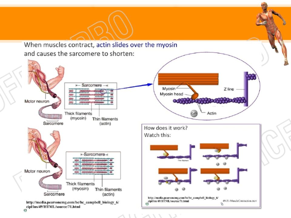
Sarcomere

24
The Sarcomere

25
11.2.6 Draw and label a diagram to show the structure of a sarcomere including Z lines, actin filaments, myosin filaments with heads, and the resultant light and dark bands.

27
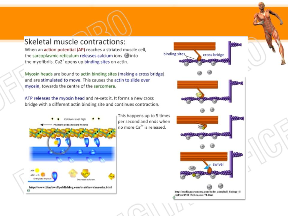
11.2.7 Explain how skeletal muscle contracts, including the release of a=calcium ions from the sarcoplasmic reticulum, the formation of cross-bridges, the sliding of actin and myosin filaments, and the use of ATP to break cross-bridges and re-set myosin heads.

29
11.2.8 Analyse electron micrographs to find the state of contraction of muscle fibres.

30
Electron micrograph showing contraction of one sarcomere

Similar presentations