Download presentation
Presentation is loading. Please wait.

1
ASSESSMENT OF A CASE OF AMENORRHEA

2
Prof. Ashis Kumar Mukhopadhyay
Professor, G & O Medical Superintendent-cum-Vice Principal CSS College of Obstetrics & Gynaecology, Kolkata National Chairperson, Medical Education Committee of FOGSI

3
AMENORRHEA Amenorrhea is the absence or abnormal cessation of the menses. A patient is diagnosed with primary amenorrhea if she has not reached menarche by age 14 without the appearance of secondary sex characterististics and by the age 16 with secondary sex characteristics. She meets the criteria for secondary amenorrhea if established menses have ceased for longer than 6 months or at least 3 of the previous cycle intervals.

4
Concealed (Cryptomenorrhoea)
Types Physiological Pathological Primary --Before puberty Secondary pregnancy Lactation Menopause Concealed (Cryptomenorrhoea) Congenital Acquired Real (True) Primary Secondary
 Congenital. Acquired. Real (True) Primary. Secondary.")
5
Five basic factors for normal menstruation
Normal female chromosome 46 XX Co-ordination of H-P-O axis Responsive Endometrium Patent outflow tract Ancillary glands like Thyroid and Adrenal

6
Causes Disorders of H-P-O axis Hypogonadotropic Hypogonadism
Constitutional delayed puberty Kallman’s syndrome CNS tomours like craniopharyngioma Isolated FSH deficiency Hypergonadotropic Hypogonadism Primary Ovarian Failure 17-alpha Hydroxylase deficiency Gonadal dysgenesis

7
Causes Abnormal chromosomal pattern Turner’s syndrome (45XO)
Mosaicism 45 X/ 46 XX Pure Gonadal dysgenesis (46 XX or 46 XY) Androgen Insensitivity Syndrome Partial deletion of X chromosome:- Deletion of long arm (Xq-) Streak Gonads but no somatic abnormalities Deletion of short arm (Xp-) Somatic features like Turner’s
 Androgen Insensitivity Syndrome. Partial deletion of X chromosome:- Deletion of long arm (Xq-) Streak Gonads but no somatic abnormalities. Deletion of short arm (Xp-) Somatic features like Turner’s.")
8
Causes Developmental defect of Genital tract
Imperforate Hymen Transverse Vaginal Septum Atresia of upper 1/3rd. Of vagina Complete absence of vagina Absent Uterus Dysfunction of Thyroid and Adrenal Cortex Adrenogenital syndrome Cretinism

9
Causes Metabolic disorders Systemic illness Unresponsive Endometrium
Juvenile DM Systemic illness Malnutrition Anaemia Wt. Loss Tuberculosis Unresponsive Endometrium Congenital Synaechiae (rare)
")
10
Etiology of Amenorrhea
Primary Gonadal failure (43%) Congenital absence of uterus and vagina (15%) Constitutional delay (14%) Secondary Chronic anovulation (39%) Hypothyroidism / hyperprolactinemia(20%) Weight loss/anorexia(16%)
 Congenital absence of uterus and vagina (15%) Constitutional delay (14%) Secondary Chronic anovulation (39%) Hypothyroidism / hyperprolactinemia(20%) Weight loss/anorexia(16%)")
11
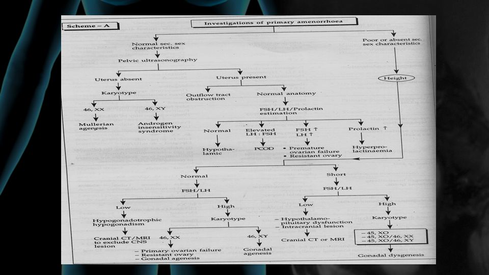
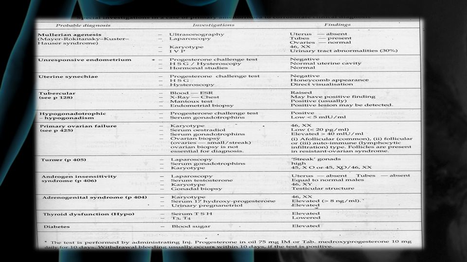
THE ASSESSMENT

13
abnormal hormonal stimulation
Primary amenorrhea breasts have developed FSH Level no high low vagina no yes congenital uterovaginal agenesis imperforate hymen complete transverse vaginal septum Pubic hair complete androgen insensitivity syndrome (CAIS) Estrogenized the (MPA) challenge + - Chromosome Analysis abnormal ovaries abnormal hormonal stimulation of normal ovaries
 Estrogenized. the (MPA) challenge. + - Chromosome Analysis. abnormal ovaries. abnormal hormonal stimulation. of normal ovaries.")
15
Secondary Amenorrhea

16
Incidence 1% of women of reproductive age.

17
The most common cause of secondary amenorrhea in reproductive age women is pregnancy and this should always be excluded by physical exam and laboratory testing for the pregnancy hormone - HCG.

18
History A good history can reveal the etiologic diagnosis in up to 85% of cases of amenorrhea.

19
History Galactorrhoea Hot flashes, breast atrophy and decreased libido
Certain medications A large amount of weight loss or gain Anorexia nervosa Cushing's disease and hypothyroidism Sheehan's syndrome. Asherman's syndrome Amenorrhoea following cervical conization Following discontinuation of oral contraception Psychological dysfunction or emotional stress Family history of apparent genetic anomalies

20
Physical examination Height, Weight and nutrition
Growth and development Signs of androgen excess Endocrinal stigma The breast exam may reveal galactorrhoea Estrogen deficiency may be suggested on pelvic exam by a smooth vagina that lacks the normal rugae (wrinkles) and a dry endocervix with no mucous. Size of pelvic organs.
 and a dry endocervix with no mucous. Size of pelvic organs.")
21
What we will do next?

22
If the history and physical exam are suggestive of a certain etiology :
for the sake of efficiency and cost-effectiveness, the workup can sometimes be more directed. ( in 85% of cases .)
")
23
Some patients will not demonstrate any obvious etiology for their amenorrhea on history and physical exam. These patients can be worked up in a logical manner using a stepwise approach.

24
The first tests to perform after pregnancy is ruled out are :
a progesterone withdrawal test TSH (thyroid stimulating hormone) prolactin level.
 prolactin level.")
25
-VE Preg.test TSH ,PROLACTIN’, Prog.challenge test
withdrawal bleeding anovulation without withdrawal bleeding compromised outflow tract. -ve.est, progest. challenge test Normal FSH HSG OR hysteroscopy asherman hypoestrogenic +ve.est,progest.challenge test FSH norm. Repeat+serum ,est.level hypothalamic-pituitary failure FSH>30-40 2wk repeat PROF

26
-VE Preg.test anovulation PROF
TSH ,PROLACTIN’, Prog.challenge test without withdrawal bleeding withdrawal bleeding hypoestrogenic compromised outflow tract. anovulation +ve.est,progest.challenge test -ve.est,progest .challenge test 2wk FSH>30-40 FSH norm. Normal FSH Repeat+serum ,est.level repeat HSG OR hysteroscopy asherman hypothalamic-pituitary failure PROF

27
Ovarian failure (premature menopause)
chromosomal anomalies If the woman is under 30, a karyotype should be performed to rule out any mosaicism involving a Y chromosome. If a Y chromosome is found the gonads should be surgically excised. autoimmune disease it is prudent to screen for thyroid, parathyroid, and adrenal dysfunction Laboratory evidence of autoimmune phenomenon is much more prevalent than clinically significant disease

28
Autoimmune Related Dysfunction
The most common association is with thyroid disease, but the parathyroids and adrenals can also be affected. Several studies have shown laboratory evidence of immune problems in about 15-40% of women with premature ovarian failure. In general, ovarian biopsy is not indicated in patients with premature ovarian failure since no clinically useful information will be obtained.

29
Hypothalamic-pituitary failure
Patients who do not bleed after the progestin challenge but do after estrogen/progestin and have normal or low FSH and LH levels

30
Hypothalamic-pituitary failure
Some medications (e.g. phenothiazines) as well as extremes of weight loss, stress or exercise can cause this type of secondary amenorrhea. A pituitary or hypothalamic tumor would be a rare finding in these patients who were all screened with prolactin levels at the beginning of the diagnostic evaluation. However, if there is no cause apparent from the history, it would be prudent to obtain a baseline CT (or MRI) evaluation of the sellar region to rule out a space occupying lesion.
 as well as extremes of weight loss, stress or exercise can cause this type of secondary amenorrhea. A pituitary or hypothalamic tumor would be a rare finding in these patients who were all screened with prolactin levels at the beginning of the diagnostic evaluation. However, if there is no cause apparent from the history, it would be prudent to obtain a baseline CT (or MRI) evaluation of the sellar region to rule out a space occupying lesion.")
31
Hypothalamic-pituitary failure
Patients with normal prolactin levels and normal imaging studies have hypothalamic amenorrhea of uncertain etiology. If the amenorrhea and lack of withdrawal bleeding persists, prolactin levels should be measured annually since a small microadenoma could be present that is escaping laboratory and radiographic detection.

32
Hypothalamic-pituitary failure
In this condition, as well as in the other hypothalamic amenorrhea situations, the patients can be significantly hypo estrogenic (a low estrogen situation similar to menopause). If the state is persistent, hormone replacement therapy should be considered for protection against osteoporosis. One approach is to get an estradiol level and if it is less than 30 pg/ml, counsel the patient that hormonal replacement therapy is indicated
. If the state is persistent, hormone replacement therapy should be considered for protection against osteoporosis. One approach is to get an estradiol level and if it is less than 30 pg/ml, counsel the patient that hormonal replacement therapy is indicated.")
Similar presentations


(6)(7)>")


>")


