Download presentation
Presentation is loading. Please wait.

1
SEXUAL REPRODUCTION AND DEVELOPMENT

2
SEXUAL REPRODUCTION Sexual reproduction requires two different parent cells from two separate organisms or from two sexually different parts of a single organism. Sexual reproduction produces off springs that are genetically different from either parent. In simple organisms, sexual reproduction involves transport of genetic material from one organism to another. In more complex organisms, two special sex cells, called gametes are needed. (pg: 417)
")
3
TYPES OF GAMETES l) ISOGAMY
It is a condition in which the sexual cells (gametes) are of the same form and size. Many algae and some fungi have isogamous gametes. Ex: Ulotrix
 are of the same form and size. Many algae and some fungi have isogamous gametes. Ex: Ulotrix.")
4
TYPES OF GAMETES ll) HETEROGAMY
It is a condition of having differently sized male and female gametes. A) ANISOGAMY The union of morphologically unlike motile gametes. Oedogonium sp. (green algae)
 ANISOGAMY. The union of morphologically unlike motile gametes. Oedogonium sp. (green algae)")
5
TYPES OF GAMETES B) OOGAMY
The union of unlike gametes, usually a large non-motile female gamete and a small motile male gamete.

6
EXCEPTIONAL CASES IN SEXUAL REPRODUCTION
A) Conjugation The type of sexual process most commonly found among simple organisms is called conjugation. In conjugation, a cytoplasmic bridge forms between two cells, and an exchange or transfer of nuclear material takes place through the bridge. Ex: Paramecium, bacteria
 Conjugation. The type of sexual process most commonly found among simple organisms is called conjugation. In conjugation, a cytoplasmic bridge forms between two cells, and an exchange or transfer of nuclear material takes place through the bridge. Ex: Paramecium, bacteria.")
7
EXCEPTIONAL CASES IN SEXUAL REPRODUCTION
B) Hermaphroditism
 Hermaphroditism.")
8
B) Hermaphroditism In some animals sexes are not separate. Instead, each individual has both testes and ovaries. These organisms are called hermaphrodite. Hermaphroditism is usually found among animals that move slowly or those that are attached to surface. Ex: earthworms, flatworms, snails, hydras, sponges and some flowering plants.

9
B) Hermaphroditism Even though hermaphroditic organisms can produce both eggs and sperm, self-fertilization is rare. Instead, these organisms exchange sperm with another individual of the same species.

10
B) Hermaphroditism ! Self fertilization is seen in flatworms but not in earth worms. ! Reproductive organs of sponges are not found at a particular site of the organism. Some cells found in different places of the organism produce eggs and sperms.

11
C) Parthenogenesis mitosis honey Male bee(n) Sperm cells (n)
worker (2n) Sterile female honey Male bee(n) Sperm cells (n) fertilization royal jelly (2n) female Queen (2n) meiosis Egg cells (n) Queen (2n) meiosis No fertilization Drone (n) parthenogenesis
 Sterile female. honey. Male bee(n) Sperm cells (n) fertilization. royal jelly. (2n) female. Queen (2n) meiosis. Egg cells (n) Queen (2n) meiosis. No fertilization. Drone (n) parthenogenesis.")
12
C) Parthenogenesis The development of an unfertilized egg into adult animal without fusion with sperm is called parthenogenesis. In nature it takes place in many insects, including bees, wasps and certain ants. For example, in bees, the queen bee mates only once. She can then produce either unfertilized eggs or fertilized eggs. The unfertilized eggs become male drones while the fertilized eggs become female workers or queen.

13
SEXUAL REPRODUCTION IN ANIMALS
External Fertilization Internal Fertilization The gametes fuse outside of the body The gametes fuse inside the body of female

14
EXTERNAL FERTILIZATION

15
EXTERNAL FERTILIZATION
In external fertilization, the eggs are in the environment outside the body of the female. Takes place in animals that live in water. Ex: Fish (but not sharks), many amphibians To overcome the hazards of external fertilization, large numbers of eggs and sperm are released. Embryo inside the fertilized egg develops in aquatic environment.
, many amphibians. To overcome the hazards of external fertilization, large numbers of eggs and sperm are released. Embryo inside the fertilized egg develops in aquatic environment.")
16
EXTERNAL FERTILIZATION

17
INTERNAL FERTILIZATION

18
INTERNAL FERTILIZATION
Fertilization within the body of the female is called internal fertilization. It is found most often in animals that reproduce on land and also found in some aquatic animals, such as sharks. Internal fertilization requires a specialized sex organ to carry sperm from the body of the male into the body of the female. Less number of eggs are produced After fertilization, either the zygote is enclosed in a protective shell and released by the female, or it remains and develops within a special part of the female’s body.

19
Metamorphosis In some animals that produce large number of eggs, embryo hatches into nymph or larvae before it completes its development. Nymphs or larvae complete their development at the outside and become adults. This series of changes is called metamorphosis.

20
Metamorphosis

21
REPRODUCTION IN FISH AND AMPHIBIANS
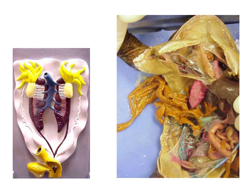
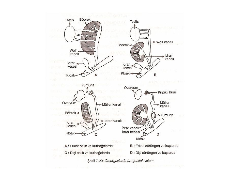
Eggs are not covered with a hard shell, instead they are surrounded by a jellylike substance. Eggs that are produced in the ovaries of females pass to the Müller’s duct through the ciliated funnel (kirpikli huni) and then released out of the body from the cloaca (kloak). Wolf’s duct carries both sperms and excretory materials to the cloaca in males.
 and then released out of the body from the cloaca (kloak). Wolf’s duct carries both sperms and excretory materials to the cloaca in males.")
22
REPRODUCTION IN FISH AND AMPHIBIANS
Egg Kidney Testes Ciliated funnel Ovary Muller’s duct Kidney Wolf’s duct Urinary bladder Ureter Urinary bladder Cloaca Cloaca Male fish and amphibian urogenital system Female fish and amphibian urogenital system

25
REPRODUCTION IN REPTILES AND BIRDS
Internal fertilization is mostly seen in reptiles and birds. Embryo found in the fertilized egg completes its development inside the egg. In females, eggs are transferred to the cloaca through the Muller’s duct. Inside the Muller’s duct egg white and shell are formed. Wolf’s duct carries only sperms in males. Egg shells of birds are harder than reptiles. Fertilization occurs in Muller’s duct. Excretory substances are transported from a separate duct to the cloaca.

26
REPRODUCTION IN REPTILES AND BIRDS
Testes Ovary Ciliated funnel Muller’s duct Wolf’s duct Kidney Egg Ureter Ureter Urinary bladder Urinary bladder Cloaca Cloaca Female Reptile and Bird Urogenital system Male Reptile and Bird Urogenital system

27
Bird Reproductive System

29
After fertilization, the development of chordates can be
oviparous, ovoviviparous or viviparous. In oviparous species, which include most fishes and amphibians and all birds, the eggs develop outside the mother’s body. Embryo obtains nutrients from the egg.

30
In ovoviviparous animals, such as sharks, the eggs develop within the mother’s body and the embryos receive nutrients from the yolk in the egg.

31
The developing embryos of viviparous animals, including most mammals, obtain nutrients directly from the mother’s body.

32
EXTRAEMBRYONIC MEMBRANES IN REPTILES AND BIRDS
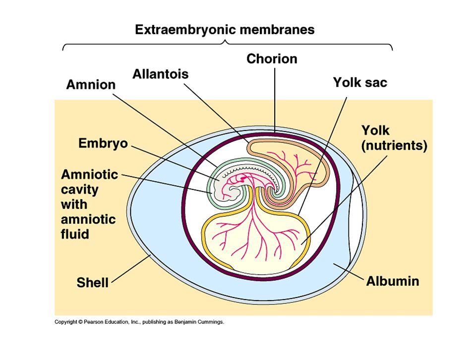
Embryonic membranes formed during the development of reptiles and birds are; Chorion (serosa) Allantois Amnion Yolk sac (Vitellus sac)
 Allantois. Amnion. Yolk sac (Vitellus sac)")
34
EXTRAEMBRYONIC MEMBRANES IN REPTILES AND BIRDS
CHORION: The outermost membrane lines the inside of the shell and surrounds the embryo and other three membranes. ***It aids in the exchange of gases between the embryo and the environment. ***In mammals, chorion is involved in the formation of placenta.

35
EXTRAEMBRYONIC MEMBRANES IN REPTILES AND BIRDS
2. ALLANTOIS: is a saclike structure that grows out of the digestive tract of the embryo. So that it aids in the exchange of gases and collection of waste materials. *** In mammals, waste materials pass from embryo to the mother, therefore allantois is degenerated. (körelmiştir)
")
36
EXTRAEMBRYONIC MEMBRANES IN REPTILES AND BIRDS
3. AMNION: The amniotic fluid within the sac acts as a cushion to protect it from shocks. *** During developmental stages of fish and frogs, amnion sac is not formed. *** In mammals, amnion sac is involved in the formation of umbilical cord (göbek bağı).
.")
37
EXTRAEMBRYONIC MEMBRANES IN REPTILES AND BIRDS
EMBRİYO ZARLARI 4. YOLK SAC (VITELLUS SAC): It surrounds the yolk (vitellus) and it is the source of food for the embryo. *** Yolk sac of birds and reptiles are larger than fish and frogs. *** As there is little amount of vitellus in the eggs of mammals, yolk sac is not developed as much. The mother meets the needs of the embryo.
: It surrounds the yolk (vitellus) and it is the source of food for the embryo. *** Yolk sac of birds and reptiles are larger than fish and frogs. *** As there is little amount of vitellus in the eggs of mammals, yolk sac is not developed as much. The mother meets the needs of the embryo.")
38
REPRODUCTION IN MAMMALS
NONPLACENTAL MAMMALS Pouched mammals (Marsupials) (Keseli memelilerde) *Some internal development of the embryo takes place in the uterus, but no placenta is formed. *The young animal crawls into a pouch on the outside of the mother’s body after birth and attaches itself to a mammary gland. *Development is completed in the pouch. Ex: kangaroo, opossum (keseli ayı) Egg-laying mammals Gagalı memelilerde (platypus, ornithorenk) *The egg of egg-laying mammals contain a large amount of yolk. *The embryo completes its development outside the body of the mother and feeds upon the mother’s milk.
 (Keseli memelilerde) *Some internal development of the embryo takes place in the uterus, but no placenta is formed. *The young animal crawls into a pouch on the outside of the mother’s body after birth and attaches itself to a mammary gland. *Development is completed in the pouch. Ex: kangaroo, opossum (keseli ayı) Egg-laying mammals. Gagalı memelilerde (platypus, ornithorenk) *The egg of egg-laying mammals contain a large amount of yolk. *The embryo completes its development outside the body of the mother and feeds upon the mother’s milk.")
39
Pouched mammals

40
Egg-laying mammals

41
REPRODUCTION IN MAMMALS
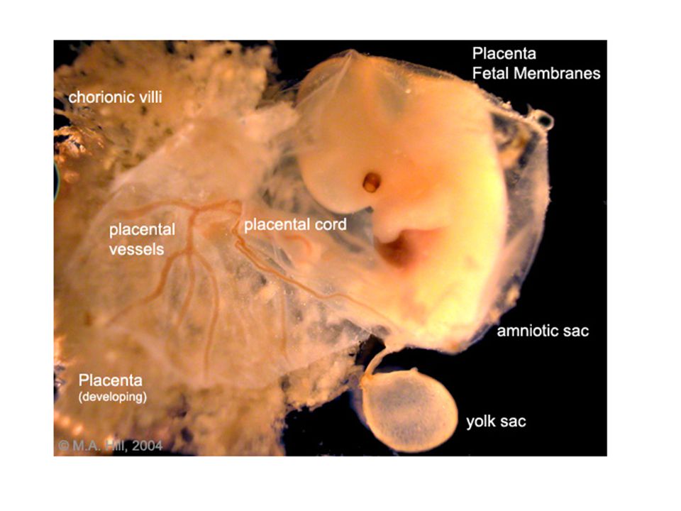
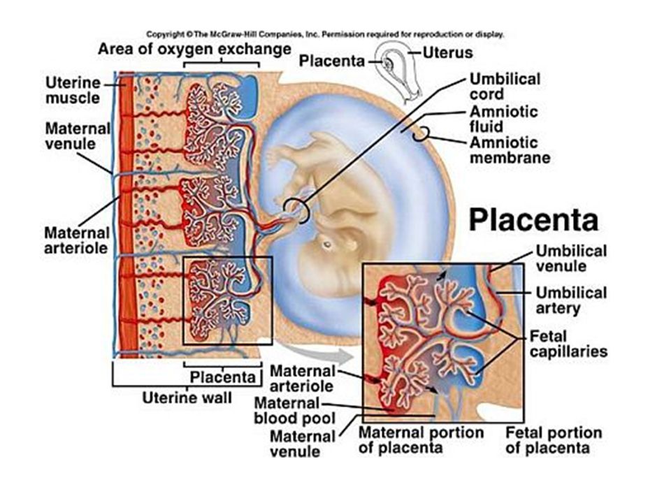
PLACENTAL MAMMALS Most mammals, including humans, are placental mammals. (Ex: hedgehog (kirpi), bat, whale, horse, donkey, elephant, seal, sheep) In these mammals, the blood vessels of the embryo’s circulatory system are in close contact with the mother’s circulatory system. This contact takes place in a specialized structure called the placenta. The placenta allows the exchange of nutrients and wastes between the embryo and the mother.
, bat, whale, horse, donkey, elephant, seal, sheep) In these mammals, the blood vessels of the embryo’s circulatory system are in close contact with the mother’s circulatory system. This contact takes place in a specialized structure called the placenta. The placenta allows the exchange of nutrients and wastes between the embryo and the mother.")
42
In humans, the chorion is the outermost extraembryonic membranes.
Small fingerlike projections called chorionic villi form on the outer surface of the chorion and extend into the uterine lining. ( Chorionic Villus + uterus wall= Placenta)
")
43
(Umbilical cord = vitellus sac + allantois)
In the human, the yolk sac and the allantois develop into the umbilical cord. This rope like structure connects the developing fetus to the placenta. This structure contains blood vessels that transport waste materials out of the embryo’s body. Veins inside the umbilical cord carry oxygen and nutrients to the embryo. (Umbilical cord = vitellus sac + allantois)
")
47
THE MALE REPRODUCTIVE SYSTEM

48
SPERMATOGENESIS Is the production of sperm in testes by meiotic division from immature sex cells called spermatogonia (2n). In humans, after a male matures sexually , there is a continual development of some spermatogonia into functional sperm. @

49
1. Spermatogonium increases in size to become a primary spermatocyte.
2. The primary spermatocyte undergoes the first meiotic division, forming secondary spermatocytes. 3. Each secondary spermatocyte undergoes second meiotic division, forming 4 haploid spermatids. 4. Each of the spermatids develops into a mature sperm with flagellum inside the epididimys.

51
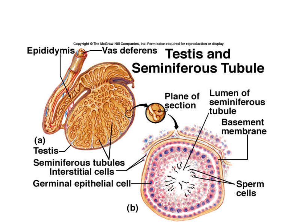
THE MALE REPRODUCTIVE SYSTEM
The male gonads are testes. The testes make sperm cells, and the male sex hormone testosterone. Testosterone causes development of the secondary sex characters, such as, body hair, muscle development and a deep voices. Secondary sex characteristics develop during adolescence and are not involved in reproduction.

52
THE MALE REPRODUCTIVE SYSTEM
Each testis is made up of coiled tubes called seminiferious tubules. ( tubules in each testis) Immature sperm are made in these tubes and then pass to the epididymis, a storage area on the upper rear part of each testis. In the epididymis, the sperm mature and leave through the vas deferens, a tube that leads upward from each testis. The two vas deferens empty into the urethra. Urethra is both the passageway for sperm and the excretion of urine.
 Immature sperm are made in these tubes and then pass to the epididymis, a storage area on the upper rear part of each testis. In the epididymis, the sperm mature and leave through the vas deferens, a tube that leads upward from each testis. The two vas deferens empty into the urethra. Urethra is both the passageway for sperm and the excretion of urine.")
55
THE MALE REPRODUCTIVE SYSTEM
In human male, urethra passes through the penis to the outside of the body. As sperm enter the urethra, the seminal vesicles, Cowper’s gland and prostate gland all secrete seminal fluid into the urethra. The mixture of sperm and fluid is called semen.

57
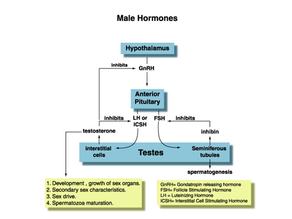
HORMONAL CONTROL OF THE MALE REPRODUCTIVE SYSTEM
FSH (Follicle-stimulating hormone): In males it controls the production of sperm cells in testes by stimulating the seminiferious tubules. LH (Luteinizing hormone): It stimulates the testes and makes them secrete testosterone FSH, LH are secreted from pituitary gland (hipofiz) and testosteron is secreted from testes.
: In males it controls the production of sperm cells in testes by stimulating the seminiferious tubules. LH (Luteinizing hormone): It stimulates the testes and makes them secrete testosterone. FSH, LH are secreted from pituitary gland (hipofiz) and testosteron is secreted from testes.")
59
FEMALE REPRODUCTIVE SYSTEM

60
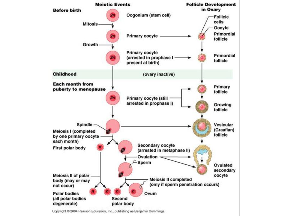
OOGENESIS Oogenesis is the production of eggs in the ovary.
Eggs develop in the ovary from immature cells called oogonia (2n) (sing:oogonium). Oogonium is surrounded by a follicle, a sac of cells within which the mature egg develops. During the early development of the female, the oogonia divide many times by mitosis to form a supply of oogonia. Each human female is born with all the oogonia she will ever have.
 (sing:oogonium). Oogonium is surrounded by a follicle, a sac of cells within which the mature egg develops. During the early development of the female, the oogonia divide many times by mitosis to form a supply of oogonia. Each human female is born with all the oogonia she will ever have.")
61
OOGENESIS Before birth and by the third month of development of a human female, oogonia within the baby’s ovaries begin to develop into cells called primary oocytes. Meiosis stops at that point until the female reaches to sexual maturity. Then, once a month in women, one of these primary oocytes finishes meiosis and develops into an egg.

62
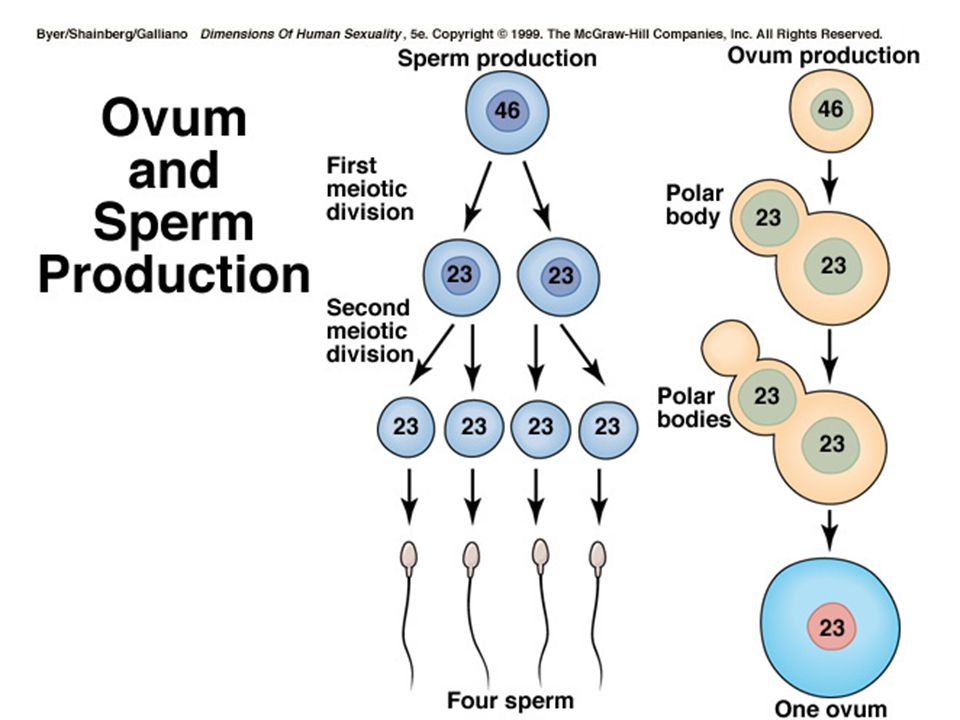
OOGENESIS When the first meiotic division takes place in the primary oocyte, the cytoplasm of the cell divides unequally. One of the daughter cells is large and called secondary oocyte (n). The other small daughter cell is called first polar body (n). (l. kutup hücresi) During the second meiotic division, the secondary oocyte divides unequally into a large cell called ootid and another polar body. The first polar body also divides into two polar bodies. (ll. kutup hücresi) The ootid grows into a mature egg(n). The polar bodies break apart and die.
. The other small daughter cell is called first polar body (n). (l. kutup hücresi) During the second meiotic division, the secondary oocyte divides unequally into a large cell called ootid and another polar body. The first polar body also divides into two polar bodies. (ll. kutup hücresi) The ootid grows into a mature egg(n). The polar bodies break apart and die.")
68
COMPARISON OF EGG AND SPERM
Round and unable to move Contains nucleus and stored food in the form of yolk Larger than sperm SPERM is made up of head,a middle piece and flagellum head contains nucleus and acrosome that contains enzymes to make sperm penetrate the egg Middle piece contains mitochondria

69
THE FEMALE REPRODUCTIVE SYSTEM
The female gonads are the ovaries. The ovaries makes eggs and secrete the female sex hormone estrogen. Estrogen causes the development of female secondary sex characteristics such as breasts, a broadened pelvis, and distribution of body fat. Estrogen plays an important role in menstrual cycle.

70
THE FEMALE REPRODUCTIVE SYSTEM
There are two ovaries. Each ovary contains about tiny egg sacs called follicles. In each follicle, there is an immature egg. When an egg matures, its follicle moves to the surface of the ovary. The follicle than breaks, releasing the egg. This process is called ovulation.

71
THE FEMALE REPRODUCTIVE SYSTEM
Near each ovary, but not connected to it, is an oviduct or Fallopian tube with a funnel-like opening. Cilia lining the oviduct, transports the released egg into the tube. In the oviduct, the egg may be fertilized if any sperm are peresent.

72
THE FEMALE REPRODUCTIVE SYSTEM
From the oviduct, egg passes into the uterus. If the egg has been fertilized, it finishes its development in th uterus attached to it’s wall. The neck of the uterus is called cervix and it opens into the vagina (birth canal), which leads to the outside of the body. Unlike the male, in the mature female, the urinary and reproductive tarcts are completely separete.
, which leads to the outside of the body. Unlike the male, in the mature female, the urinary and reproductive tarcts are completely separete.")
75
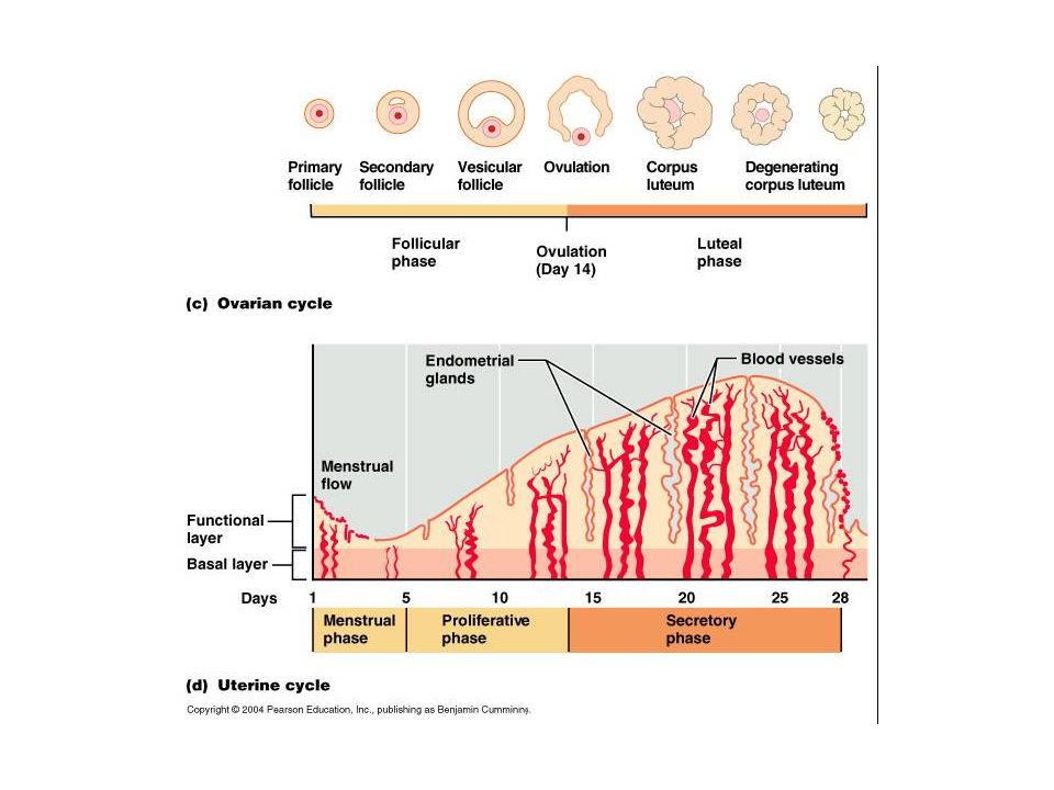
MENSTRUAL CYCLE @ In human female, a mature egg develops and leaves one of the ovaries about every 28 days. At this time uterus wall thickens and is prepared to accept a fertilized egg. If the egg is not fertilized, the uterine wall breaks down and along with the unfertilized egg passes from the body. Then another egg matures and uterine wall builds up again. This cycle is called menstrual cycle.

76
STAGES OF MENSTRUAL CYCLE
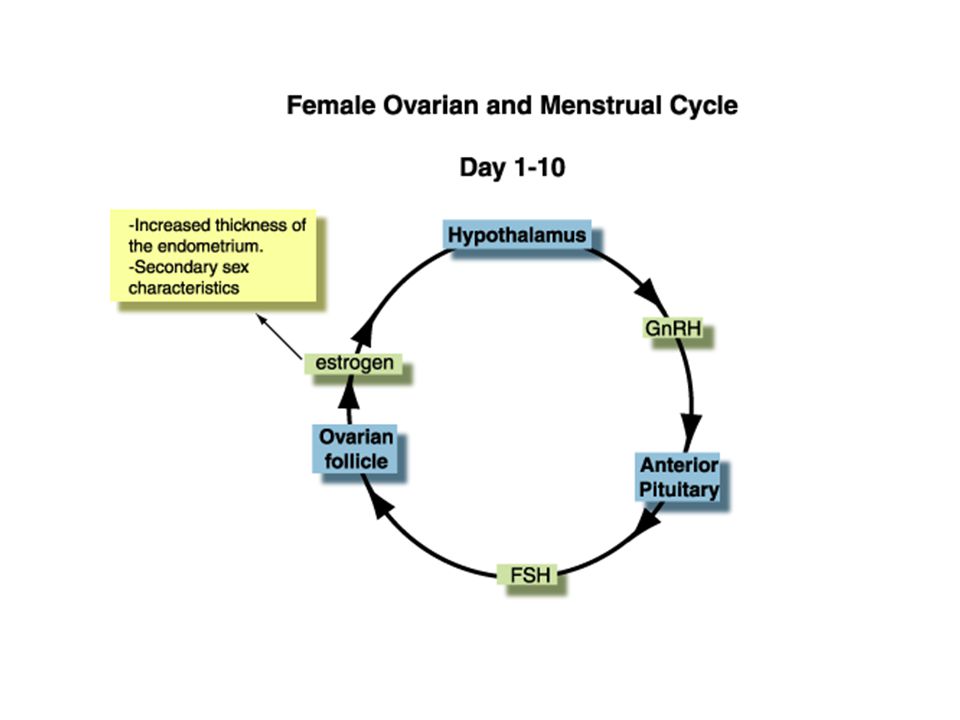
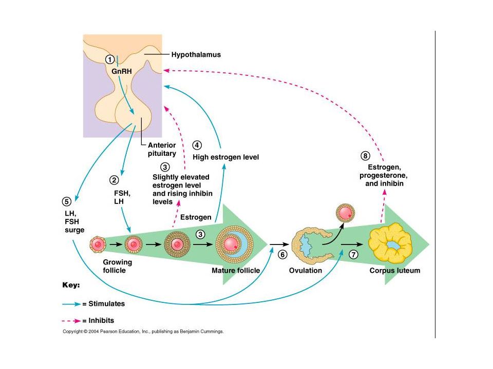
FOLLICLE STAGE: The pituitary gland secretes FSH (Follicle stimulating hormone), which causes several follicles in the ovary to begin developing. Usually one follicle matures. As the follicle develops, it secretes estrogen. The estrogen stimulates the uterine lining to thicken with mucus to prepare for pregnancy. This stage lasts 10 to14 days.
, which causes several follicles in the ovary to begin developing. Usually one follicle matures. As the follicle develops, it secretes estrogen. The estrogen stimulates the uterine lining to thicken with mucus to prepare for pregnancy. This stage lasts 10 to14 days.")
78
STAGES OF MENSTRUAL CYCLE
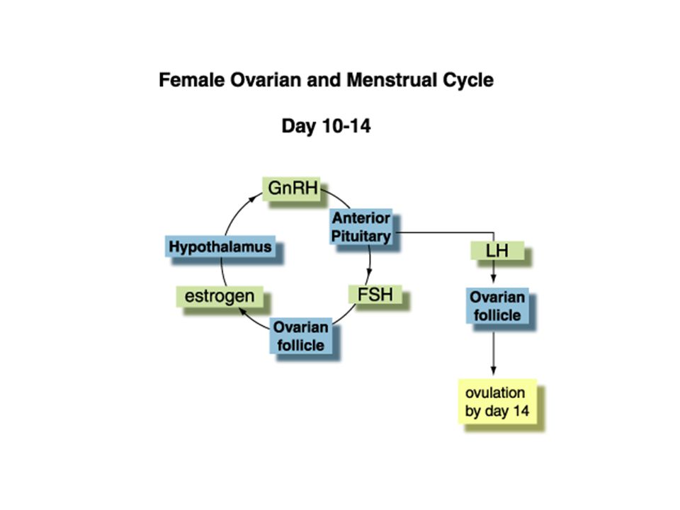
2. OVULATION: A high level of estrogen in the blood causes the pituitary to decrease the secretion of FSH and begin the secretion of luteinizing hormone (LH). When the concentration of LH reaches a certain level, ovulation takes place. The follicle breaks and releases the mature egg.
. When the concentration of LH reaches a certain level, ovulation takes place. The follicle breaks and releases the mature egg.")
80
STAGES OF MENSTRUAL CYCLE
3. CORPUS LUTEUM: After ovulation, LH causes the broken follicle to fill with cells, forming a yellow body called the corpus luteum. The corpus luteum begins to secrete progesterone, which brings about the continued growth of the uterine lining. Progesterone also stops the development of new follicles in the ovary by inhibiting the release of FSH. Fertilized egg attaches to the wall of uterus by progesterone hormone. The corpus luteum stage lasts 10 to 14 days.

82
STAGES OF MENSTRUAL CYCLE
4. MENSTRUATION: If fertilization does not occur, secretion of LH decreases, and the corpus luteum breaks down. This causes a decrease in the level of progesterone. As the progesterone level decreases, uterine wall breaks down. Layers of wall, unfertilized egg, and small amount of blood pass out of the body. This is called menstruation. It lasts about 3-5 days. During menstruation, estrogen level decreases. The pituitary secretes more FSH and a new follicle starts to mature.

83
@

84
@1 @2

88
HORMONAL CONTROL OF THE MALE REPRODUCTIVE SYSTEM
Hormones produced by hypothalamus called releasing factors (RF), control the release of hormones from the pituitary gland. Pituitary gland secretes; FSH (follicle stimulating hormone) Luteinizing hormone (LH) Luteotrophic hormone (prolactin) **Secretions of pituitary effects directly the ovary and ovary stimulates uterus activities.
, control the release of hormones from the pituitary gland. Pituitary gland secretes; FSH (follicle stimulating hormone) Luteinizing hormone (LH) Luteotrophic hormone (prolactin) **Secretions of pituitary effects directly the ovary and ovary stimulates uterus activities.")
89
Luteotrophic hormone (Prolactin)
It maintains the continuity of corpus luteum, and the secretion of progesterone and estrogen. It stimulates the secretion of milk by the mammary glands of the female after she gives birth. It is also known as prolactin.

90
During pregnancy, placenta secretes a hormone which acts like estrogen and progesterone and corpus luteum continues the secretion of progesterone. During pregnancy, progesterone helps uterine activities continue until the childbirth. After birth, placenta passes out of the body and secretion of progesterone stops. Thus, uterine walls break down and return to it’s position before birth.

91
OXYTOCIN: It is secreted from the pituitary gland, and stimulates contractions of the uterus muscles during childbirth along with the estrogen. After childbirth, it helps milk that is secreted from the mammary glands of the female, fill the milk channels.

94
PITUITARY GLAND HORMONES HORMONES SECRETED FROM OVARIES
FSH: stimulates the development of egg cells in follicles LH: causes the release of egg cells from ovaries of female (ovulation ) After ovulation, it causes the broken follicle to form corpus luteum. LTH: it maintains the continuity of corpus luteum. Also stimulates the secretion of milk ESTROGEN: Stimulates the development of female rep. system and secondary sex characteristics It increases the no. of ciliated epithelium cells in fallopian tubules 3. Increases the amount of blood and tissue fluid in uterus PROGESTRON: It stimulates the development of uterus It ıncreases the amount of glycogen and fat inside the fallopian tubule It helps embryo attach to the wall of uterus
 After ovulation, it causes the broken follicle to form corpus luteum. LTH: it maintains the continuity of corpus luteum. Also stimulates the secretion of milk. ESTROGEN: Stimulates the development of female rep. system and secondary sex characteristics. It increases the no. of ciliated epithelium cells in fallopian tubules. 3. Increases the amount of blood and tissue fluid in uterus. PROGESTRON: It stimulates the development of uterus. It ıncreases the amount of glycogen and fat inside the fallopian tubule. It helps embryo attach to the wall of uterus.")
95
PITUITARY GLAND HORMONES HORMONES SECRETED FROM TESTES
FSH: controls the production of sperm LH: It stimulates testes to secrete testosteron TESTOSTERON: 1.Stimulates development of male rep. system and secondary sex characteristics 2. Stimulates development of sperms

Similar presentations