Download presentation
Presentation is loading. Please wait.

1
Muscles to Identify Friends don’t let friends use anabolic steroids!

2
Naming Skeletal Muscles
A. Location of muscle B. Shape of muscle C. Relative size D. Direction of fibers E. Number of origins F. Location of attachments G. Action

3
A. Location of muscle – bone or body region associated with the muscle
Temporalis Frontalis Occipitalis

4
B. Shape of muscle Trapezius Rhomboid major Deltoid

5
C. Relative size Gluteus medius Gluteus minimus Gluteus maximus

6
D. Direction of fibers External oblique Rectus abdominis Internal
Transversus abdominis

7
E. Number of origins Biceps brachii

8
G. Action

9
Muscles of the Head and Neck

10
Point of Origin= Medial wall of the orbit
Orbicularis Oculi Point of Origin= Medial wall of the orbit Point of Insertion= Circular path around the orbit Action= Closes eyes
11
Point of Origin= Mandible and Maxilla
Orbicularis Oris Point of Origin= Mandible and Maxilla Point of Insertion= Corners of the mouth Action= Closes mouth; purse lips; kissing

12
Point of Origin= Superior temporal fossa
Temporalis Point of Origin= Superior temporal fossa Point of Insertion= Coronoid process of the mandible Action= Closes jaw and elevates the mandible
13
Point of Origin= Manubrium (Sternum) and Clavicle
Sternocleidomastoid Point of Origin= Manubrium (Sternum) and Clavicle Point of Insertion= Mastoid process (Temporal bone) Action= Flexion of neck and rotation of head
 and Clavicle. Point of Insertion= Mastoid process (Temporal bone) Action= Flexion of neck and rotation of head.")
14
F. Location of attachments
Sternocleidomastoid

18
Anterior Shoulder and Thorax Muscles

19
Point of Insertion= Lateral lip of intertubercular sulcus
Pectoralis Major Point of Origin= Medial and anterior clavicle, lateral manubrium, and lateral sternal body Point of Insertion= Lateral lip of intertubercular sulcus Action= Adduction and Internal rotation of the shoulder
20
Abdominal Muscles

21
Point of Origin= Ribs 5-12 (at an angle)
External Oblique Point of Origin= Ribs 5-12 (at an angle) Point of Insertion= Anterior iliac crest and lateral to the rectus abdominis Action= Flexion and rotation of vertebral column
23
Point of Origin= Pubic crest and pubic symphysis
Rectus Abdominis Point of Origin= Pubic crest and pubic symphysis Point of Insertion= 5th-7th costal cartilages Action= Flexion of the vertebral column and posterior tilt of the pelvis
24
Posterior Shoulder and Thorax Muscles

25
Point of Origin= Supraspinous fossa
Supraspinatus Point of Origin= Supraspinous fossa Point of Insertion= Greater tubercle of the humerus Action= Abduction of the shoulder

26
Point of Origin= Infraspinous fossa
Infraspinatus Point of Origin= Infraspinous fossa Point of Insertion= Greater tubercle of the humerus Action= External (lateral) rotation of the shoulder
 rotation of the shoulder.")
28
Point of Origin= Transverse processes C1-C4
Levator Scapulae Point of Origin= Transverse processes C1-C4 Point of Insertion= Superior angle of the scapula Action= elevation of the scapula

29
Point of Insertion= Clavicle, acromion and spine (of the scapula)
Trapezius Point of Origin= Occipital bone and the spinous processes of C7 through T12 Point of Insertion= Clavicle, acromion and spine (of the scapula) Action= Elevation and depression of the scapula
 Action= Elevation and depression of the scapula.")
30
Posterior Shoulder and Thorax Muscles

31
Point of Insertion= Intertubercular sulcus
Latissimus Dorsi Point of Origin= Spinous processes of T6-L5, the iliac crest, the sacrum and ribs 9-12.nous Point of Insertion= Intertubercular sulcus Action= Adduction of the humerus

32
Point of Origin= Clavicle, acromion and spine of the scapula
Deltoid Point of Origin= Clavicle, acromion and spine of the scapula Point of Insertion= Deltoid tuberosity Action= Abduction of the shoulder

33
Anterior and Posterior Deltoid

34
© 2014 Pearson Education, Inc.
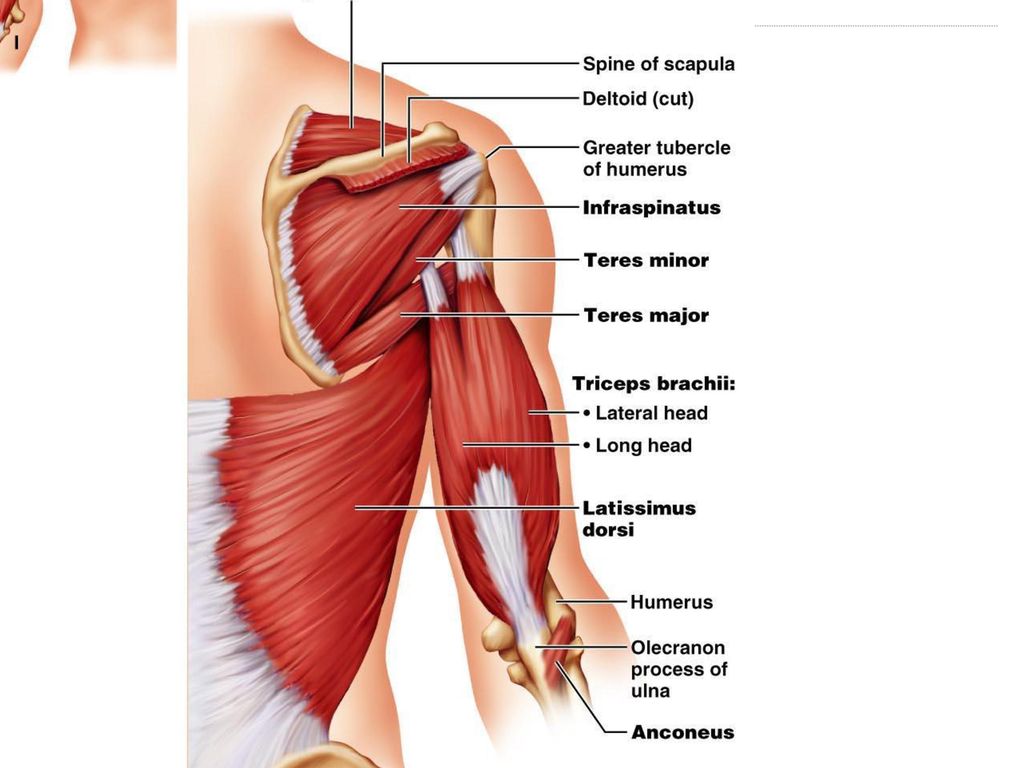
Figure 10.15a–b Muscles crossing the shoulder and elbow joints, causing movements of the arm and forearm, respectively. Clavicle Supraspinatus* Spine of scapula Deltoid (cut) Greater tubercle of humerus Deltoid Infraspinatus* Sternum Teres minor* Pectoralis major Teres major Coracobrachialis Triceps brachii: Triceps brachii: Lateral head Lateral head Long head Long head Medial head Biceps brachii Latissimus dorsi Brachialis Brachio- radialis Humerus Olecranon of ulna Anconeus Anterior view Posterior view © 2014 Pearson Education, Inc. 34
 Greater tubercle. of humerus. Deltoid. Infraspinatus* Sternum. Teres minor* Pectoralis. major. Teres major. Coracobrachialis. Triceps brachii: Triceps brachii: Lateral head. Lateral head. Long head. Long head. Medial head. Biceps brachii. Latissimus dorsi. Brachialis. Brachio- radialis. Humerus. Olecranon. of ulna. Anconeus. Anterior view. Posterior view. © 2014 Pearson Education, Inc. 34.")
35
Point of Origin= Distal, anterior surface of the humerus
Brachialis Point of Origin= Distal, anterior surface of the humerus Point of Insertion= Coronoid process of the ulna Action=Flexion of the elbow

36
Point of Insertion= Radial tuberosity
Biceps Brachii Point of Origin= Long head: superior margin of the glenoid fossa Short head: the coracoid process of the scapula Point of Insertion= Radial tuberosity Action= Flexion of the elbow

37
Point of Insertion= Olecranon
Triceps Brachii Point of Origin= ong Long head: the inferior margin of the Glenoid fossa Lateral head: the lateral, posterior surface of the humerus Medial head: the posterior surface of the humerus Point of Insertion= Olecranon Action= Extension of the elbow

38
© 2014 Pearson Education, Inc.
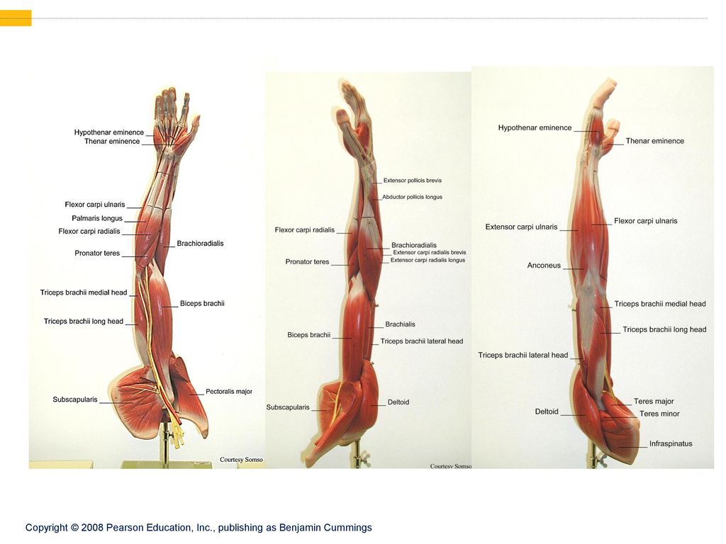
Figure 10.16a Muscles of the anterior fascial compartment of the forearm acting on the right wrist and fingers. Superficial transverse ligament of palm Palmar aponeurosis Flexor retinaculum Flexor digitorum superficialis Pronator quadratus Flexor pollicis longus Flexor carpi ulnaris Palmaris longus Extensor carpi radialis longus Flexor carpi radialis Brachio- radialis Medial epicondyle of humerus Pronator teres Tendon of biceps brachii Medial head of triceps brachii Biceps brachii © 2014 Pearson Education, Inc. 38

39
© 2014 Pearson Education, Inc.
Figure 10.17a Muscles of the posterior fascial compartment of the right forearm acting on the wrist and fingers. Extensor expansion Tendons of extensor digitorum Extensor pollicis longus brevis Abductor pollicis longus Extensor digitorum Extensor carpi radialis brevis radialis longus carpi radialis brevis and longus Extensor indicis Extensor digiti minimi ulnaris Flexor carpi ulnaris Anconeus Insertion of triceps brachii Brachioradialis © 2014 Pearson Education, Inc.

40
© 2014 Pearson Education, Inc.
Figure 10.20a Anterior and medial muscles promoting movements of the thigh and leg. 12th thoracic vertebra 12th rib Quadratus lumborum Psoas minor Iliac crest Psoas major Iliopsoas Iliacus 5th lumbar vertebra Anterior superior iliac spine Tensor fasciae latae Pectineus Adductor longus Sartorius Gracilis Quadriceps femoris • Rectus femoris Adductor magnus • Vastus lateralis • Vastus medialis Tendon of quadriceps femoris Patella Patellar ligament © 2014 Pearson Education, Inc.

41
Figure 10.21a Posterior muscles of the right hip and thigh.
Gluteus medius Gluteus maximus Adductor magnus Gracilis Iliotibial tract Long head Biceps femoris Short head Hamstrings Semitendinosus Semimembranosus © 2014 Pearson Education, Inc.

42
Figure 10.22a Muscles of the anterior compartment of the right leg.
Fibularis longus Tibia Gastrocnemius Tibialis anterior Extensor digitorum longus Soleus Extensor hallucis longus Fibularis tertius Superior and inferior extensor retinacula Extensor hallucis brevis Extensor digitorum brevis © 2014 Pearson Education, Inc.

43
Figure 10.24a Muscles of the posterior compartment of the right leg.
Plantaris Medial head Gastrocnemius Lateral head Tendon of gastrocnemius Calcaneal tendon Medial malleolus Lateral malleolus Calcaneus Superficial view of the posterior leg. © 2014 Pearson Education, Inc.

44
Figure 10.24b Muscles of the posterior compartment of the right leg.
Lateral head (cut) Plantaris Gastrocnemius Popliteus Medial head (cut) Head of fibula Soleus Tendon of plantaris Fibularis longus Fibularis brevis Tendon of tibialis posterior The gastrocnemius has been removed to show the soleus immediately deep to it. © 2014 Pearson Education, Inc.
 Plantaris. Gastrocnemius. Popliteus. Medial. head (cut) Head of. fibula. Soleus. Tendon of. plantaris. Fibularis. longus. Fibularis. brevis. Tendon of. tibialis. posterior. The gastrocnemius has been. removed to show the soleus. immediately deep to it. © 2014 Pearson Education, Inc.")
45
Point of Insertion= Tibial tuberosity
Rectus Femoris Point of Origin= AIIS Point of Insertion= Tibial tuberosity Action= Extension of the knee joint

46
Point of Origin= Greater trochanter
Vastus Lateralis Point of Origin= Greater trochanter Point of Insertion= Tibial tuberosity Action= Extension of the knee joint

47
Point of Origin= Intertrochanteric line
Vastus Medialis Point of Origin= Intertrochanteric line Point of Insertion= Tibial tuberosity Action= Extension of the knee joint

48
Point of Origin= Inferior pubic ramus
Gracilis Point of Origin= Inferior pubic ramus Point of Insertion= Medial condyle of the tibia Action= Adduction of the hip and flexion of the knee

49
Point of Insertion= Medial and anterior tibia
Sartorius Point of Origin= ASIS Point of Insertion= Medial and anterior tibia Action= Hip flexion and medial rotation of the knee

50
Point of Origin= Lateral surface of the ilium
Gluteus Medius Point of Origin= Lateral surface of the ilium Point of Insertion= Greater trochanter Action= Abduction of the hip

51
Point of Origin= lateral surface of the ilium
Gluteus Maximus Point of Origin= lateral surface of the ilium Point of Insertion=Gluteal tuberosity and iliotibial (IT) band Action=Extension and abduction of thigh at the hip
 band. Action=Extension and abduction of thigh at the hip.")
52
Point of Origin= Lateral condyle of the tibia
Tibialis Anterior Point of Origin= Lateral condyle of the tibia Point of Insertion= Medial cuneiform and 1st metatarsal Action= Dorsiflexion and inversion of the foot

53
Point of Origin= Medial and lateral condyles of the femur
Gastrocnemius Point of Origin= Medial and lateral condyles of the femur Point of Insertion= Posterior calcaneus Action= Plantar flexion

Similar presentations


>")


 evaluationsStudent Evaluation of Course and Instructor (SECI) evaluations Please do.>")
 (b) (e) (d) (g) (f) (c) Circular (orbicularis oris) (b) Convergent (pectoralis major) (c) Parallel.>")


