Download presentation
Presentation is loading. Please wait.

1
Chapter 11 Muscles Exam 1 will cover sections 11.1-11.4

2
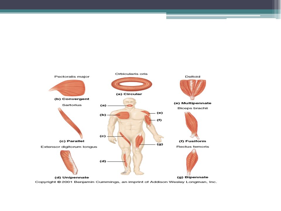
Section 1 Fascicle Arrangement Fascicles: bundles of muscle fibers in skeletal muscle fibers in each fascicle are parallel, but fascicle arrangement can vary ▫parallel muscles- fascicles are parallel to long axis of muscle (biceps brachii), when muscle fibers contract, muscle shortens by 30 % ▫convergent muscles- fascicles extend over a broad area & meet (converge) at the attachment site, (pectoralis muscles)
, when muscle fibers contract, muscle shortens by 30 % ▫convergent muscles- fascicles extend over a broad area & meet (converge) at the attachment site, (pectoralis muscles)")
3
▫Pennate muscles- (feather) fascicles form a shared angle w/ tendon contracted muscle doesn’t move tendon as far as parallel muscles-fibers pull @ angle contain more muscle fibers than parallel muscle, so they produce more tension ▫Circular muscles- (sphincter) arranged around openings when contracted, diameter decreases
 fascicles form a shared angle w/ tendon contracted muscle doesn’t move tendon as far as parallel muscles-fibers angle contain more muscle fibers than parallel muscle, so they produce more tension ▫Circular muscles- (sphincter) arranged around openings when contracted, diameter decreases")
5
11.2 Classes of Levers Nature and site of muscle connection determine force, speed, and range of motion ▫lever (bone) moves when the applied force (AF) is great enough to overcome the load (L) or resistance (R) that would prevent the movement ▫in the body, each joint acts as a fulcrum, and muscles provide the applied force ▫the load can vary (weight of limb, object held, or entire body)
 moves when the applied force (AF) is great enough to overcome the load (L) or resistance (R) that would prevent the movement ▫in the body, each joint acts as a fulcrum, and muscles provide the applied force ▫the load can vary (weight of limb, object held, or entire body)")
6
▫levers can change direction of applied force distance & speed of movement effective strength of applied force Classes of levers ▫first class: fulcrum in the middle (teeter totter) ▫second class: load in the middle (wheel barrow), small force can move a larger weight, at the expense of speed and distance
 ▫second class: load in the middle (wheel barrow), small force can move a larger weight, at the expense of speed and distance")
7
▫third class: most common in body, force is between the load and the fulcrum, speed and distance traveled are increased at the expense of effective force (muscles must generate 6X the tension to support the load)
")
8
11.3 Muscle Origins & Insertions Ends of skeletal muscle are attached to structures that limit their motion (bone, cartilage, connective tissue) ▫origin-the place where fixed end of muscle attaches ▫insertion-the site where the moveable end attaches to another structure ▫action- movement produced when muscle contracts
 ▫origin-the place where fixed end of muscle attaches ▫insertion-the site where the moveable end attaches to another structure ▫action- movement produced when muscle contracts")
9
Actions Actions are described ▫by bone or region affected (flexion of forearm) ▫by joint involved (flexion of elbow) ▫based on functions agonist (prime mover)- contraction responsible for producing specific movement, ex: biceps brachii muscle produces flexion of elbow antagonist- muscle whose action opposes the action of an agonist, ex: triceps brachii extends the elbow, going against the biceps brachii
 ▫by joint involved (flexion of elbow) ▫based on functions agonist (prime mover)- contraction responsible for producing specific movement, ex: biceps brachii muscle produces flexion of elbow antagonist- muscle whose action opposes the action of an agonist, ex: triceps brachii extends the elbow, going against the biceps brachii")
10
synergist- helps a larger agonist work efficiently, may provide pull near insertion or may stabilize near origin; useful in start of motion fixator- a synergist that stabilizes origin of agonist by preventing movement at another joint

11
11.4 Descriptive terms Locational terms ▫regional terms are common; abdominis- abdomen, capitis- head, femoris- femur, etc (PG 342) Origin and Insertion ▫first part of name indicates the origin, second part, the insertion ex: sternocleidomastoid originates at sternum, inserts at the clavicle (cleido)
 Origin and Insertion ▫first part of name indicates the origin, second part, the insertion ex: sternocleidomastoid originates at sternum, inserts at the clavicle (cleido)")
12
Fascicle organization ▫Rectus: straight, parallel muscles that run along long axis of body (rectus abdominis) ▫transverse/oblique: muscles have fibers that run at an angle to long axis of body (external obliques) Position ▫externus/superficialis: visible at the body surface ▫internus/profundus:deeper muscles ▫Intrinsic: located within an organ
 ▫transverse/oblique: muscles have fibers that run at an angle to long axis of body (external obliques) Position ▫externus/superficialis: visible at the body surface ▫internus/profundus:deeper muscles ▫Intrinsic: located within an organ")
13
Structural Characteristics number of tendons (biceps brachii, triceps brachii) shape- muscles named after their shape: trapezius, deltoid(triangle), rhomboid length and size: ▫ longus (long), longissimus (longest) ▫teres (long & round), brevis (short) ▫magnus (large), major (bigger), or maximus (biggest) ▫minor (small), or minimus (smallest)
 shape- muscles named after their shape: trapezius, deltoid(triangle), rhomboid length and size: ▫ longus (long), longissimus (longest) ▫teres (long & round), brevis (short) ▫magnus (large), major (bigger), or maximus (biggest) ▫minor (small), or minimus (smallest)")
14
Stop! Test 1 will cover 11.1-11.4

15
11.5 Muscles of facial expression Orbicularis oculi ▫Sphincter muscle of eyelid ▫O: frontal & maxillary bones ▫I: tissue of eyelid ▫Action: closes eye ▫Nerve: facial / CN VII (11.6)
")
16
Muscles of facial expression Zygomaticus ▫O: zygomatic bone ▫I: corners of mouth ▫Action: smiling ▫Nerve: facial / CN VII (11.6) Major and Minor
 Major and Minor")
17
Muscles of mastication Masseter ▫O: zygomatic arch ▫I: angle & ramus of mandible ▫Action: elevate mandible ▫Nerve: trigeminal nerve / CN V (foramen ovale) (11.7a) Masseter
 (11.7a) Masseter")
18
Muscles of mastication Temporalis ▫O: temporal fossa ▫I: coronoid process of mandible ▫Action: elevate and retract mandible ▫Nerve: trigeminal n. / CN V (11.7a)
.")
19
Extrinsic tongue muscles Styloglossus ▫O: styloid process of temporal bone ▫I: tongue ▫Action: retract & elevate tongue ▫Nerve: hypoglossal n. / CN XII (11.7c) Styloglossus
 Styloglossus.")
20
Extrinsic tongue muscles Hyoglossus ▫O: hyoid bone ▫I: tongue ▫Action: depresses tongue ▫Nerve: hypoglossal n. / CN XII (11.7c) Hyoid bone
 Hyoid bone.")
21
Muscles of neck & throat Digastric ▫two muscle bellies with an intermediate tendon attached to the hyoid bone ▫O: inferior margin of mandible (anterior belly); mastoid process of temporal bone (posterior belly) ▫I: hyoid bone ▫Action: elevate hyoid, stabilize hyoid, depress mandible ▫Nerve: trigeminal n. / CN V (anterior belly), facial n. / CN VII (posterior belly) (11.8a) Anterior belly Posterior belly
, facial n. / CN VII (posterior belly) (11.8a) Anterior belly Posterior belly.")
22
Muscles of neck & throat Stylohyoid ▫O: styloid process of temporal bone ▫I: hyoid bone ▫Action: elevate & retract hyoid, swallow ▫Nerve: facial n. / CN VII (11.7c) Stylohyoid
 Stylohyoid.")
23
Muscles of neck & throat Sternohyoid ▫O: manubrium & medial end of clavicle ▫I: hyoid bone ▫Action: depress hyoid (11.8a) Sternohyoid
 Sternohyoid")
24
Muscles of neck & throat Pharyngeal constrictors ▫O: mandible, pterygoid process, hyoid, laryngeal cartilages ▫I: posterior medial raphe of pharynx ▫Action: peristaltic contraction / swallow (11.8b) Superior Middle Inferior
 Superior Middle Inferior")
25
Muscles of neck & throat Sternocleidomastoid ▫O: manubrium & medial clavicle ▫I: mastoid process of temporal bone ▫Action: head & neck flexion, rotate head contralaterally ▫Nerve: accessory n. / CN XI (11.8a)
.")
26
Muscles of neck & throat Sternocleidomastoid

27
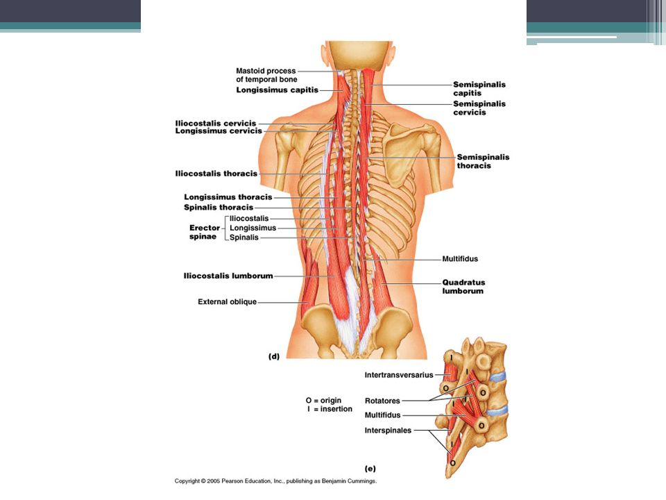
Back muscles Spinalis ▫O: spines of upper lumbar and lower thoracic vertebrae ▫I: spines of upper thoracic and cervical vertebrae ▫Action: extends vertebral column (11.9d)
")
29
Deep thorax muscles External & internal intercostals ▫O&I: inferior & superior border of ribs ▫Action: inspiration and expiration (11.10a) Internal
 Internal")
30
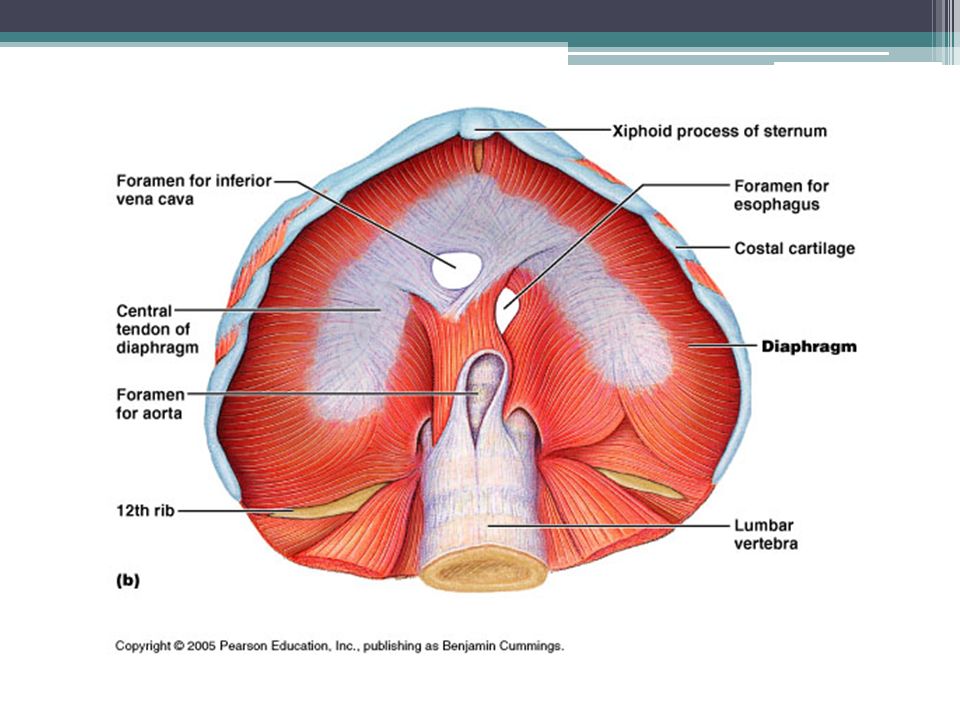
Deep thorax muscles Diaphragm ▫O: inferior internal surface of rib cage & sternum, inferior costal cartilages, lumbar vertebrae ▫I: central tendon ▫Action: flattens on contraction (inspiration) ▫Nerve: phrenic nerves (11.10b) Xiphoid process
 ▫Nerve: phrenic nerves (11.10b) Xiphoid process")
32
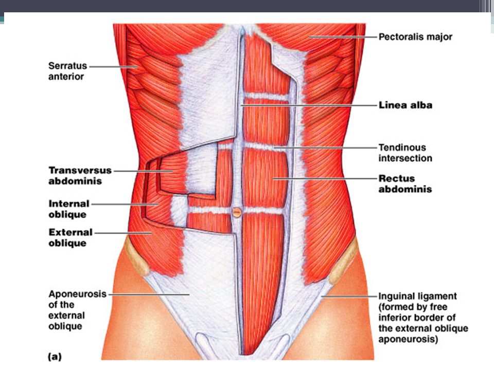
Abdominal wall muscles Rectus abdominis ▫O: pubic crest & symphysis ▫I: xiphoid process and costal cartilages of ribs 5-7 ▫Action: flex & rotate lumbar vertebrae, fix & depress ribs, stabilize pelvis during walking, increase intraabdominal pressure ▫Note: 3 tendinous insertions, aponeurosis & linea alba (11.11ab)
")
35
Section 11.6 Appendicular Muscles Position & stabilize pectoral & pelvic girdles Move upper & lower limbs Split into 2 major groups: ▫Muscles of shoulder & upper limb ▫Muscles of pelvis & lower limb

36
Differences in function: ▫Pectoral girdle has muscular connections with axial skeleton, acts as a shock absorber Example: can use hands when you run ▫Pelvic girdle transfers weight from axial muscles to appendicular skeleton, needs more bony support

37
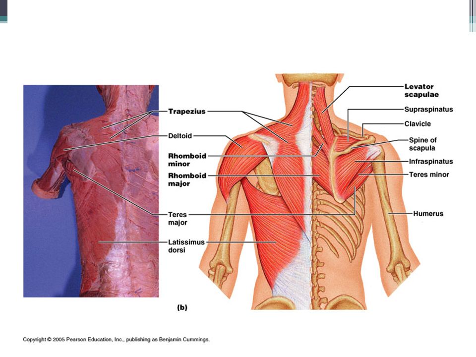
I. Shoulder & Upper Limb A.Muscles that position the pectoral girdle 1.Trapezius- covers back & parts of the neck *reaches the base of the skull *inserts on the clavicles and spines of the scapulae *regions can contract independently so the action varies *superficial

38
Superificial thorax muscles Trapezius ▫I: spine of scapula, clavicle ▫Action: stabilize, elevate, adduct, depress scapula, extend head (11.13b)
")
39
B. Rhomobid *adducts (retracts) scapula * attaches to cervical and thoracic vertebrae *inserts at the scapula *deep C. Levator scapulae *elevates scapula * deep
 scapula * attaches to cervical and thoracic vertebrae *inserts at the scapula *deep C. Levator scapulae *elevates scapula * deep.")
41
Superficial thorax muscles

42
D. Serratus anterior- on the chest *fan shaped *originates on chest side of ribs *inserts on the scapula *protracts scapula, swings shoulder forward (pushing) *superficial
 *superficial.")
43
E. Subclavius *deep chest muscle * originates on rib 1 *inserts on inferior border of clavicle * depresses & protracts clavicle & shoulder F. Pectoralis Minor * originates on ribs 3-5 *inserts on coracoid process of scapulae *complements contraction of subclavius, downward rotation of shoulder

44
Superficial thorax muscles Pectoralis minor ▫O: anterior surface of ribs 3-5 ▫I: coracoid process of scapula ▫Action: pulls scapula anterior & inferior (11.13a)
")
45
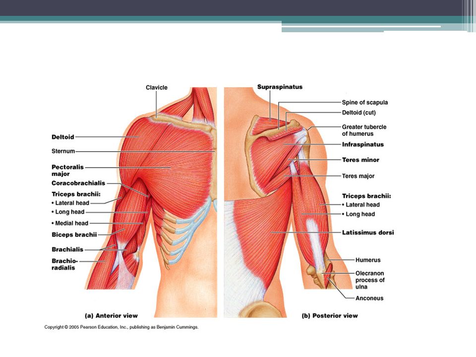
B. Muscles that move the arm 1. deltoid *originates on clavicle & scapulae *inserts on humerus *major abductor of arm (away from body) 2. supraspinatus * superior to posterior portion of deltoid (under trapezius) *helps w/start of abduction movement
 2. supraspinatus * superior to posterior portion of deltoid (under trapezius) *helps w/start of abduction movement.")
46
3. Subscapularis *located on anterior side of scapula * rotates arm medially 4. Teres major *helps rotate arm medially

47
5. Infraspinatus *located on the posterior side of the scapula, deep to deltoid *located below the scapular spine * rotates arm laterally (away from body) 6. Teres minor *aides in lateral rotation
 6. Teres minor *aides in lateral rotation.")
48
7. Rotator cuff muscles Act to stabilize head of humerus in glenoid cavity and prevent dislocation, can be a frequent site of sports injuiries Supraspinatus Infraspinatus Teres minor Subscapularis Remember SITS

49
8.Coracobrachialis * small muscle * only muscle attached to scapula that flexes & adducts humerus 9. Pectoralis major * originates on ribs 2-6 cartilage,body of sternum, and inferior portion of clavicle * inserts on greater tubercle of humerus * flexes, adducts, and medially rotates humerus 10. Latisumus dorsi *extends between the thoracic vertebrae & lesser tubercle of humerus * extends arm

51
C. Muscles that move the forearm & hand(move the elbow & wrist) Most muscles that insert on the forearm originate on the humerus ▫2 exceptions: biceps brachii & long head of triceps brachii 1.Biceps brachii * on anterior side of body * short & long head * originate on scapula, insert on radius * flexes & supinates arm (bends elbow)
 Most muscles that insert on the forearm originate on the humerus ▫2 exceptions: biceps brachii & long head of triceps brachii 1.Biceps brachii * on anterior side of body * short & long head * originate on scapula, insert on radius * flexes & supinates arm (bends elbow).")
52
2. Triceps brachii *lateral, long, & median heads, all insert on olecranon process of ulna * located on posterior side of humerus * extends forearm (straightens elbow) 3. Brachialis * deep muscle, located on anterior side of elbow * flexes the elbow *originates on humerus, inserts on ulna
 3. Brachialis * deep muscle, located on anterior side of elbow * flexes the elbow *originates on humerus, inserts on ulna.")
53
4. Brachioradialis * superficial, located along anterior thumb side of forearm * originates on humerus, inserts on radius * flexes elbow 5. Anconeus * extends elbow (assists triceps) * located on posterior side of elbow
 * located on posterior side of elbow.")
55
6. Flexor carpi radialis Flexor carpi ulnaris Palmaris longus * all contribute to flexion at wrist * anterior side * radials abducts *ulnaris adducts (11.15a) Anterior view radialis ulnaris
 Anterior view radialis ulnaris.")
56
7. Extensor carpi radialis Extensor carpi ulnaris *extends wrist *on posterior side * radials abducts * ulnaris adducts

57
D. Muscles that move the hand and fingers 1. Muscles of forearm end before reaching the wrist, only tendons cross to ensure mobility a. Tendons of wrist pass through synovial tendon sheaths to reduce friction 2. Fine control of hand involves small intrinsic muscles that originate on the carpal & metacarpals a. only tendons extend across distal joints of fingers b. extensor & flexor retinaculum-bands of connective tissue that hold tendons in place

58
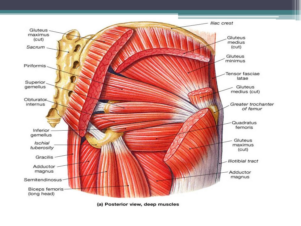
Video 4 II. Muscles of the Pelvis & lower limb A. muscles that move the thigh 1. gluteus maximus *largest, most posterior, superficial *originates at sacrum & coccyx *shares insertion w/ tensor fasciae latae muscle, both insert on iliotibial tract - band of collagen fibers extending from thigh to tibia, acts as brace *produces extension & lateral rotation of hip

59
2. Gluteus medius gluteus minimus *deep to maximus *medius originates on crest of illium *minimus originates below crest of illium * both insert on greater trochanter of femur * adbuct and medially rotate hip 3. Lateral rotators * deep posterior muscles * 6 muscles in all, piriformis & obturator are dominant * all aid in lateral rotation at hip

61
4. Adductors *all originate on ramus of pubis *most insert on femur *include adductor magnus, longus, brevis, pectineus & gracilis * most perform hip flexion & adduction (use these when riding horse) * pulled groin refers to tear in one of these muscles
 * pulled groin refers to tear in one of these muscles.")
62
5. Iliopsoas muscle * made of psoas major & iliacus muscle *locate on anterior interior side of pelvis * Are powerful hip flexor muscles

64
Hip & knee muscles Quadriceps femoris ▫O: anterior inferior iliac spine, superior margin of acetabulum, greater trochanter, shaft of femur ▫I: patella and tibial tuberosity via patellar tendon ▫Action: extend knee, flex thigh

65
Hip & knee muscles Hamstrings ▫O: ischial tuberosity, shaft of femur ▫I: lateral & medial condyles and shaft of tibia, head of fibula, lateral condyle femur ▫Action: extend thigh, flex knee ▫Makes touching toes hard

66
Leg muscles Tibialis anterior ▫O: lateral condyle and shaft of tibia ▫I: tarsal and first metatarsal bones ▫Action: dorsiflexion

67
Leg muscles Gastrocnemius ▫O: medial & lateral condyles of femur ▫I: calcaneus via Achilles tendon ▫Action: plantar flexion, flex knee

Similar presentations