Download presentation
Presentation is loading. Please wait.

1
Dermatopathology Kimiko Suzue, MD PhD October 25 and 27, 2011

3
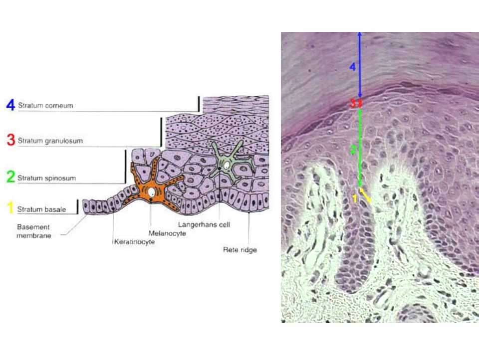
Dermis Papillary dermis - the thin upper layer - of the dermis - lies directly below and interdigitates with the epidermal rete ridges. The papillary dermis is composed of loosely interwoven collagen (Collagen III) Thicker reticular dermis with its coarser and horizontally running bundles of collagen (Collagen I)
 Thicker reticular dermis with its coarser and horizontally running bundles of collagen. (Collagen I)")
4
Verruca vulgaris Verrucous/papillomatous epidermal hyperplasia
Hyperkeratosis Prominent keratohyaline granules Koilocytes

5
Verruca vulgaris

6
Verruca plantaris (plantar wart)
Point out black dots in these warts, ie thrombosed capillaries

7
Verruca vulgaris (common warts)
")
8
Seborrheic Keratosis Small keratin-filled cysts (horn cysts)
Down-growth of keratin into main tumor mass (pseudo-horn cysts) Benign proliferation of basaloid keratinocytes
 Benign proliferation of basaloid keratinocytes.")
9
Actinic Keratosis Solar elastosis Parakeratosis (arrow) Atypia
(dermis looks purplish/sun damgae) Parakeratosis (arrow) Atypia (nuclei in bottom portion of epidermis are hyperchromatic/enlarged) Inflammation Not full-thickness atypia
 Parakeratosis (arrow) Atypia. (nuclei in bottom portion of epidermis are hyperchromatic/enlarged) Inflammation. Not full-thickness atypia.")
10
Actinic Keratosis

11
SCC in situ - Histopathology
Highly atypical cells at all levels of the epidermis Abundant mitoses, crowding of nuclei, pleomorphism, ± necrosis Cells keratinize and stain for keratin Bowen disease

12
Invasive SCC Endophytic growth of atypical epithelium with patchy lymphocytic infiltrate Cells have broken through basement membrane Haphazardly oriented lobules of varying shapes & sizes within the dermis Squamous pearls: whorled aggregates of parakeratotic horn Mild to severe cytologic atypia

13
Basal Cell Carcinoma - Histopathology
Nodular masses or cords of darkly staining cells with high nucleus to cytoplasm ratio In 90% of cases, a connection to the epidermis can be seen

14
Basal Cell Carcinoma

15
Malignant Melanoma CLINICAL FEATURES: A B C D E Asymmetry
Border irregularity Color variegation Diameter > 0.6 cm Evolution

16
Superficial spreading melanoma

17
Superficial spreading melanoma

19
Acral Lentiginous Melanoma

20
Malignant Melanoma HISTOPATHOLOGY: Asymmetry Architectural disorder
Single-cell proliferation, not just nests Pagetoid spread: melanocytes above basal layer Cytologic atypia, usually severe

21
Malignant melanoma

22
Tumor Thickness as Sole Predictor of Outcome 10 years after Definitive Therapy of Primary Melanoma
Breslow Thickness (mm) Survival (%) ≤ –88 1.01– –79 2.01– –64 > –54
 Survival (%) ≤1 83– –2 64– –4 51–64. >4 32–54.")
23
Psoriasis

24
Psoriasis-Morphology
Acanthosis- marked epidermal thickening Overlying parakeratotic scale Auspitz sign-Vessels in dermal papillae bleed eaily when scale is removed Munro microabscess-neutrophil microabscess in parakeratotic stratum corneum

25
Psoriasis

26
Psoriasis

Similar presentations


>")


 Pigment Changes (2) Nodules (2) Purpura (1) Blisters (4) Systemic.>")


