Download presentation
Presentation is loading. Please wait.

1
3 parasitic zoonoses Arthropooda
Dr. Raad H.H.

2
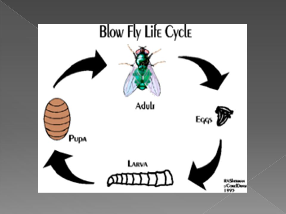
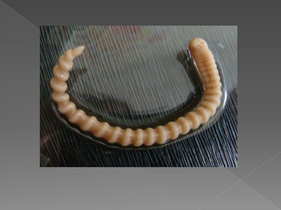
Myiasis Screwworm

4
Myiasis as "the infestation of live human and vertebrate animals with dipterous larvae, which at least for a period, feed on the host's dead or living tissue, liquid body substances, or ingested food".

5
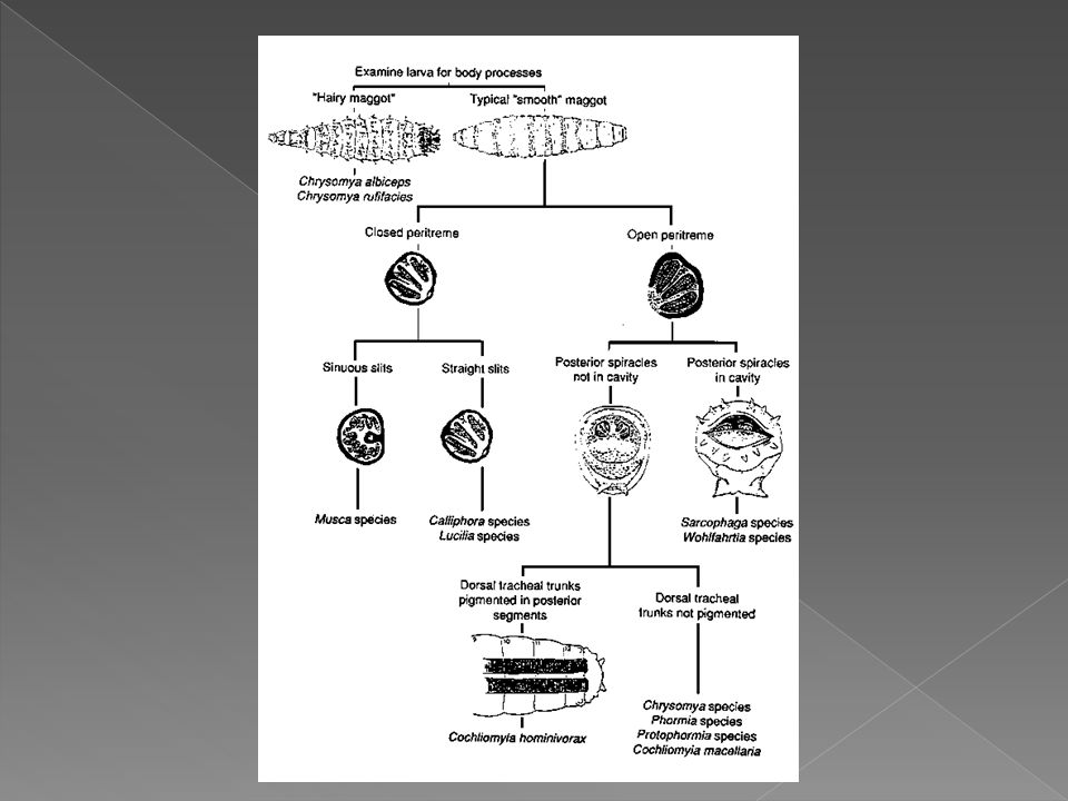
The classical description of myiasis is according to the part of the host that is infected. This is the classification used by. For example: dermal sub-dermal Cutaneous Creeping, where larvae burrow through or under the skin Furuncular, where a larva remains in one spot, causing a boil-like lesion nasopharyngeal nose, sinuses or pharynx. Ophthalmic or ocular in or about the eye Auricular in or about the ear gastric, rectal, or intestinal/enteric for the appropriate part of the digestive system urogenital .

6
Another aspect is the relationship between the host and the parasite
Another aspect is the relationship between the host and the parasite . Thus the myiasis is described as either: Obligatory, where the parasite cannot complete its life cycle without its parasitic phase, which may be Specific, Semispecific, or Opportunistic. Facultative, incidental, or accidental, where it is not essential to the life cycle of the parasite; perhaps a normally free-living larva accidentally gained entrance to the host.

7
Accidental myiasis commonly is enteric, resulting from swallowing eggs or larvae with one's food. The effect is called pseudomyiasis. One traditional cause of pseudomyiasis was the eating of maggots of cheese flies in cheeses such as Stilton Musca domestica (housefly) Fannia spp. (latrine flies) Eristalis tenax (rat-tailed maggots) Muscina spp
 Fannia spp. (latrine flies) Eristalis tenax (rat-tailed maggots) Muscina spp.")
8
Vectors in humans: There are three main fly families causing economically important myiasis in livestock and also, occasionally, in humans: Calliphoridae (blowflies) Oestridae (botflies) Sarcophagidae (fleshflies) Other families occasionally involved are: Anisopodidae Piophilidae Stratiomyidae Syrphidae
 Oestridae (botflies) Sarcophagidae (fleshflies) Other families occasionally involved are: Anisopodidae. Piophilidae. Stratiomyidae. Syrphidae.")
9
Specific myiasis: Caused by flies that need a host for larval development Dermatobia hominis (human botfly) Cordylobia anthropophaga (tumbu fly) Oestrus ovis (sheep botfly) Hypoderma spp. (cattle botflies or ox warbles) Gasterophilus spp. (horse botfly) Cochliomyia hominivorax (new world screwworm fly) Chrysomya bezziana (old world screwworm fly) Auchmeromyia senegalensis (Congo floor maggot) Cuterebra spp. (rodent and rabbit botfly)
 Oestrus ovis (sheep botfly) Hypoderma spp. (cattle botflies or ox warbles) Gasterophilus spp. (horse botfly) Cochliomyia hominivorax (new world screwworm fly) Chrysomya bezziana (old world screwworm fly) Auchmeromyia senegalensis (Congo floor maggot) Cuterebra spp. (rodent and rabbit botfly)")
10
Semispecific myiasis Caused by flies that usually lay their eggs in decaying animal or vegetable matter, but that can develop in a host if open wounds or sores are present Lucilia spp. (green-bottle fly) Cochliomyia spp. (screw-worm fly) Phormia spp. (black-bottle fly) Calliphora spp. (blue-bottle fly) Sarcophaga spp. (flesh fly or sarcophagids) Flesh flies, or sarcophagids, members of the family Sarcophagidae, can cause intestinal myiasis in humans if the females lay their eggs on meat or fruit.
 Cochliomyia spp. (screw-worm fly) Phormia spp. (black-bottle fly) Calliphora spp. (blue-bottle fly) Sarcophaga spp. (flesh fly or sarcophagids) Flesh flies, or sarcophagids, members of the family Sarcophagidae, can cause intestinal myiasis in humans if the females lay their eggs on meat or fruit.")
13
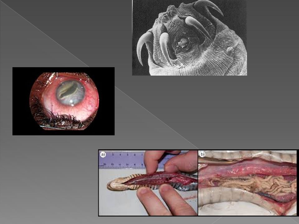
Pentastomiasis

17
Tungiasis Tunga penetrans

18
Tungiasis or related species.
is an infestation by the burrowing flea Tunga penetrans or related species. The flea has many common names, being known in various locations as the chigger flea, sand flea, chigoe, jigger, nigua, pigue, or le bicho de pe (see the image below). Painful infections with T penetrans can cause significant morbidity. Tungiasis was first reported in crewmen who sailed with Christopher Columbus. The flea is indigenous to the West Indies/Caribbean/Central America region, but it has spread to Africa, India, Pakistan, and South America. To reproduce, the flea requires a warm-blooded host. In addition to humans, reservoir hosts include pigs, dogs, cats, cattle, sheep, horses, mules, rats, mice, and other wild animal
. Painful infections with T penetrans can cause significant morbidity. Tungiasis was first reported in crewmen who sailed with Christopher Columbus. The flea is indigenous to the West Indies/Caribbean/Central America region, but it has spread to Africa, India, Pakistan, and South America. To reproduce, the flea requires a warm-blooded host. In addition to humans, reservoir hosts include pigs, dogs, cats, cattle, sheep, horses, mules, rats, mice, and other wild animal.")
19
Jigger flea chigoe flea

20
Dermatitis due to Acrid mites Cheyletiella

21
Dermanyssidae

22
Rodent Mites The tropical rat mite, Ornithonyssus bacoti
The house mouse mite, Liponyssoides sanguineus

23
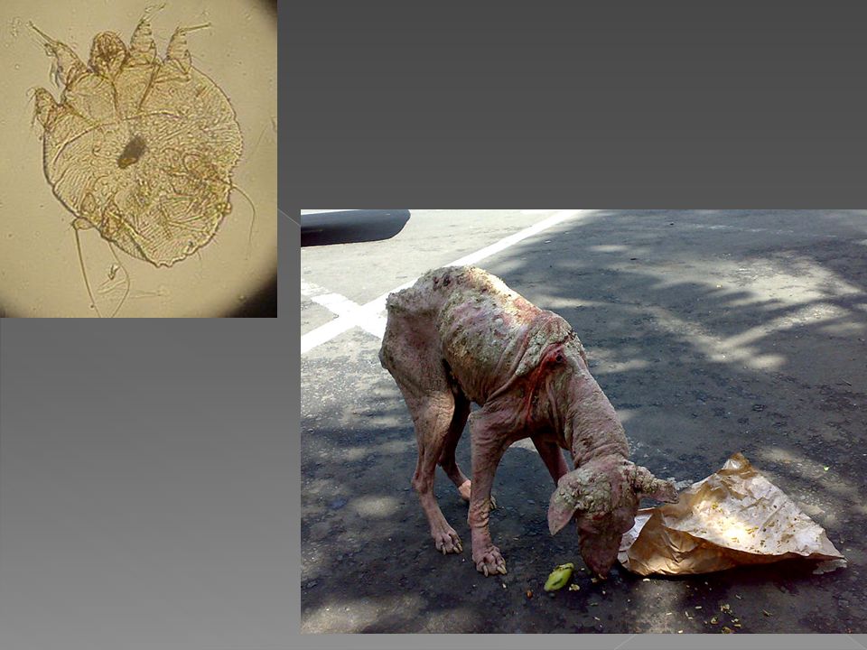
Scabies

25
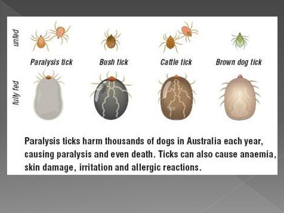
Tick paralysis Tick paralysis is believed to be due to toxins found in the tick's saliva that enter the bloodstream while the tick is feeding. The two ticks most commonly associated with North American tick paralysis are the Rocky Mountain wood tick (Dermacentor andersoni) and the American dog tick (Dermacentor variabilis); however, 43 tick species have been implicated in human disease around the world. Most North American cases of tick paralysis occur from April to June, when adult Dermacentor ticks emerge from hibernation and actively seek hosts. In Australia, tick paralysis is caused by the tick Ixodes holocyclus. Prior to 1989, 20 fatal cases were reported in Australia
 and the American dog tick (Dermacentor variabilis); however, 43 tick species have been implicated in human disease around the world. Most North American cases of tick paralysis occur from April to June, when adult Dermacentor ticks emerge from hibernation and actively seek hosts. In Australia, tick paralysis is caused by the tick Ixodes holocyclus. Prior to 1989, 20 fatal cases were reported in Australia.")
Similar presentations


 LARVA FREE LIVING, ADULT PARASITIC MOSQUITO larvae live in water, feed on microorganisms MOSQUITO adults.>")


 Brown dog tick (Rhipicephalus sanguineus) Rocky.>")


