Download presentation
Presentation is loading. Please wait.

1
LECTURE 35 DR FARHAT AAMIR LECTURER ANATOMY
POPLITEAL FOSSA

2
POPLITEAL FOSSA At the end of this lecture the students should be able to: Describe boundaries of the popliteal fossa. Describe the contents of the popliteal fossa. Identify clinical application.

3
REFERENCES Clinical anatomy by regions 9th edition (Pages 476 – 479).
Gray’s anatomy for student, 2ed edition (Pages 584 – 585).
.")
4
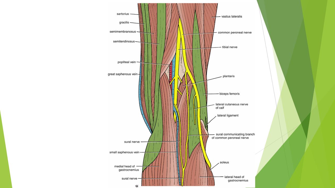
POPLITEAL FOSSA The popliteal fossa is a diamond-shaped inter muscular space situated at the back of the knee. The fossa is most prominent when the knee joint is flexed. CONTENTS. Popliteal vessels The small saphenous vein The common peroneal and tibial nerves The posterior cutaneous nerve of the thigh. The genicular branch of the obturator nerve Connective tissue Lymph nodes.

5
BOUNDARIES OF POPLITEAL FOSSA
Laterally: above: The biceps femoris below: lateral head of the gastrocnemius and plantaris Medially: above: The semimembranosus and semitendinosus below: medial head of the gastrocnemius

6
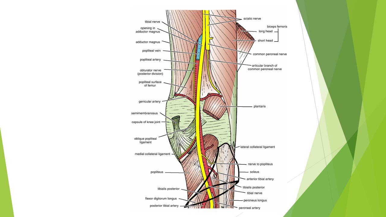
The anterior wall or floor of the fossa is formed by the popliteal surface of the femur, the posterior ligament of the knee joint, and the popliteus muscle The roof is formed by skin, superficial fascia, and the deep fascia of the thigh.

7
POPLITEUS MUSCLE The popliteus muscle plays a key role in the movements of the knee joint. Origin: From the lateral surface of the lateral condyle of the femur by a rounded tendon. Insertion: The fibers pass downward and medially and are attached to the posterior surface of the tibia, above the soleal line. Nerve supply: Tibial nerve Action: Medial rotation of the tibia on the femur or, if the foot is on the ground, lateral rotation of the femur on the tibia. The latter action occurs at the commencement of flexion of the extended knee, and its rotatory action slackens the ligaments of the knee joint; this action is sometimes referred to as unlocking the knee joint.

8
POPLITEAL ARTERY The popliteal artery is deeply placed and enters the popliteal fossa through the opening in the adductor magnus, as a continuation of the femoral artery. It ends at the level of the lower border of the popliteus muscle by dividing into anterior and posterior tibial arteries. Relations Anteriorly: The popliteal surface of the femur, the knee joint, and the popliteus muscle. Posteriorly: The popliteal vein and the tibial nerve, fascia, and skin Branches The popliteal artery has muscular branches and articular branches to the knee.

12
POPLITEAL ANEURYSM The pulsations of the wall of the femoral artery against the tendon of adductor magnus at the opening of the adductor magnus is thought to contribute to the cause of popliteal aneurysms.

13
SEMIMEMBRANOSUS BURSA SWELLING
Semimembranosus bursa swelling is the most common swelling found in the popliteal space. It is made tense by extending the knee joint and becomes flaccid when the joint is flexed. It should be distinguished from a Baker's cyst, which is centrally located and arises as a pathologic (osteoarthritis) diverticulum of the synovial membrane through a hole in the back of the capsule of the knee joint.
 diverticulum of the synovial membrane through a hole in the back of the capsule of the knee joint.")
14
POPLITEAL VEIN The popliteal vein is formed by the junction of the venae comitantes of the anterior and posterior tibial arteries at the lower border of the popliteus muscle on the medial side of the popliteal artery. As it ascends through the fossa, it crosses behind the popliteal artery so that it comes to lie on its lateral side. It passes through the opening in the adductor magnus to become the femoral vein. Tributaries Veins that correspond to branches given off by the popliteal artery Small saphenous vein, which perforates the deep fascia and passes between the two heads of the gastrocnemius muscle to end in the popliteal vein.

15
Arterial Anastomosis Around the Knee Joint
To compensate for the narrowing of the popliteal artery, which occurs during extreme flexion of the knee, around the knee joint is a profuse anastomosis of small branches of the femoral artery with muscular and articular branches of the popliteal artery and with branches of the anterior and posterior tibial arteries.

16
Popliteal Lymph Nodes About six lymph nodes are embedded in the fatty connective tissue of the popliteal fossa. They receive superficial lymph vessels from the lateral side of the foot and leg They also receive lymph from the knee joint and from deep lymph vessels accompanying the anterior and posterior tibial arteries.

17
TIBIAL NERVE It is the larger terminal branch of the sciatic nerve.
It arises in the lower third of the thigh. It runs downward through the popliteal fossa, lying first on the lateral side of the popliteal artery, then posterior to it, and finally medial to it The popliteal vein lies between the nerve and the artery throughout its course. The nerve enters the posterior compartment of the leg by passing beneath the soleus muscle. BRANCHES 1. Cutaneous: The sural nerve descends between the two heads of the gastrocnemius muscle and is usually joined by the sural communicating branch of the common peroneal nerve Muscular branches supply both heads of the gastrocnemius and the plantaris, soleus, and popliteus 2. Articular branches supply the knee joint.

18
COMMON PERONEAL NERVE It is the smaller terminal branch of the sciatic nerve It arises in the lower third of the thigh. It runs downward through the popliteal fossa, closely following the medial border of the biceps muscle It leaves the fossa by crossing superficially the lateral head of the gastrocnemius muscle. It then passes behind the head of the fibula, winds laterally around the neck of the bone, pierces the peroneus longus muscle, and divides into two terminal branches: the superficial peroneal nerve and the deep peroneal nerve As the nerve lies on the lateral aspect of the neck of the fibula, it is subcutaneous and can easily be rolled against the bone. Branches Cutaneous: The sural communicating branch The lateral cutaneous nerve of the calf Muscular branch to the short head of the biceps femoris muscle, Articular branches to the knee joint

19
COMMON PERONEAL NERVE INJURY
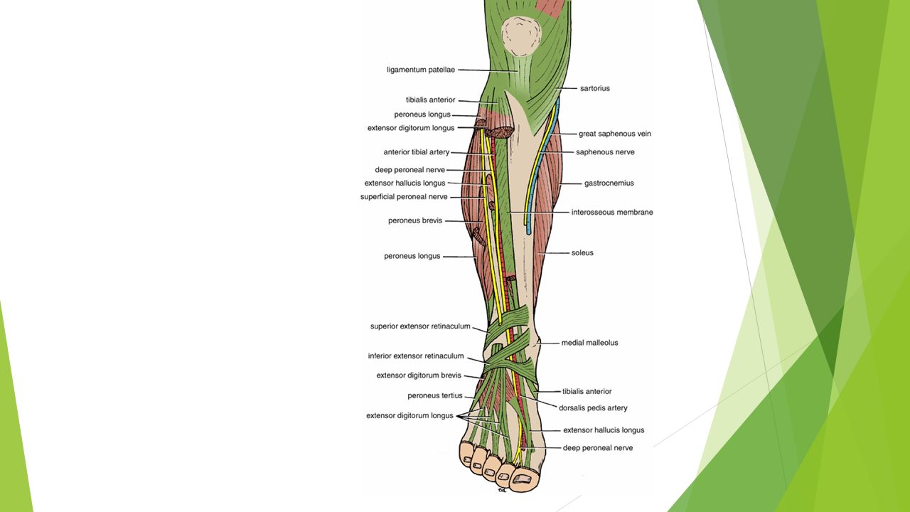
The common peroneal nerve is extremely vulnerable to injury as it winds around the neck of the fibula. At this site, it is exposed to direct trauma or is involved in fractures of the upper part of the fibula. Injury to the common peroneal nerve causes footdrop.

Similar presentations


.>")

>")


 Joint>")