Download presentation
Presentation is loading. Please wait.

1
Pregnancy, Growth, Development and Genetics
Prenatal Development & Care

2
Fun facts: Did you know some infants are delivered in the amniotic sac? It’s termed “Veiled Birth” or caulbearer There are some very rare cases of multiple ovulation, i.e., the release of several eggs at the same time. 3 Triplets 4 Quadruplets 5 Quintuplets

3
Fertilization When a male sperm meets a female egg (oocyte)
Only one sperm cell can fertilize an egg. Occurs after ovulation. aka Conception result in: Zygote Before implantation Blastocyst After implantation Embryo Implantation to about 8 weeks in the womb Fetus After 8 Weeks in the womb

4
Fertilization: Multiple Births
Fraternal Twins (Dizygotic) Two eggs released & fertilized by two sperm cells Different genetic makeup May or may not be the same gender Identical Twins (Monozygotic) Single egg fertilized by a single sperm cell Divides forming two embryos Shares a single placenta Genetically identical, same gender, similar appearance
 Two eggs released & fertilized by two sperm cells. Different genetic makeup. May or may not be the same gender. Identical Twins (Monozygotic) Single egg fertilized by a single sperm cell. Divides forming two embryos. Shares a single placenta. Genetically identical, same gender, similar appearance.")
6
Early Embryonic Development

7
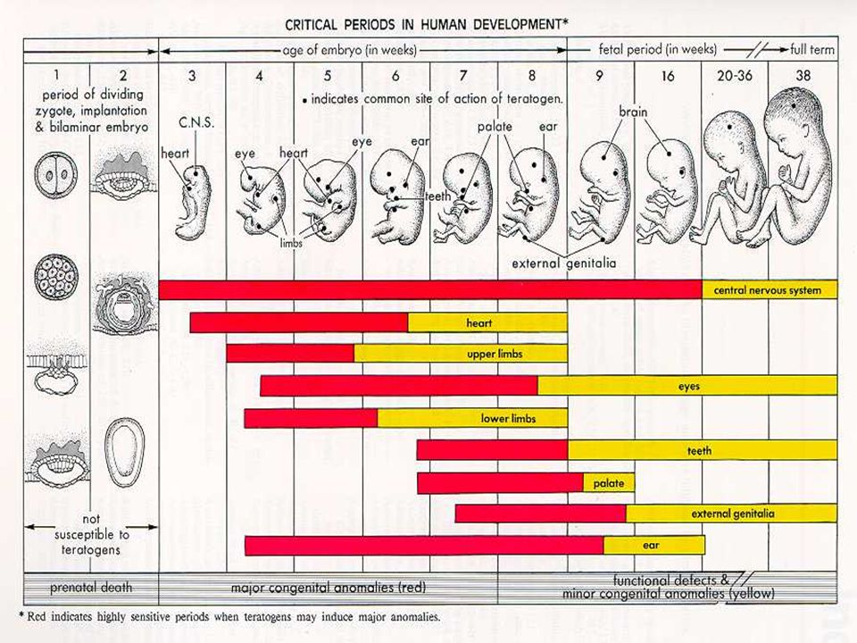
Embryonic Growth During this period: The offspring is termed Embryo
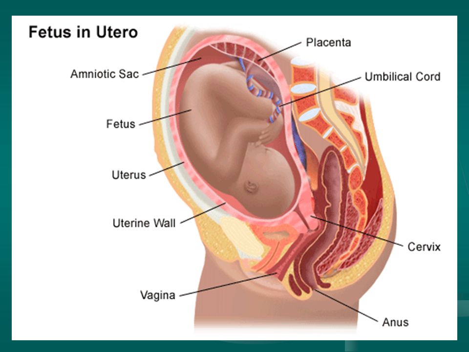
Until the end of the 8th wk, when the basic structural form of the human body is recognizable During this period: The amniotic sac forms- Thin filled membrane that surrounds and protects the embryo -insulates the embryo from temperature changes Umbilical Cord- Rope-like structure that connects the embryo and the mother’s placenta Placenta- Thick blood-rich tissues that lines the wall of the uterus during pregnancy and nourishes the embryo Begins to form in the 4th week of pregnancy Does Not allow blood to mix between mother and child Provides nutrients Main internal organs develop Major external body structures appear

9
Development of Internal Organs via Tissue Layers
Three tissue layers form: 1st layer-Ectodermal cells Nervous system, sense organs, skin, hair, nails and mouth 2nd layer-Mesodermal cells Bones, muscles & connective tissue Blood vessels & blood Internal reproductive organs & kidneys 3rd layer-Endodermal Cells Linings of Respiratory and digestive systems Urinary bladder & urethra

10
Fetal Development Fetal Stage begins at the end of the 8th wk and last until birth. About nine full months, that is divided into three periods called trimesters. First Trimester 0-14 weeks Second Trimester 14-28 weeks Third Trimester 29-40 weeks

11
Fetal Development First Trimester 0-2 weeks 3-8 weeks 9-14 weeks
Zygote floats freely for two days Spinal cord grows faster than rest of the body Brains, ears, and arms begin developing Heart begins to beat 3-8 weeks Embryo is about one inch long at eight weeks Mouth, nostrils, eyelids, hands, fingers, feet, and toes begin to form Nervous system can respond to stimuli * stimulus-something that causes a response Cardiovascular system is fully functional 9-14 weeks Fetus develops human profile Sex organs, eyelids, fingernails and toenails develop By 12 weeks the fetus makes crying motions and may suck it’s thumb

12
Fetal Development Second Trimester 15-20 weeks 21-28 weeks
Can blink it’s eyes Body grows Growth of head slows Limbs reach full proportion Eyebrows and eyelashes develop Fetus becomes more active (gasping kicking, etc) 21-28 weeks Fetus can hear conversations Cycle of waking and sleeping Gains weight Fetus is a foot long, weighs a little more then one pound Can be born at 24 weeks, but will need special care if this occurs
 weeks. Fetus can hear conversations. Cycle of waking and sleeping. Gains weight. Fetus is a foot long, weighs a little more then one pound. Can be born at 24 weeks, but will need special care if this occurs.")
13
Fetal Development Third Trimester (final) 29-40 weeks
Fetus can use all five senses Passes water from the bladder From ingesting amniotic fluid through the mouth Some fetuses begin to dream Baby weighs 6-9 pounds Ready to be born

15
Labor and Delivery Labor Signs of Labor
Series of continuous, progressive contractions of the uterus which help the cervix to open (dilate) and to thin (efface), allowing the fetus to move through the birth canal. Usually starts two weeks before/after due date Signs of Labor Bloody show Contractions Rupture of amniotic sac (water break)
 and to thin (efface), allowing the fetus to move through the birth canal. Usually starts two weeks before/after due date. Signs of Labor. Bloody show. Contractions. Rupture of amniotic sac (water break)")
17
Stages of Birth Pregnancy ends w/ the birth process, labor. Stage 1-
Initial phase-fetus position before birth Active phase- dilation of the cervix (10cm) Stage 2- Expulsion of the fetus Stage 3- expulsion of the placenta (afterbirth)
 Stage 2- Expulsion of the fetus. Stage 3- expulsion of the placenta (afterbirth)")
18
Stages Explained The first phase of the first stage of labor is called the latent phase when contractions are becoming more frequent (usually 5 to 20 minutes apart) and somewhat stronger. However, discomfort is minimal. The cervix dilates (opens approximately three or four centimeters) and effaces (thins out). Some women may not recognize that they are labor if their contractions are mild and irregular. The latent phase is usually the longest and least intense phase of labor. The mother-to-be is usually admitted to the hospital during this phase. Pelvic exams are performed to determine the dilatation of the cervix. The second phase of the first stage (active phase) is signaled by the dilatation of the cervix from 4 to 7 centimeters. Contractions become longer, more severe, and more frequent (usually 3 to 4 minutes apart). The third phase is called transition and is the last phase. the cervix dilates from 8 to 10 centimeters. Contractions are usually very strong, lasting 60 to 90 seconds and occurring every few minutes. Most women feel the urge to push during this phase. In most cases, the active and transition phases are shorter than the latent phase.
 and somewhat stronger. However, discomfort is minimal. The cervix dilates (opens approximately three or four centimeters) and effaces (thins out). Some women may not recognize that they are labor if their contractions are mild and irregular. The latent phase is usually the longest and least intense phase of labor. The mother-to-be is usually admitted to the hospital during this phase. Pelvic exams are performed to determine the dilatation of the cervix. The second phase of the first stage (active phase) is. signaled by the dilatation of the cervix from 4 to 7 centimeters. Contractions become longer, more severe, and more frequent (usually 3 to 4 minutes apart). The third phase is called transition and is the last phase. the cervix dilates from 8 to 10 centimeters. Contractions are usually very strong, lasting 60 to 90 seconds and occurring every few minutes. Most women feel the urge to push during this phase. In most cases, the active and transition phases are shorter than the latent phase.")
19
Delivery Labor Complications Fetal Meconium Abnormal Fetal heart rate
Meconium in the amniotic fluid Abnormal Fetal heart rate Normal range is beats per minute might increase if the fetus is in distress Abnormal position of the fetus during birth A physician may try to deliver, attempt to turn before delivery or perform a C-section

20
Labor and Delivery In some cases, the vaginal opening does not stretch enough to accommodate the fetus. If there is a risk of tearing, the physician may perform an episiotomy – an incision through the vaginal wall and the perineum (the area between the thighs, extending from the anus to the vaginal opening) to help deliver the fetus. Cesarean Section (C-Section): Surgical incision is made through the abdominal wall and uterus. There are several conditions which may make having a baby by cesarean section more likely. previous cesarean section, fetal distress, abnormal delivery presentation, a labor that fails to progress or does not progress normally, placental complications, twins or other multiples
 to help deliver the fetus. Cesarean Section (C-Section): Surgical incision is made through the abdominal wall and uterus. There are several conditions which may make having a baby by cesarean section more likely. previous cesarean section, fetal distress, abnormal delivery presentation, a labor that fails to progress or does not progress normally, placental complications, twins or other multiples.")
Similar presentations


 Usually one egg once a month All about timing!!! › Uterus must be ready when egg gets there in.>")


