Download presentation
Presentation is loading. Please wait.

1
The Reproductive System
Chapter 19 The Reproductive System

2
19-1: Reproductive System
Produces, stores, nourishes, & transports male & female reproductive cells (gametes) Includes: Gonads Ducts for receiving/transporting gametes Accessory glands & organs External genitalia
 Includes: Gonads. Ducts for receiving/transporting gametes. Accessory glands & organs. External genitalia.")
3
Male reproductive system
Gonads (testes) secrete sex hormones (androgens) Spermatozoa (sperm) transported in semen Female reproductive system Gonads (ovaries) release one oocyte (egg) per month Sperm fuse with the oocyte through fertilization Uterus supports developing embryo
 secrete sex hormones (androgens) Spermatozoa (sperm) transported in semen. Female reproductive system. Gonads (ovaries) release one oocyte (egg) per month. Sperm fuse with the oocyte through fertilization. Uterus supports developing embryo.")
4
19-1 Checkpoint Define gamete.
List the basic components of the reproductive system. Define gonads.

5
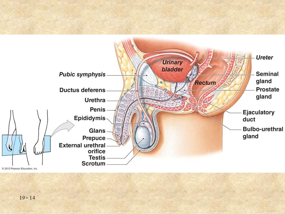
19-2: Male Reproductive System
Male reproductive tract delivers sperm Epididymis, ductus deferens, ejaculatory duct, urethra Male accessory organs add secretions Seminal glands, prostate gland, bulbourethral gland External genitalia Scrotum & penis

7
The Testes Testes hang within scrotum
Muscles contract testes closer to body Testis divided into lobules containing seminiferous tubules Sperm produced in tubules enter epididymis Spaces between tubules produce androgens (testosterone)
")
9
Spermatogenesis Begins with mitosis Continues with meiosis
Spermatogonia undergo mitosis; become spermatocytes Continues with meiosis Spermatocytes produce spermatids Each spermatid contains 23 chromosomes Finishes with spermiogenesis Spermatid matures into spermatozoon (sperm cell) Takes about 9 weeks
 Takes about 9 weeks.")
10
Anatomy of Spermatozoon
Head contains chromosomes Covered by acrosome—contains enzymes to penetrate surface of egg Middle piece contains mitochondria to provide energy Tail moves sperm cell

12
Male Reproductive Tract
Epididymis Sperm cells transported to epididymis from seminiferous tubules Sperm mature, travel to ductus deferens Sperm leaving epididymis are immobile Must undergo capacitation to become motile

13
Ductus Deferens (Vas Deferens)
Ascends into abdominal cavity, descends behind bladder Peristalsis pushes sperm in ductus deferens Sperm can be stored for several months Ductus deferens joins seminal gland to create ejaculatory duct Duct empties into urethra Vasectomy cuts ductus deferens

15
The Accessory Glands Generate fluid components of semen
Seminal Glands (Vesicles) Contribute about 60% of semen volume: Fructose—energy for sperm Prostaglandins—stimulate male & female smooth muscle contractions Slightly alkaline fluid—neutralizes acids in vagina Contact with secretions causes sperm to start beating flagella
 Contribute about 60% of semen volume: Fructose—energy for sperm. Prostaglandins—stimulate male & female smooth muscle contractions. Slightly alkaline fluid—neutralizes acids in vagina. Contact with secretions causes sperm to start beating flagella.")
16
Prostate Gland Surrounds urethra as it leaves the bladder Produces fluids that add to seminal fluids Bulbourethral Glands Secrete mucus to neutralize acids in male urethra, lubricate tip of penis

18
Fluid that contains sperm & secretions of accessory glands
Semen Fluid that contains sperm & secretions of accessory glands Ejaculation expels 2 – 5 mL of ejaculate containing: Sperm cells (sperm count—20 million – 100 million/mL) Seminal fluid (secretions) Enzymes (dissolves vaginal secretions)
 Seminal fluid (secretions) Enzymes (dissolves vaginal secretions)")
19
The External Genitalia
Penis introduces semen into vagina & conducts urine through urethra Processes don’t occur at the same time Regions of penis: Root—attaches penis to body wall Body (shaft)—contains erectile tissue Glans—surrounds urethral opening Prepuce (foreskin) surrounds tip of penis Removed through circumcision
—contains erectile tissue. Glans—surrounds urethral opening. Prepuce (foreskin) surrounds tip of penis. Removed through circumcision.")
20
Penis contains three columns of erectile tissue
2 columns of corpora cavernosa; 1 column of corpus spongiosum Increased blood flow to tissues generate erection Erectile dysfunction (ED) can result from hormonal, vascular, or nerve damage
 can result from hormonal, vascular, or nerve damage.")
22
Hormones & Male Reproductive Function
Follicle-stimulating hormone (FSH) & luteinizing hormone (LH) Promote spermatogenesis & spermiogenesis Testosterone Stimulates bone & muscle growth Enlarges reproductive organs Establishes & maintains male secondary sex characteristics (hair growth, lower voice)
 & luteinizing hormone (LH) Promote spermatogenesis & spermiogenesis. Testosterone. Stimulates bone & muscle growth. Enlarges reproductive organs. Establishes & maintains male secondary sex characteristics (hair growth, lower voice)")
23
19-2 Checkpoint List the male reproductive structures.
On a warm day, would the tissues surrounding the scrotum be contracted or relaxed? Why? What happens when the arteries within the penis dilate? What effect would low FSH levels have on sperm production?

24
19-3: Female Reproductive System
Produces sex hormones, gametes, supports & nourishes developing embryo Ovaries, uterine tubes, uterus, vagina, external genitalia Female accessory glands release secretions into female reproductive tract

26
The Ovaries Paired ovaries responsible for:
Producing female gametes (ova) Secreting female hormones (estrogens & progestins) Each ovary is flattened, almond-shaped Supported in abdominal cavity by ovarian ligament
 Secreting female hormones (estrogens & progestins) Each ovary is flattened, almond-shaped. Supported in abdominal cavity by ovarian ligament.")
28
Oogenesis Oogenesis begins before birth, accelerates at puberty, ends with menopause Oogenesis occurs each month between puberty & menopause Oogonia form before birth Primary oocytes undergo meiosis during fetal development (contain 23 chromosomes) Mature into secondary oocyte during puberty & released each month
 Mature into secondary oocyte during puberty & released each month.")
29
Follicle Development Ovarian follicles grow oocytes & promote oogenesis Primordial follicles begin generation of primary oocyte Develop into primary follicles at puberty Primary oocyte within follicle enlarges & divides Primary follicle expands & becomes secondary follicle Follicle may then enter ovarian cycle to release oocyte

31
The Ovarian Cycle Ovarian cycle—28 day cycle divided into follicular phase & luteal phase Phases separated by ovulation Follicular phase (days 1 – 14) FSH triggers secondary follicle to form a tertiary follicle Oocyte within follicle triggered by LH to become secondary oocyte
 FSH triggers secondary follicle to form a tertiary follicle. Oocyte within follicle triggered by LH to become secondary oocyte.")
32
Ovulation (day 14) Luteal phase (days 15 – 28)
Tertiary follicle ruptures & releases secondary oocyte into uterine tubes In 1 – 2% of ovulations, more than 1 oocyte is released (multiple births) Luteal phase (days 15 – 28) Empty follicle collapses & forms corpus luteum No fertilization: corpus luteum breaks down & new ovarian cycle begins Fertilization: corpus luteum remains & prevents another ovarian cycle
 Luteal phase (days 15 – 28) Empty follicle collapses & forms corpus luteum. No fertilization: corpus luteum breaks down & new ovarian cycle begins. Fertilization: corpus luteum remains & prevents another ovarian cycle.")
34
The Uterine Tubes Uterine tubes (Fallopian tubes/oviducts) connect ovaries to uterus Infundibulum partially surrounds ovarian edge Fimbriae (projections) draw ovulated egg into uterine tube Uterine tube peristalsis draws oocyte into uterine cavity Fertilization must occur within hours of ovulation
 draw ovulated egg into uterine tube. Uterine tube peristalsis draws oocyte into uterine cavity. Fertilization must occur within hours of ovulation.")
35
The Uterus Provides protection & nutrition for developing embryo & fetus Regions of uterus Body—largest division of uterus Cervix—bottom of uterus; opening leads into uterine cavity Uterine wall made of inner endometrium, muscular myometrium & covered by perimetrium

37
The Uterine Cycle Uterine cycle (menstrual cycle)—repeating changes in endometrium Menarche—first uterine cycle; menopause—last uterine cycle Average uterine cycle is 28 days; can be longer or shorter First phase: menses (days 1 – 7) Endometrial lining degenerates due to low progesterone & estrogen Lining shed as menstruation
 Endometrial lining degenerates due to low progesterone & estrogen. Lining shed as menstruation.")
38
Second phase: proliferative phase (days 7 – 14)
Rising estrogen triggers growth & thickening of endometrial lining Third phase: secretory phase (days 15 – 28) Endometrium prepares for developing embryo Declining hormone levels trigger menses Last day of uterine cycle occurs with menstruation Begins first day of next cycle
 Endometrium prepares for developing embryo. Declining hormone levels trigger menses. Last day of uterine cycle occurs with menstruation. Begins first day of next cycle.")
39
The Vagina Extends from uterus to body’s exterior
Vaginal walls contain blood vessels & smooth muscle Hymen—tissue that partially blocks vaginal entrance Functions: Passageway for menstrual fluids Receives penis during sexual intercourse Passes fetus during childbirth

40
The External Genitalia
Vulva—area containing external female genitalia External genitalia includes: Labia minora surround vaginal opening Clitoris composed of erectile tissue Vestibular glands lubricate vagina Labia majora conceal vaginal structures

42
The Mammary Glands Mammary glands nourish infants by producing & secreting milk Lactation—milk production Each breast contains mammary gland within subcutaneous fat Mammary gland opens to surface at the nipple Areola—area surrounding nipple Mammary gland composed of lobes that release milk through lactiferous duct

44
Breast Cancer Caused by tumors in the mammary glands
Leading cause of death in women 35 – 45; most common in women older than 50 12% of women develop breast cancer in their lifetime Risk factors: Family history of breast cancer First pregnancy after age 30 Early first period or late menopause

45
Hormones & Female Reproductive Cycle
Circulating hormones coordinate ovarian & uterine cycles Hormones & the Follicular Phase FSH stimulates follicular growth; follicles stimulate estrogen release Estrogen Stimulates bone & muscle growth Establishes female secondary sex characteristics (breast development, fat deposit)
")
46
Hormones & the Luteal Phase
Estrogen levels rise as follicle matures Day 14: estrogen peaks, LH levels surge & trigger ovulation Hormones & the Luteal Phase Corpus luteum releases progesterone to prepare uterus for pregnancy Stimulates endometrial lining If no pregnancy, corpus luteum breaks down, progesterone levels fall, endometrial lining is shed

48
19-3 Checkpoint Name the female reproductive structures.
How would a blockage of the uterine tubes affect a woman’s ability to conceive? Which layer of the uterus is shed during menstruation? What changes would you expect to observe in the ovarian cycle if the LH surge did not occur?

49
19-4: Male & Female Sexual Function
Sexual arousal stimulates nervous system & leads to increased blood flow, causing an erection Emission—peristalsis pushes semen & fluid into urethra Ejaculation releases semen through orgasm Impotence—inability to achieve or maintain erection

50
Female Sexual Function
Arousal stimulates nervous system & leads to increased blood flow to clitoris Vaginal surfaces increase lubrication Female orgasm includes peristalsis of uterine & vaginal walls May cause semen to be drawn into uterus

51
19-4 Checkpoint List the physiological events of sexual intercourse in both sexes, and indicate those that occur in males but not in females.

52
19-5: Functional Changes Due to Age
Menopause Time after which ovulation & menstruation stop; typically between ages 45 – 55 2 million oocytes present at birth, only a few hundred thousand present at puberty, only a few hundred ovulated Decreased estrogen levels, “hot flashes,” osteoporosis common

53
Male Climacteric Male climacteric—declining reproductive function in males (similar to menopause) Testosterone levels decrease Sperm production continues throughout most of male’s life

54
19-5 Checkpoint Define menopause. What is the male climacteric?

55
Sexually Transmitted Diseases
Sexually transmitted diseases (STDs)—infections transferred through sexual activity Most common cause of reproductive disorders Gonorrhea & syphilis used to be most common STDs; numbers are rising again Currently herpes, HPV, chlamydia most common STDs
—infections transferred through sexual activity. Most common cause of reproductive disorders. Gonorrhea & syphilis used to be most common STDs; numbers are rising again. Currently herpes, HPV, chlamydia most common STDs.")
56
Many STDs go unnoticed in early stages, especially in women
Symptoms of STDs: Burning sensation during urination Vaginal / penile discharge Sores, blisters, bumps or rashes STDs cause pelvic inflammatory disease in females; can damage the uterus Abstinence is the best way to prevent STDs

57
Birth Control Methods Contraceptive methods try to avoid fertilization following sexual intercourse Coitus interruptus Withdrawal of the penis prior to ejaculation Least effective means of contraception Rhythm method Abstinence before & after ovulation High rates of pregnancy—ovulation can be hard to calculate

58
Hormonal contraceptives
Mechanical barriers Prevent sperm cells from entering female; reduce the spread of STDs Include spermicidal jelly or cream Ex: condom, diaphragm, cervical cap Hormonal contraceptives Manipulate female hormonal cycle to prevent ovulation Nearly 100% effective if used correctly Ex: contraceptive pills, Depo-Provera, Norplant, NuvaRing, “morning after” pill

59
Intrauterine device (IUD)
Small plastic device surgically implanted in the uterus Prevents oocyte implantation for up to 5 years Sterilization Male—vasectomy; ductus deferens cut, sperm unable to pass through reproductive tract Female—tubal ligation; uterine tubes cut, ova unable to pass through reproductive tract Both are considered permanent

Similar presentations