Download presentation
Presentation is loading. Please wait.

1
CHAPTER 3 CELL STRUCTURE & FUNCTION Pictures from Essentials of Anatomy & Physiology, Third Edition PowerPoint by Laurie Forsythe

2
Overview Be sure you know all the cell structures & functions

3
Cell Membrane Barrier Regulates exchange with environment Sensitivity – receptors Supports – connections between cells & with in cells

4
Membrane Structure Membrane lipids – phospholipid bilayer –CO 2 & O 2 can cross –Water soluble can’t cross Membrane proteins – Table 3-2, function as –Receptors – bind to extracellular cmpds –Channels – lets ions thru lipids (Ca +, etc) –Carriers - transports solutes to inside of cell –Enzymes – intestinal tract –Recognition – itself from other cells –Anchoring – fibers attach to other cells
 –Carriers - transports solutes to inside of cell –Enzymes – intestinal tract –Recognition – itself from other cells –Anchoring – fibers attach to other cells")
5
Membrane Transport – Table 3-3 1.Diffusion – osmosis 2.Filtration 3.Carrier-mediated (facilitated diffusion & active transport) 4.Vesicular transport (endocytosis & exocytosis) Go over each individually
 4.Vesicular transport (endocytosis & exocytosis) Go over each individually")
6
1.Diffusion Movement from an area of high conc to low conc Small inorganic ions, lipid-soluble materials

7
Osmosis Diffusion of water across the cm Movement of water molecules toward solution containing a higher solute concentration

8
Know Isotonic – no net movement of water Hypo (below) – water into the cell, hemolysis Hyper (above) – water out of cell, crenation
 – water into the cell, hemolysis Hyper (above) – water out of cell, crenation")
9
3.Carrier Mediated Transport – 2 types 1.Facilitated – no energy needed, carrier proteins transport from high conc to low conc (glucose, amino acids)
")
10
2.Active – requires energy, carrier proteins transport substances regardless of concentration (ion pumps)
")
11
4.Vesicular Transport Endocytosis – creation of vesicle to come into the cell (nutrients, pathogens) Exocytosis – vesicle created inside the cell & discharges contents into extra-cellular fluid (hormones, mucus, waste products)
 Exocytosis – vesicle created inside the cell & discharges contents into extra-cellular fluid (hormones, mucus, waste products)")
12
Cytoplasm – cytosol & organelles Organelles – structures that perform specific functions to cell structure, maintenance & metabolism, bounded by membranes to separate from cytoplasm Cytosol – intracellular fluid –contains dissolved nutrients, ions soluble & insoluble proteins & waste products –Higher conc of K ions & lower conc of Na ions (opposite of extracellular fluid –High conc of dissolved proteins (enzymes), consistency varies
, consistency varies")
13
Cell Membrane & Nonmembraneous Organelles

14
Cytoskeleton Protein filaments & tubules that give strength & flexibility Microfilaments – –made of actin & interact with myosin to produce cell movement Microtubules –gives strength & rigidity, –anchor organelles –forms spindles during cell division

15
Microvilli Finger-like projections of cm – increase surface area Common in cells that absorb material from extracellular fluids – kidneys & digestive tract

16
Cytoskeleton

17
Centrioles Centrioles – (animal cells) create spindle fibers, not found in heart, muscle, rbc, neurons Cilia – longer extensions of cm, undergo movement (require ATP) to move fluids, secretions across cell surface. (respiratory tract, fallopian tubes) Flagella – like cilia, but longer, move cells thru fluid (require ATP), only in sperm in humans
 Flagella – like cilia, but longer, move cells thru fluid (require ATP), only in sperm in humans.")
18
Ribosomes Manufacture proteins via mRNA Made of rRNA and proteins In all cells, number depends on cell type & activity (more in liver cells that fat cells) Free – scattered thru cytoplasm, make proteins that are used by the cell Fixed, on ER – make proteins that will be exported form cell
 Free – scattered thru cytoplasm, make proteins that are used by the cell Fixed, on ER – make proteins that will be exported form cell")
19
Membraneous Organelles

20
Endoplasmic Reticulum (ER)
")
21
Endoplasmic Reticulum (rough & smooth) Connected to nucleus 3 functions 1.Synthesis – proteins, carbohydrates, lipids 2.Storage – of molecules or absorbed materials from cytosol 3.Transport – materials travel within cell without leaving the ER
 Connected to nucleus 3 functions 1.Synthesis – proteins, carbohydrates, lipids 2.Storage – of molecules or absorbed materials from cytosol 3.Transport – materials travel within cell without leaving the ER")
22
Smooth Endoplasmic Reticulum SER – synthesis of Phospholipids, cholesterol & carbohydrates (cm) Steroid hormones, testosterone & estrogen (sex hormones) And storage of glycogen in skeletal muscle & liver cells
 Steroid hormones, testosterone & estrogen (sex hormones) And storage of glycogen in skeletal muscle & liver cells")
23
Rough Endoplasmic Reticulum Transport proteins after they are made – pinch off into transport vesicles & transferred to Golgi apparatus Amt of REF:SER varies depending on the type of cell: –Pancreas – high RER:low SER –Testes, ovaries – low RER: high SER

24
Glogi Apparatus Major functions: Make & package secretions (enzymes – secretory vesicles to outside of cell - exocytosis) Renewal or modification of cm Packaging of special enzymes for use in cytosol (lysosomes)
 Renewal or modification of cm Packaging of special enzymes for use in cytosol (lysosomes)")
25
Golgi Apparatus

26
Lysosomes Lyso = break down soma = body Digestive enzymes – cleanup & recycle within cell – made in Golgi apparatus Activated when they fuse with cm of damaged organelles Fuse with endocytosis vesicles (bacteria) Autolysis - in damaged or dead cells, lysosome membranes disintegrate – releasing enzymes into cytosol – “suicide packets”
 Autolysis - in damaged or dead cells, lysosome membranes disintegrate – releasing enzymes into cytosol – suicide packets")
27
Perioxisomes Smaller than lysosomes, different group of enzymes Breakdown fatty acids & other organic cmpds (catabolic rxs) In all cells, most abundant in metabolically active cells (liver)
 In all cells, most abundant in metabolically active cells (liver)")
28
Mitochondria Aerobic metabolism Glycolysis – in cytosol, breaks down glucose (6 C) into pyruvic acid (3 C) Pyruvic acid absorbed into mitochondria to produce ATP Number of mito depends on energy demands of cell – none in rbc, 20% of volume of active liver cells
 into pyruvic acid (3 C) Pyruvic acid absorbed into mitochondria to produce ATP Number of mito depends on energy demands of cell – none in rbc, 20% of volume of active liver cells")
29
Mitochondria

30
Nucleus Contains DNA Control center of cell

31
Genetic Code Adenine pairs w/Thymine Guanine pairs w/Cytosine Each replication DNA molecule has one original strand of DNA & one new strand of DNA

32
Chromosome Structure Contain strands of DNA coiled tightly Loosely coiled DNA is known as chromatin Cells have 23 pairs of chromosomes

33
Chromosomes

34
Cell Cycle Consists of interphase, mitosis or meiosis, and cytokinesis Mitosis occurs in body (somatic) cells Meiosis occurs in sex (gametes) cells
 cells Meiosis occurs in sex (gametes) cells")
35
Interphase Prophase: nuclear envelope disappears & chromosomes become visible Interphase: longest stage; chromosomes replicated during this stage

36
Mitosis Metaphase: chromosomes line up in middle on spindle fibers Anaphase: Sister chromatids separate to opposite poles; spindle fibers pull them apart Telophase: chromosomes continue to move to opposite poles; nuclear envelope reforms

37
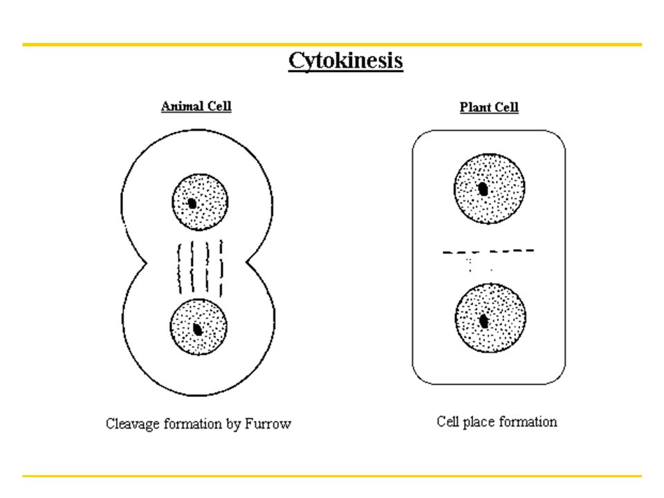
Cytokinesis Cytoplasm divides or splits In animals = cleavage furrowing or pinching off of the cytoplasm to form two daughter cells In plants = forms a cell plate between two daughter cells

Similar presentations


>")


 Relationship.>")


