Download presentation
Presentation is loading. Please wait.

1
Neoplasia

2
Fig. 2 Hanahan and Weinberg Cell 100:57 Signaling Networks in Cancer

3
Background The tumor is a common disease all over the world. In many countries especially developed countries, malignant tumor has become the first or second leading cause of death. Although many research works focused on oncology and great progress has been made in understanding tumors in the past decades, the morbidity and mortality rate of malignant tumor is increasing. The underlying causes include air pollution, pressure, excess weight, unhealthy lifestyle, ageing population and so on.

4
Cancer: General Etiology and Pathogenesis

5
How Cancer Arises Cancer cells violate the civic rules that govern normal cells by not responding to go-signals for proliferation and stop-signals for reproduction. Cancer cells descend from a common ancestral cell: clonal origin. But at some point one of the off-springs mutate that becomes worse with more mutation, and finally the accumulated mutated cells disobey all civic controls of normal cells in a tissue, becoming invasive and malignant. Since mutations occur at the gene level, that is, DNA molecules that reside in the nuclei of the cells, most human cancer can be traced there.

6
Proto-oncogenes from neighboring cells produce growth factors that encourage cell growth during cell cycle by producing growth- stimulatory signals. Mutation in these genes may cause cells divide without any signal from outside. One example is mutated ras gene. A quarter of all human tumors have mutated ras gene. Similarly myc gene family if abnormal causes leukemia, lymphomas. Receptors on the recipient cells that bind proto-oncogene growth factors may also mutate and stimulate cell growth. Thus, in breast cancer, Erb-B2 receptors behave abnormally. Tumor suppressor genes that control unrestricted growth of cells and inhibit cell growth. If they stop working, cancer cells grow wild and uninterrupted. Example, pRB, P53 (tumor-suppressor gene protein), TGF-beta (inhibits cell growth)
, TGF-beta (inhibits cell growth).")
8
Cancer results from mutations in genes that control cell division Mutations in two types of genes can cause cancer – Oncogenes – Proto-oncogenes normally promote cell division – Mutations to oncogenes enhance activity – Tumor-suppressor genes – Normally inhibit cell division – Mutations inactivate the genes and allow uncontrolled division to occur Copyright © 2009 Pearson Education, Inc.

9
Cancer results from mutations in genes that control cell division Oncogenes – Promote cancer when present in a single copy – Can be viral genes inserted into host chromosomes – Can be mutated versions of proto-oncogenes, normal genes that promote cell division and differentiation – Converting a proto-oncogene to an oncogene can occur by – Mutation causing increased protein activity – Increased number of gene copies causing more protein to be produced – Change in location putting the gene under control of new promoter for increased transcription Copyright © 2009 Pearson Education, Inc.

10
Approximately 100 oncogenes have been identified. Examples of Oncogenes The rasK oncogene is found in 25% of lung cancers, 50% of colon cancers, and 90% of pancreatic cancers. The rasN oncogene is associated with leukemias and lymphomas. These oncogenes stimulate growth even when there is no growth factor produced.

11
Cancer results from mutations in genes that control cell division ( Tumor-suppressor genes) Proteins produced by some tumor-suppressor genes stop the cell cycle when DNA becomes damaged. If the DNA cannot be repaired, these proteins cause the cell to kill itself (called apoptosis). For example, a peeling sunburn is due to apoptosis. The DNA has become too badly damaged to be repaired, so the cells kill themselves. This prevents cancer from developing from the mutations. Copyright © 2009 Pearson Education, Inc.
. For example, a peeling sunburn is due to apoptosis. The DNA has become too badly damaged to be repaired, so the cells kill themselves. This prevents cancer from developing from the mutations. Copyright © 2009 Pearson Education, Inc..")
12
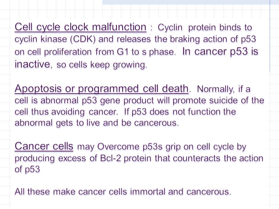
A tumor-suppressor gene called p53 stops the cell cycle when damage has occurred to DNA and it stimulates repair enzymes to repair the DNA. It produces a protein that combines with the cyclin-kinase complex and inactivates it, thus preventing the cell from dividing. If the DNA cannot be repaired, the p53 protein stimulates apoptosis. When tumor-suppressor genes don't function properly, they may not be able to prevent abnormal cell division in mutated cells and the result could be cancer. The most frequent cause of new cancer is a mutation in the p53 gene. It is involved in an estimated 60% of all cancers including cancers of the breast, lung, liver, skin, prostate, bladder, cervix, and colon. Breast cancer prognosis is associated with a tumor suppressor gene called p27.

13
BRCA1 appears to encode a tumor suppressor protein. Mutations that affect the function of this protein cause increased rates of cell division and a predisposition towards the development of malignancy. Several BRCA1 mutations, including point mutations, deletions, and insertions, have been identified that may contribute to loss of tumor suppressor function.

14
Mutation within the gene Hyperactive growth- stimulating protein in normal amount Proto-oncogene DNA Multiple copies of the gene Gene moved to new DNA locus, under new controls Oncogene New promoter Normal growth- stimulating protein in excess Normal growth- stimulating protein in excess

15
Mutated tumor-suppressor gene Tumor-suppressor gene Defective, nonfunctioning protein Normal growth- inhibiting protein Cell division under control Cell division not under control

16
Multiple genetic changes underlie the development of cancer Four or more somatic mutations are usually required to produce a cancer cell One possible scenario for colorectal cancer includes – Activation of an oncogene increases cell division – Inactivation of tumor suppressor gene causes formation of a benign tumor – Additional mutations lead to a malignant tumor Copyright © 2009 Pearson Education, Inc.

17
1 Colon wall Cellular changes: DNA changes: Oncogene activated Increased cell division Tumor-suppressor gene inactivated Growth of polyp Second tumor- suppressor gene inactivated Growth of malignant tumor (carcinoma) 2 3
 2 3")
18
Chromosomes 1 mutation Normal cell 4 mutations 3 mutations 2 mutations Malignant cell

19
Faulty proteins can interfere with normal signal transduction pathways Path producing a product that stimulates cell division Product of ras proto-oncogene relays a signal when growth hormone binds to receptor Product of ras oncogene relays the signal in the absence of hormone binding, leading to uncontrolled growth Copyright © 2009 Pearson Education, Inc.

20
Faulty proteins can interfere with normal signal transduction pathways Path producing a product that inhibits cell division – Product of p53 tumor-suppressor gene is a transcription factor – p53 transcription factor normally activates genes for factors that stop cell division – In the absence of functional p53, cell division continues because the inhibitory protein is not produced Copyright © 2009 Pearson Education, Inc.

21
Growth factor Protein that Stimulates cell division Translation Nucleus DNA Target cell Normal product of ras gene Receptor Relay proteins Transcription factor (activated) Hyperactive relay protein (product of ras oncogene) issues signals on its own Transcription
 Hyperactive relay protein (product of ras oncogene) issues signals on its own Transcription")
22
A model for p53 function

23
Growth-inhibiting factor Protein that inhibits cell division Translation Normal product of p53 gene Receptor Relay proteins Transcription factor (activated) Nonfunctional transcription factor (product of faulty p53 tumor-suppressor gene) cannot trigger transcription Transcription Protein absent (cell division not inhibited)
 Nonfunctional transcription factor (product of faulty p53 tumor-suppressor gene) cannot trigger transcription Transcription Protein absent (cell division not inhibited)")
24
How many people like to drink diet soda?

25
Background: Aspartame (L-phenylalanine N-L- ·-aspartyl-1-methyl ester) is an artificial sweetener with widespread applications. Previously published results have shown that among rats receiving aspartame a significant increase of lymphoreticular neoplasms, brain tumours and transitional cell tumours occurred. The aim of our short- term experiment was to investigate the biological effect of aspartame consumption by determining the expressions of key oncogenes and a tumour suppressor gene. Materials and Methods: After one week per os administration of various doses of aspartame to CBA/CA female mice, p53, c-myc, Ha-ras gene expression alterations were determined in individual organs. Results: The results showed an increase in gene expressions concerning all the investigated genes especially in organs with a high proliferation rate: lymphoreticular organs, bone-marrow and kidney. Conclusion: Aspartame has a biological effect even at the recommended daily maximum dose.

26
Let’s review… Definition Structure Characteristics of Tumors Nomenclature Differentiation and Anaplasia Growth, Local Invasion and Metastasis Difference Between Benign and Malignant tumors Effects of Tumors on the Hosts Precancerous Lesions, Dysplasia, and Carcinoma in situ Brief Introduction of Common Neoplasms

27
Definition of Neoplasm A neoplasm is an abnormal mass of tissue, it’s growth exceeds and is uncoordinated with that of the normal tissue and persist in the same excessive manner after cessation of the stimuli which evoke the change. (Dr. RA Willis)
.")
28
Definition of Neoplasm At the molecular level, neoplasm is disorder of growth regulatory genes ( the activation of proto- oncogenes and the inactivation of tumor suppressor genes ). It develops in a multistep fashion, such that different neoplasms, even of the same histological type, may show different genetic changes.

29
Features of Neoplasm 1.Excessive cellular proliferation; 2.Lack of responsiveness to control mechanisms; 3.Lack of dependence on the continued presence of the stimulus.

30
Structure Characteristics of Tumors The gross appearance of tumor is varied. It is usually related to histogenesis, site and biologic behavior.

31
Structure Characteristics of Tumors lipoma Hepatocellular carcinoma Color of tumor:

32
Structure Characteristics of Tumors Two basic components of all the tumors: 1.Parenchyma – the parenchyma is made up of proliferating neoplastic cells and largely determines the biologic behavior of the tumor. In addition, the classification, nomenclature and histological diagnosis are also made according to the parenchymal cells. 2.Supporting stroma – the supporting stroma is made up of connective tissue, blood vessels, and possibly lymphatics.

33
parenchyma supporting stroma

34
Nomenclature Basic principle: Neoplasms are named according to binomial system denoting their histogenic origin of the parenchymal component and the biologic behavior.

35
Nomenclature Benign tumors: “ ~ oma”, e.g., fibroma; lipoma Malignant tumors: “ ~ carcinoma, ~ sarcoma”

36
thyroid adenomacolonic adenoma leiomyoma of uterus fibroadenoma of breast

37
heptocellular carcinoma Squamous cell carcinomaadenocarcinoma of colon osteosarcoma of bone

38
Tumor Cell Characteristics

39
Differentiation and Anaplasia Neoplasm differentiation denotes the degree to which a neoplasm cell resembles the normal mature cells of the tissue both morphologically and functionally. What is neoplasm differentiation?

40
Differentiation and Anaplasia Benign tumors are usually well differentiated. They resemble closely their normal counterpart. Malignant tumors, on the other hand, show variable degree of differentiation. Malignant tumors that are composed of undifferentiated cells are said to be “anaplastic”,that means no morphological resemblance to normal tissue.

41
Differentiation and Anaplasia lack of differentiation; literally means ‘to form backward,’ implying a ‘reverse differentiation’ of mature normal cells. For cancers, it does not represent reverse differentiation. It means lack of differentiation. What is anaplasia?

42
Genetic Instability Malignant tumors will have an abnormal number of chromosomes

43
Growth, Local Invasion and Metastasis The growth rate of neoplastic cells varies greatly and is one of its chief factors that serves to distinguish benign from malignant. In a general rule, the degree of malignancy of a neoplasm is correlated with its growth rate: the more rapid the growth, the more malignant the neoplasm. Rate of growth and malignancy:

44
Growth, Local Invasion and Metastasis Nearly all benign tumors grow as cohesive expansile masses that remain localized to their site of origin. a.Benign tumors grow slowly and usually develop a fibrous capsule keeping the tumor as a discrete, readily palpable and easily movable mass that can be excised. b.When a benign tumor arises in a epithelial or mucosal surface, the tumor grow away from the surface, often forming a polypoid. Invasion ( Infiltration)
.")
45
Lipoma Here is a benign lipoma on the serosal [si'rəusəl] surface of the small intestine. It has the characteristics of a benign neoplasm: it is well circumscribed, slow growing, and resembles the tissue of origin (fat).
![Lipoma Here is a benign lipoma on the serosal [si rəusəl] surface of the small intestine.](http://images.slideplayer.com/35/10331462/slides/slide_45.jpg "It has the characteristics of a benign neoplasm: it is well circumscribed, slow growing, and resembles the tissue of origin (fat)..")
46
Growth, Local Invasion and Metastasis The growth of cancers, in contrast, is accompanied by infiltration, invasion, and destruction of the surrounding tissue. a.In general, malignant tumors are lack of a well defined cleavage plane and usually exhibit local invasiveness or infiltration that make it difficult to be excised. b.Malignant tumors on epithelial or mucosa surface may form a protrusion in the early stages, but eventually invade the underlying normal tissue. Invasion ( Infiltration)
.")
47
Squamous cell carcinoma of lung Malignant neoplasms are also characterized by the tendency to invade surrounding tissues. Here, a lung cancer is seen to be spreading along the bronchi into the surrounding lung.

48
hepatic adenoma hepatocellular carcinoma Here is a small hepatic adenoma that shows how well- demarcated an benign neoplasm is. In contrast, this hepatocellular carcinoma is not as well circumscribed (note the infiltration of tumor off to the lower right) nor as uniform in consistency. It is also arising in a cirrhotic (nodular) liver.
 nor as uniform in consistency. It is also arising in a cirrhotic (nodular) liver..")
49
Metastasis Metastasis is to form a second neoplastic mass through transfer of the neoplastic cells from the first neoplasm to a distant site on separate from the original tumor. What is metastasis of neoplasms?

50
Metastasis 1.Lymphatogenous metastasis - The most common pathway for initial dissemination of carcinomas, but sarcomas may also use this route. 2.Hematogenous metastasis - This route is typical of sarcomas but is also seen with carcinoma.. 3.Metastasis in body cavities ( seeding ) - Direct seeding of body cavities or surface (exfoliation and implantation on peritoneum, pleura, subarachnoid) Routes of metastasis
 - Direct seeding of body cavities or surface (exfoliation and implantation on peritoneum, pleura, subarachnoid) Routes of metastasis.")
51
Sentinal Node Initial node to which the primary tumor drains

52
characteristics BenignMalignant DifferentiationWell differentiatedRange from well differentiate to undifferentiated Rate of growthSlow growth over a period of years Rapid growth, sometimes erratic Type of growthExpansileProgressive infiltration, invasion, and destruction of surrounding tissue Separated fromYes, has fibrous capsule composed of stroma of native tissue Poorly separated MetastasisNoYes Effect on hostOften insignificantSignificant, fever, anemia, infections, etc. RecurrenceRareOften Cell shapeMonomorphicPleomorphic Tumor giant cells Nuclear chromatin NormalInreased, hyperchromatic; Peripheral clumping NucleoliNot prominentProminent, irregular shape

53
Precancerous Lesions A premalignant or precancerous lesion is an abnormality in a tissue area which is a just a step away from cancer. a.Not all precancerous lesions change to cancer, but most have potential to become malignant. b.It is important to recognize precancerous lesions because surgical excision is curative. What is precancerous lesions?

54
Dysplasia Dysplasia is an abnormality of both differentiation and maturation. This term should be restricted to abnormalities of cell growth with the characteristics as following: a.Increased size of the nucleus, (absolute and relative to the amount of cytoplasm) b.Hyperchromatism c.Abnormal chromatin distribution (coarse clumping) d.Nuclear membrane is thickneng and wrinkling. e.In squamous epithelium, mitotic figures appear in many layers. What is dysplasia?
 b.Hyperchromatism c.Abnormal chromatin distribution (coarse clumping) d.Nuclear membrane is thickneng and wrinkling. e.In squamous epithelium, mitotic figures appear in many layers. What is dysplasia .")
55
Carcinoma in situ The term carcinoma in situ refers to an epithelial neoplasm exhibiting all the malignant cellular features. But it has not yet invaded with through the epithelial basement membranes separating it from potential route of metastasis. It is only at this very early stage the excision of the tumor will guarantee a cure. So detection of carcinoma in situ is very important. In clinical practice, detection of carcinoma at the in situ stage, or detection of precancerous lesions is the aim of population screening programs for cervical, breast and some other carcinoma. Through these popular screening, many lives have been saved. What is carcinoma in situ?

56
Etiology of Cancer

57
Cancer Associated Genes

58
Oncogenes

59
Tumor Suppressor Genes

60
Tumor Cell Transformaton

61
Other factors… Heredity Hormones Radiation Obesity Immunologic Carcinogens Viral Agents

Similar presentations


>")


 - gene location. Cystic Fibrosis (CF): Molecular defect.>")
