Download presentation
Presentation is loading. Please wait.

1
BIOL 2261 CHARACTERISTICS FEATURS OF BACTERIAL CELL CHAPTER 9 © Thomas Deerinck, NCMIR / Science Photo Library

2
Types of Cells Two major classes: eukaryotes & prokaryotes. Differences: the materials making up the nucleus of eukaryotic cells are separated from the rest of the cell by the nuclear membrane, whereas in prokaryotic cells these materials are not separated. All animals and plant cells are eukaryotic including fungi. Bacteria, cyanobacteria and the mycoplasmas are prokaryotic.

3
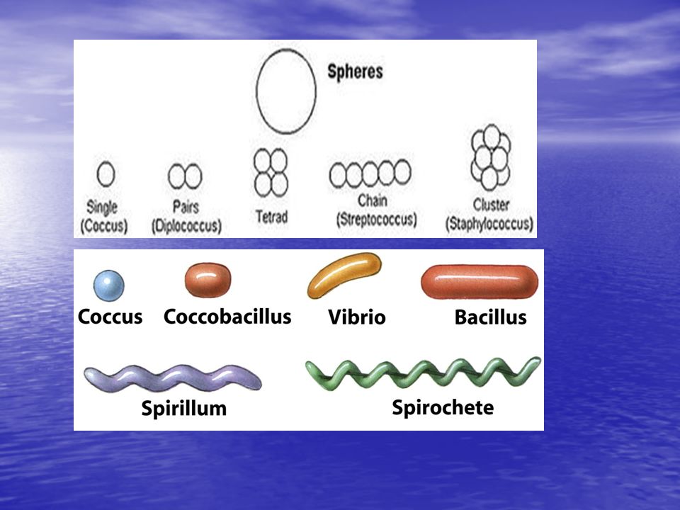
Size, Shape, and Arrangement of Bacterial cells Cocci (s., coccus) – spheres –diplococci (s., diplococcus) – pairs –streptococci – chains –staphylococci – grape-like clusters –tetrads – 4 cocci in a square –sarcinae – cubic configuration of 8 cocci
 – spheres –diplococci (s., diplococcus) – pairs –streptococci – chains –staphylococci – grape-like clusters –tetrads – 4 cocci in a square –sarcinae – cubic configuration of 8 cocci")
4
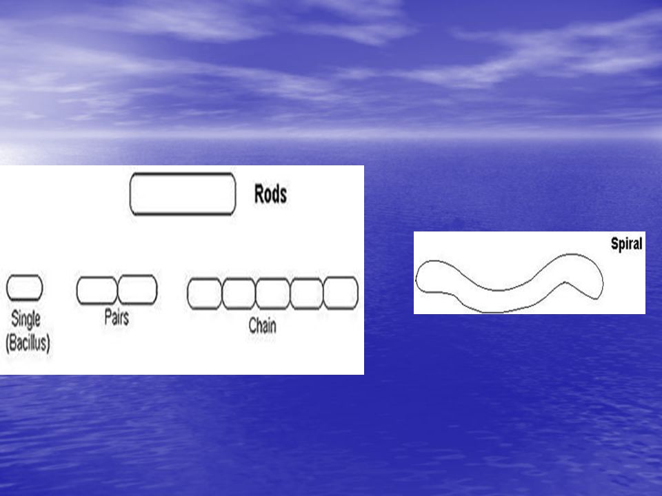
……Size, Shape, and Arrangement Bacilli (s., bacillus): – rods –coccobacilli – very short rods –vibrios – resemble rods, comma shaped spirilla (s., spirillum) – rigid helices spirilla (s., spirillum) – rigid helices spirochetes – flexible helices spirochetes – flexible helices mycelium – network of long, multinucleate filaments Check on line lab Manual for Bacterial shapes) mycelium – network of long, multinucleate filaments Check on line lab Manual for Bacterial shapes)
: – rods –coccobacilli – very short rods –vibrios – resemble rods, comma shaped spirilla (s., spirillum) – rigid helices spirilla (s., spirillum) – rigid helices spirochetes – flexible helices spirochetes – flexible helices mycelium – network of long, multinucleate filaments Check on line lab Manual for Bacterial shapes) mycelium – network of long, multinucleate filaments Check on line lab Manual for Bacterial shapes)")
7
……Size, Shape, and Arrangement –Sizes: Typically ~ 0.1 - 20 m (with some exceptions) Typically ~ 0.1 - 20 m (with some exceptions) Typical coccus: ~ 1 m (e.g. Staphylococcus) Typical coccus: ~ 1 m (e.g. Staphylococcus) Staphylococcus Typical short rod: ~ 1 x 5 m (e.g. E. coli) Typical short rod: ~ 1 x 5 m (e.g. E. coli)E. coliE. coli Barely within the best resolution of a good compound light microscope Barely within the best resolution of a good compound light microscopecompound light microscopecompound light microscope
 Typical coccus: ~ 1 m (e.g. Staphylococcus) Staphylococcus Typical short rod: ~ 1 x 5 m (e.g. E. coli) Typical short rod: ~ 1 x 5 m (e.g. E. coli)E. coliE. coli Barely within the best resolution of a good compound light microscope Barely within the best resolution of a good compound light microscopecompound light microscopecompound light microscope.")
8
Bacterial Shapes

9
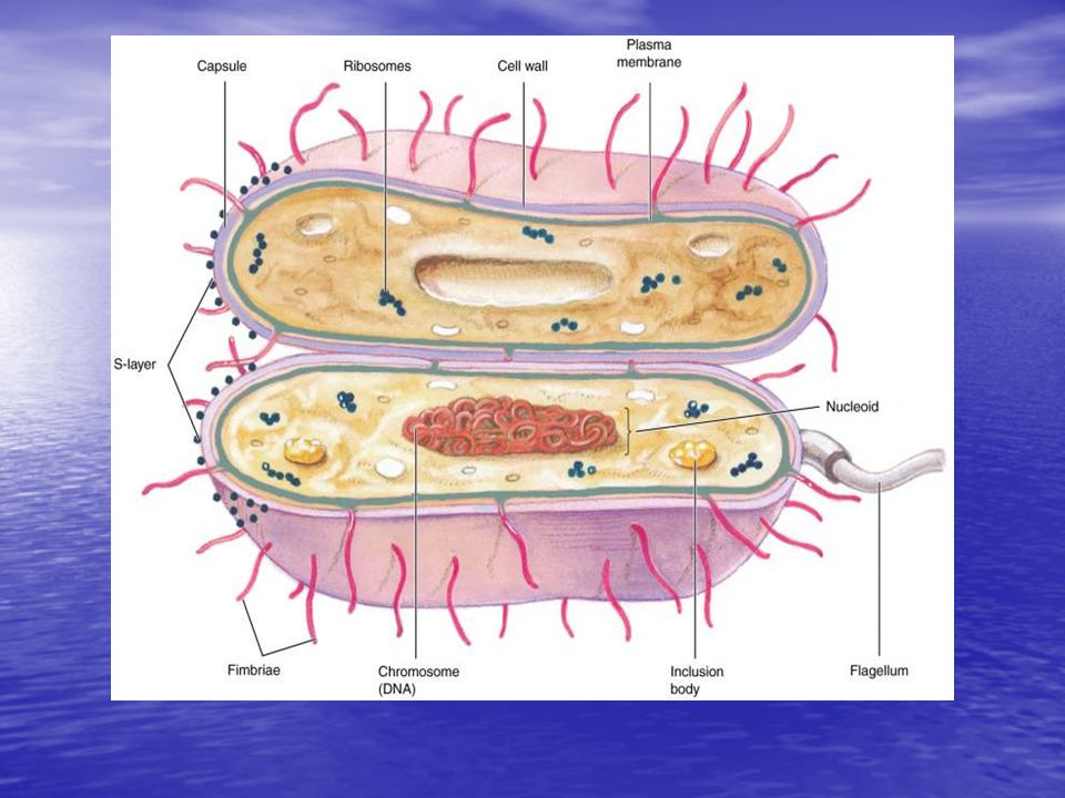
Cell Structure of Procaryotes Prokaryotic cells The constituents of a typical bacterium are as follows: Bacterial Cell Wall and Capsule – bacteria are surrounded by a cell wall, which not only contains polysaccharide but also contains protein and lipid. Bacterial Cell Wall and Capsule – bacteria are surrounded by a cell wall, which not only contains polysaccharide but also contains protein and lipid. In some bacteria, the cell wall is surrounded by the capsule. In some bacteria, the cell wall is surrounded by the capsule. The cell wall and capsule provide shape and form to the bacterium and also acts as a physical barrier between the bacterium and its environment. The cell wall and capsule provide shape and form to the bacterium and also acts as a physical barrier between the bacterium and its environment. Nucleoids – in bacteria the nuclear material is concentrated in a region called the nucleoid within the cytoplasm. Nucleoids – in bacteria the nuclear material is concentrated in a region called the nucleoid within the cytoplasm.

10
Typical Prokaryotic Cell

11
A typical Bacterial Cell

13
….Cell Structure There is no membrane-bound nucleus in prokaryotes. There is no membrane-bound nucleus in prokaryotes. Instead the DNA is located within a specialized region of the cytoplasm of the cell called the nucleoid region. Instead the DNA is located within a specialized region of the cytoplasm of the cell called the nucleoid region. There is no nuclear membrane surrounding the nucleoid. There is no nuclear membrane surrounding the nucleoid. Bacterial flagella – many bacteria possess one or more flagella for locomotion. Bacterial flagella – many bacteria possess one or more flagella for locomotion.

14
Bacterial Cell Walls Bacterial Cell Wall: rigid structure that lies just outside the plasma membrane (detail to continue) rigid structure that lies just outside the plasma membrane (detail to continue) The cell wall is a complex meshwork structure composed of several parts. The cell wall is a complex meshwork structure composed of several parts.

15
…..Bacterial Cell Walls The primary structure of the cell wall is peptidoglycan which is composed of repeating sugar molecules. The primary structure of the cell wall is peptidoglycan which is composed of repeating sugar molecules. –N-acetyl glucosamine (NAG) –N-acetyl muramic acid (NAM) The meshwork is held together with small peptide chains (Fig 9.3). The meshwork is held together with small peptide chains (Fig 9.3).
 –N-acetyl muramic acid (NAM) The meshwork is held together with small peptide chains (Fig 9.3). The meshwork is held together with small peptide chains (Fig 9.3)..")
16
Functions of Cell Wall provides characteristic shape to cell provides characteristic shape to cell The bacterial cell wall is a protective barrier against: The bacterial cell wall is a protective barrier against: –Osmotic pressure changes –Other environmental stresses may also contribute to pathogenicity may also contribute to pathogenicity very few procaryotes lack cell walls very few procaryotes lack cell walls The cell wall is different in Gram- positive and Gram-negative bacteria. The cell wall is different in Gram- positive and Gram-negative bacteria.

17
Gram stain-Cell Walls of Bacteria Gram Staining developed by Gram in 1884: bacteria are divided into two major groups based on the response to gram- stain procedure bacteria are divided into two major groups based on the response to gram- stain procedure –gram-positive bacteria stain purple (positive-purple-pp) –gram-negative bacteria stain pink staining reaction due to cell wall structure staining reaction due to cell wall structure
 –gram-negative bacteria stain pink staining reaction due to cell wall structure staining reaction due to cell wall structure")
19
Gram-Positive Cell Walls Component of Cell Wall Gram positive bacteria composed primarily of peptidoglycan/murein Gram positive bacteria composed primarily of peptidoglycan/murein Peptidoglycan are polymers which contains amino acids: N-acetylglucosamine and N- acetylmuramic acid and several different amino acids Peptidoglycan are polymers which contains amino acids: N-acetylglucosamine and N- acetylmuramic acid and several different amino acids Walls contain teichoic acid ( polymers of glycerol or ribitol joined by phosphate groups) Walls contain teichoic acid ( polymers of glycerol or ribitol joined by phosphate groups)
 Walls contain teichoic acid ( polymers of glycerol or ribitol joined by phosphate groups)")
20
Gram-Positive Cell Walls Teichoic Acid Teichoic Acid – There are two forms Wall teichoic acids – go part way through the wall. Wall teichoic acids – go part way through the wall. Lipoteichoic acids – go completely through the wall and link to the plasma membrane. Lipoteichoic acids – go completely through the wall and link to the plasma membrane. – Both forms protrude above the wall, which gives the bacterial cell a negative charge.

21
Gram Positive and Gram negative

22
..Gram-Positive Cell Walls M Protein M Protein –This is a virulence factor. –It protrudes from the cell wall. –It is required for infection. –It is highly susceptible to mutations

23
..Gram-Positive Cell Walls Mycolic Acid Mycolic Acid –This is found in the Mycobacterium species. –It consists of a waxy lipid incorporated into the cell wall. –It makes cells extremely resistant to environmental stress. –It acts as a barrier against antibiotics and host defenses.

24
..Gram-Positive Cell Walls The periplasmic space lies between plasma membrane and cell wall and is smaller than that of gram-negative bacteria The periplasmic space lies between plasma membrane and cell wall and is smaller than that of gram-negative bacteria as such periplasm has relatively few proteins as such periplasm has relatively few proteins enzymes secreted by gram-positive bacteria are called exoenzymes enzymes secreted by gram-positive bacteria are called exoenzymes

25
Gram Positive Cell Wall Gram Positive Cell Wall

26
Gram-Negative Cell Walls consist of a thin layer of peptidoglycan surrounded by an outer membrane consist of a thin layer of peptidoglycan surrounded by an outer membrane outer membrane composed of lipids, lipoproteins, and lipopolysaccharide (LPS) outer membrane composed of lipids, lipoproteins, and lipopolysaccharide (LPS) Lipoprotein molecules fasten the outer membrane to the peptidoglycan layer. Lipoprotein molecules fasten the outer membrane to the peptidoglycan layer. no teichoic acids no teichoic acids

27
….Gram-Negative Cell Walls The outer membrane of Gram-negative bacteria has a unique outer layer. The outer membrane of Gram-negative bacteria has a unique outer layer. –It is composed of lipopolysaccharides instead of the standard phospholipid molecules. It serves as a major barrier to the outside world for the Gram-negative cell. It serves as a major barrier to the outside world for the Gram-negative cell.

28
…..Gram Negative more complex than gram-positive walls more complex than gram-positive walls periplasmic space differs from that in gram-positive cells (occupies larger space) periplasmic space differs from that in gram-positive cells (occupies larger space) –may constitute 20-40% of cell volume –many enzymes present in periplasm
 periplasmic space differs from that in gram-positive cells (occupies larger space) –may constitute 20-40% of cell volume –many enzymes present in periplasm")
29
Gram Negative Cell Wall Gram Negative Cell Wall

30
Differences between a gram positive and gram negative cell wall.

31
Acid-fast Bacteria Acid-fast Bacteria Acid-fast Cell Walls Acid-fast Cell Walls –Many genera in the “High GC gram- positive” bacterial group contain mycolic acids, embedded in the peptidoglycan. mycolic acidsmycolic acids –Mycolic acids are a class of waxy, extremely hydrophobic lipids. –Certain genera contain very large amounts of this lipid, and are difficult to gram stain. –These genera may be identified by the “acid-fast” staining technique. “acid-fast” staining technique “acid-fast” staining technique –Includes Mycobacterium. Mycobacterium

32
Wall deficient Organisms Mycoplasmas Bacteria that are naturally have no cell walls Bacteria Includes Mycoplasma and Ureaplasma ArchaeaArchaea : Archaea Have archaea cell walls with no peptidoglycan archaea cell walls archaea cell walls

33
Cell Structures outside the Bacterial cell wall— External There are five structures that can be found outside the cell wall. No bacterium has all five of these structures

34
Capsules, Slime Layers, and S-Layers Layers of material lying outside the cell wall –capsules usually composed of polysaccharides usually composed of polysaccharides well organized and not easily removed from cell well organized and not easily removed from cell –slime layers similar to capsules except diffuse, unorganized and easily removed. similar to capsules except diffuse, unorganized and easily removed. a capsule or slime layer composed of polysaccharides can also be referred to as a glycocalyx a capsule or slime layer composed of polysaccharides can also be referred to as a glycocalyx

35
…Glycocalyx-Slime layer & Capsule

36
….Glycocalyx The glycocalyx is a sticky substance composed of polypeptides, polysaccharides or both. The glycocalyx is a sticky substance composed of polypeptides, polysaccharides or both. It is produced in the cytoplasm and secreted to the outer part of the cell wall. It is produced in the cytoplasm and secreted to the outer part of the cell wall. It provides a protective element against environmental stress. It provides a protective element against environmental stress. It can be used for nutrition. It can be used for nutrition.

37
S-layers: S-layers: If glycocalyx is loosely attached to the wall, it is called a slime layer. If glycocalyx is loosely attached to the wall, it is called a slime layer. If adhered tightly to the wall, it is called a capsule. If adhered tightly to the wall, it is called a capsule. –regularly structured layers of protein or glycoprotein. Clinical Significance of Glycocalyx: Adherence is a major part of infection (staying in), especially in respiratory and urinary tract infections. The glycocalyx is a primary factor of adherence. The glycocalyx is a primary factor of adherence.
, especially in respiratory and urinary tract infections. The glycocalyx is a primary factor of adherence. The glycocalyx is a primary factor of adherence..")
38
Functions of Capsules, Slime Layers, and S-layers protection from host defenses (e.g., phagocytosis) protection from host defenses (e.g., phagocytosis) protection from harsh environmental conditions (e.g., desiccation), chemicals or osmotic stress protection from harsh environmental conditions (e.g., desiccation), chemicals or osmotic stress attachment to surfaces attachment to surfaces facilitate motility facilitate motility –nutrient Storage
 protection from host defenses (e.g., phagocytosis) protection from harsh environmental conditions (e.g., desiccation), chemicals or osmotic stress protection from harsh environmental conditions (e.g., desiccation), chemicals or osmotic stress attachment to surfaces attachment to surfaces facilitate motility facilitate motility –nutrient Storage")
39
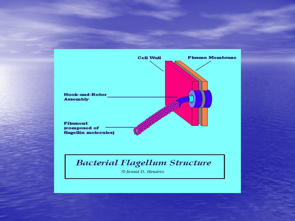
Flagella Flagella In bacteria the flagellum is a hair-like projection used for movement by rotating around its axis. Flagella are only used for movement. In bacteria the flagellum is a hair-like projection used for movement by rotating around its axis. Flagella are only used for movement. Sex pilli are projections that transfer DNA material from one bacterium to another's plasmid. Sex pilli are projections that transfer DNA material from one bacterium to another's plasmid. Pilli helps bacteria to fasten onto surfaces such as host membranes. Pilli helps bacteria to fasten onto surfaces such as host membranes.

40
…FLAGELLA CONFIGURATIONS Different species have different flagella arrangements ( types of flagella Ref pg 93).flagella arrangements Monotrichous – one flagellum located at the end of the cell Amphitrichous – two flagella, one at each end of the cell Lophotrichous – two or more flagella located at the same end of the cell Peritrichous – flagella surround the entire cell.
.flagella arrangements Monotrichous – one flagellum located at the end of the cell Amphitrichous – two flagella, one at each end of the cell Lophotrichous – two or more flagella located at the same end of the cell Peritrichous – flagella surround the entire cell.")
41
….Function & Structure of Flagella Function: –Mobility –Almost all motile bacteria are motile by means of flagella bacteria –Motile vs. non motile bacteria. Structure: A flagella consists of the following three parts: A flagella consists of the following three parts: –Filament –Hook –Basal body –Filament composed of the protein flagellin –Hook & Rotor Assembly & Permits rotational "spinning" movement

43
Chemotaxis movement towards a chemical attractant/light or away from a chemical repellent/light movement towards a chemical attractant/light or away from a chemical repellent/light concentrations of chemical attractants and chemical repellents detected by chemoreceptors on surfaces of cells concentrations of chemical attractants and chemical repellents detected by chemoreceptors on surfaces of cells

44
Pili and Fimbriae Fimbriae (sing., fimbria) –short, thin, hairlike, proteinaceous appendages up to 1,000/cell –mediate attachment to surfaces sex pili/Conjugating pilli (s., pilus): –similar to fimbriae except longer, thicker, and less numerous (1-10/cell) –required for mating
 –short, thin, hairlike, proteinaceous appendages up to 1,000/cell –mediate attachment to surfaces sex pili/Conjugating pilli (s., pilus): –similar to fimbriae except longer, thicker, and less numerous (1-10/cell) –required for mating")
45
Microbiology: A Clinical Approach © Garland Science FIMBRIAE AND PILI –Axial filaments Panel a: © Thomas Deerinck, NCMIR / Science Photo Library; Panel b: © Dennis Kunkel

46
Axial Filaments — Attachment

47
Homework Homework 1.List the clinical significance of Bacterial cell wall ( positive and negative) ( Pg 160)? 2. List the differences between a gram positive and gram negative cell wall.

48
STRUCTURES INSIDE THE BACTERIAL CELL WALL There are six major structures found inside the bacterial cell well: Plasma membrane Nuclear region Plasmids Ribosomes Inclusion bodies Endospores

49
Procaryotic Cell Membranes Cell Membranes: membranes are an absolute requirement for all living organisms. membranes are an absolute requirement for all living organisms. plasma membrane encompasses the cytoplasm plasma membrane encompasses the cytoplasm some procaryotes also have internal membrane systems some procaryotes also have internal membrane systems

50
Functions of the Plasma Membrane separation of cell from its environment separation of cell from its environment selectively permeable barrier selectively permeable barrier –some molecules are allowed to pass into or out of the cell –transport systems aid in movement of molecules detection of and response to chemicals in surroundings with the aid of special receptor molecules in the membrane detection of and response to chemicals in surroundings with the aid of special receptor molecules in the membrane

51
Cell Structures — Membrane

52
Fluid Mosaic Model of Membrane Structure

53
…..Plasma membrane

54
…PLASMA MEMBRANE

55
…Phospholipid layer polar ends polar ends –interact with water –hydrophillic nonpolar ends nonpolar ends –insoluble in water –hydrophobic

56
Phospholipid layer Phospholipid layer

57
Membrane Proteins Peripheral proteins: –loosely associated with the membrane and easily removed Integral proteins –embedded within the membrane and not easily removed ATP production occurs at the plasma membrane. ATP production occurs at the plasma membrane. The proteins associated with electron transport are located in the plasma membrane The proteins associated with electron transport are located in the plasma membrane

58
TYPES OF MEMBRANE TRANSPORT The plasma membrane regulates what enters the cell cytoplasm and what does not. There are three types of membrane transport: Osmosis- water chases the concentration of solutes (higher to lower concentration) Passive transport: 2 types: -Simple diffusion- does not require ATP (higher to lower concentration) - Facilitated diffusion- does not require ATP but uses carrier proteins called permease proteins. Active transport-Active transport requires ATP. ( Against a concentration gradient)
 Passive transport: 2 types: -Simple diffusion- does not require ATP (higher to lower concentration) - Facilitated diffusion- does not require ATP but uses carrier proteins called permease proteins. Active transport-Active transport requires ATP. ( Against a concentration gradient).")
59
Internal Structures Cytoplamic Matrix: Cytoplasm contains the nucleoid, ribosomes and inclusion bodies Cytoplasm contains the nucleoid, ribosomes and inclusion bodies lacks organelles bound by unit membranes lacks organelles bound by unit membranes composed largely of water composed largely of water is a major part of the protoplasm (the plasma membrane and everything within) is a major part of the protoplasm (the plasma membrane and everything within)
 is a major part of the protoplasm (the plasma membrane and everything within)")
60
..Cytoplasmic Matrix –Viscous aqueous suspension of proteins, nucleic acid, dissolved organic compounds, mineral salts –Network of protein fibers similar to the eukaryotic cytoskeleton.

61
Nuclear Region/The Nucleoid Nucleoid: irregularly shaped region irregularly shaped region location of chromosome location of chromosome –usually 1/cell not membrane-bound not membrane-bound

62
The Procaryotic Chromosome The Chromosomes: usually a closed circular, double- stranded DNA molecule usually a closed circular, double- stranded DNA molecule looped and coiled extensively looped and coiled extensively

63
PLASMIDS Plasmids: usually small, closed circular DNA molecules usually small, closed circular DNA molecules exist and replicate independently of chromosome exist and replicate independently of chromosome have relatively few genes present have relatively few genes present

64
…..PLASMIDS Plasmids are extra-chromosomal pieces of DNA that are separate from the main DNA structure. Some bacteria can carry more than one plasmid. Plasmids often carry genes for toxins and resistance to antibiotics. Plasmids can be transferred from one cell to another through pili during conjugation.

65
Ribosomes Ribosomes: Ribosomes: Ribosomes are nonmembrane-enclosed organelles involved in protein synthesis Ribosomes are nonmembrane-enclosed organelles involved in protein synthesis complex structures consisting of protein and RNA (ribonucleic acid) complex structures consisting of protein and RNA (ribonucleic acid) sites of protein synthesis ( translation) sites of protein synthesis ( translation) smaller than eucaryotic ribosomes smaller than eucaryotic ribosomes –procaryotic ribosomes 70S –eucaryotic ribosomes 80S Clinical Significance – The inhibition of protein synthesis is a lethal event so ribosomes are a major target for antibiotics.
 complex structures consisting of protein and RNA (ribonucleic acid) sites of protein synthesis ( translation) sites of protein synthesis ( translation) smaller than eucaryotic ribosomes smaller than eucaryotic ribosomes –procaryotic ribosomes 70S –eucaryotic ribosomes 80S Clinical Significance – The inhibition of protein synthesis is a lethal event so ribosomes are a major target for antibiotics.")
66
Cytoplasmic Inclusion Bodies: Inclusion bodies are membrane-enclosed organelles used to store important materials Granules of organic or inorganic material that are stockpiled by the cell for future use (lipids,glycogen,protein, fat globules) collectively called granules or others called vesicles. Granules of organic or inorganic material that are stockpiled by the cell for future use (lipids,glycogen,protein, fat globules) collectively called granules or others called vesicles. Granules contain glucose polymer for energy, polyphospahte granules called volutin, or iron containing vesicles- magnetosomes
 collectively called granules or others called vesicles. Granules contain glucose polymer for energy, polyphospahte granules called volutin, or iron containing vesicles- magnetosomes.")
67
Bacterial Endospores Bacterial Spores Endospores are formed through the process of sporulation. Endospores are formed through the process of sporulation. They form when a bacterium is exposed to great environmental stress. They form when a bacterium is exposed to great environmental stress. They are formed by some bacteria as dormant structures. They are formed by some bacteria as dormant structures. resistant to numerous environmental conditions e.g heat, radiation,chemicals, nutrient depletion, resistant to numerous environmental conditions e.g heat, radiation,chemicals, nutrient depletion, desiccation, and waste buildup. desiccation, and waste buildup. –Bacterial spores are NOT a reproductive structure, like plant or fungal spores. –Produced by very few genera of bacteria bacteria –Major examples Bacillus & Clostridium BacillusClostridiumBacillusClostridium

68
…endospores Sporogenesis

69
..Spore Germination Activation: –prepares spores for germination –often results from treatments like heating Germination: –spore swelling –rupture or absorption of spore coat –loss of resistance to heat and other stresses –increased metabolic activity Outgrowth: –emergence of vegetative cell

70
Spore Germination

71
Bibliography Microbiology, A clinical Approach -Danielle Moszyk-Strelkauskas-Garland Science 2010. Microbiology, A clinical Approach -Danielle Moszyk-Strelkauskas-Garland Science 2010. http://en.wikipedia.org/wiki/Scientific_method http://en.wikipedia.org/wiki/Scientific_method http://en.wikipedia.org/wiki/Scientific_method http://www.bio.mtu.edu/campbell/prokaryo.htmhttp:// molecular- biology.suite101.com/article.cfm/cell_structure http://www.bio.mtu.edu/campbell/prokaryo.htmhttp:// molecular- biology.suite101.com/article.cfm/cell_structure http://www.bio.mtu.edu/campbell/prokaryo.htmhttp:// molecular- biology.suite101.com/article.cfm/cell_structure http://www.bio.mtu.edu/campbell/prokaryo.htmhttp:// molecular- biology.suite101.com/article.cfm/cell_structure http://water.me.vccs.edu/courses/ENV108/lesson5_2.ht m http://water.me.vccs.edu/courses/ENV108/lesson5_2.ht m http://water.me.vccs.edu/courses/ENV108/lesson5_2.ht m http://water.me.vccs.edu/courses/ENV108/lesson5_2.ht m

Similar presentations


 Flagella 1) Functions in movement of the cell 2) 3 components.>")


 membrane.>")