Download presentation
Presentation is loading. Please wait.

1
Lower Limbs Lu Xiaoli Regional Anatomy & Operative Surgery
China Medical University

2
Gluteal Thigh Knee Leg Ankle Foot

3
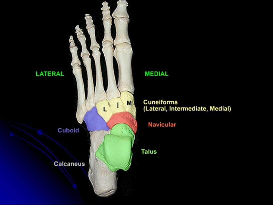
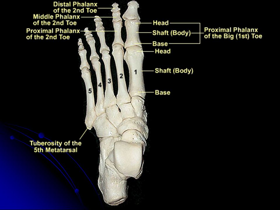
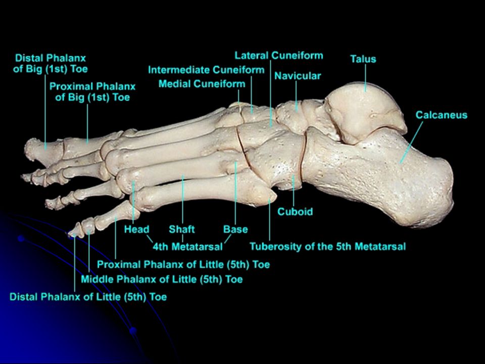
32 Bones Hip Bone (1) Femur (1) Patella (1) Tibia (1) Fibula (1)
Tarsals (8) Metatarsals (5) Proximal Phalanges (5) Intermediate Phalanges (5) Distal Phalanges (4)
 Metatarsals (5) Proximal Phalanges (5) Intermediate Phalanges (5) Distal Phalanges (4)")
4
Hip

9
Femoral Bone

13
Patella

14
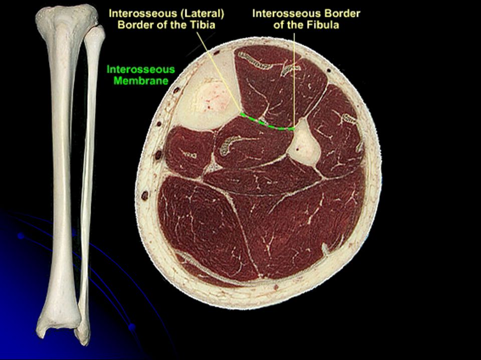
Tibia & Fibula

17
Proximal End of Left Tibia

24
Gluteal Region

25
Iliac Crest Gluteal Crease Gluteal Cleft

26
Hip Joint Superficial Layers Deep Layers Skin Superficial Fascia
Deep Fascia Muscles Suprapiriform & Infrapiriform Foramen Lesser Sciatic Foramen Hip Joint

27
Superficial Fascia the middle part is thinnest
Contains Superficial blood vessels, lymphatic vessels and cutaneous nerves

28
Bedsore (Decubitus Ulcer)
A pressure-induced ulceration of the skin occurring in persons confined to bed for long periods of time. Also called decubitus ulcer, pressure sore. Bedsore (Decubitus Ulcer)
")
29
Arteries Superficial branches of 4th lumbar artery
Inferior gluteal artery Lateral sacral artery Superior gluteal artery

30
Cutaneous Nerves branches of the subcostal nerve (T12)
dorsal rami of lumbar nerves dorsal rami of sacral nerves inferior cluneal nerves posterior femoral cutaneous nerve

31
Deep Fascia Attach to iliac crest superiorly Invest gluteus maximus
Attach to Sacral bone and coccyx medially Continuous with fascia lata laterally

32
Muscles Superficial Gluteus Maximus Tensor Fasciae Latae
Glluteus Medius Piriformis Superior Gemellus Obturator Internus Inferior Gemellus Quadratus Femoris Middle Deep Gluteus Minimus Obturator Externus

33
Gluteus Maxinus Tensor Fasciae Latae

34
Glluteus Medius Piriformis Superior Gemellus Obturator Internus Inferior Gemellus Quadratus Femoris

35
Gluteus Minimus Obturator Externus

36
Suprapiriform Foramen
greater sciatic notch Piriformis Lateral to medial: Superior gluteal n.– a.– v.

37
Infrapiriform Foramen
Piriformis Ischial spine & sacrospinous ligament Lateral to medial: sciatic n.– posterior femoral cutaneous n. – inferior gluteal n. a. & v.– internal pudendal a. & v. – pudendal n.

38
Sciatic n. Tibial n. Common Fibular n.

39
66.3% 27.3% Tibial n. Common Fibular n. 6.4%

41
Piriformis Syndrome Pain radiates down the back of the leg

42
Iliac crest Ischial tuberosity

43
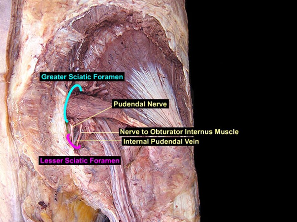
Lesser Sciatic Foramen
Boundaries front: tuberosity of ischium above: spine of ischium & sacrospinous lig. behind: sacrotuberous lig. Contents tendon of Obturator internus internal pudendal a. internal pudendal v. pudendal n.

47
Hip Anterior Posterior
it does not reach down as far as the trochanteric crest. Posterior

48
It is based on this capsular attachment that fractures of the neck of femur are classified as:
INTRACAPSULAR: within the capsular attachment. EXTRACAPSULAR: outside the attachment of the capsule Mixed

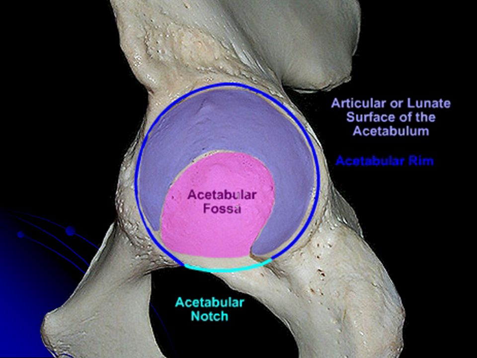
49
Intracapsular Lig. of head of femur Transverse acetabular lig.

50
Extracapsular (Anterior)
Iliofemoral lig. Pubofemoral lig.

51
Extracapsular (posterior)
Ischiofemoral lig. Zona orbicularis

52
Blood Supply Superior gluteal a. Inferior gluteal a.
Lateral circumflex femoral a. Medial Circumflex femoral a. Branches of Obturator a. 1st perforating branches of deep femoral a. Femoral nutrient a.

53
Superior gluteal a. Inferior gluteal a. 1st perforating branches of deep femoral a.

54
Superior retinacular a.
Anterior retinacular a. Inferior retinacular a. Medial Circumflex femoral a. Lateral circumflex femoral a.

55
Branches of Obturator a.

56
Femoral nutrient a.

57
If the head of the femur is dislocated postero-medially, compression of which nerve is likely to result? Femoral Lumbosacral trunk Obturator Sciatic Superior gluteal

58
What muscle passes through the lesser sciatic foramen?
Gluteus minimus Obturator internus Piriformis Quadratus femoris Superior gemellus

59
In order to avoid injury to the sciatic nerve, intramuscular injections should be given in which quadrant of the buttock? upper medial upper lateral lower medial lower lateral middle

60
Of the branches of the internal iliac artery, the one exiting from the greater sciatic foramen superior to the piriformis muscle is the: Iliolumbar artery Internal pudendal artery Lateral sacral artery Superior gluteal artery

61
Thigh

62
Anterior compartment Posterior compartment

64
Superficial iliac circumflex a.
Superficial epigastric a. External pudendal a. Superficial lateral femoral a. Superficial medial femoral a.

65
Groin Flap

66
Great Saphenous V. 70 ~80 cm femoral v. fossa ovalis
medial side of thigh medial condyles of tibia & femur medial side of leg medial malleolus dorsal venous arch of foot

67
Tributaries Superficial iliac circumflex v. Superficial epigastric v.
External pudendal v. Superficial lateral femoral v. Superficial medial femoral v. surgical ligation

68
in emergency, great saphenous vein may be opened surgically for intravenous drip for the patients suffering from insufficiency of body fluid. great saphenous vein as venous graft can be used in the coronary bypass surgery for coronary heart disease.

69
one-way valves become weak & don't close properly

71
Superficial inguinal LN
Horizontal groups Vertical groups

73
Fascia Lata Iliotibial tract Saphenous hiatus (fossa ovalis )
fascia cribrosa Falciform margin

74
Lacuna musculorum & vasorum
between inguinal lig. and hip bone, and separated by iliopectineal arch lateral: lacuna musculorum medial: lacuna vasorum

75
Lacuna musculorum Boundaries: Contents: ant.:inguinal lig. post.:ilium
med.:iliopectineal arch Contents: iliopsoas lat. femoral cutaneous n. femoral n.

77
Lacuna vasorum Boundaries: Contents: ant.:inguinal lig.
post.:pectineal lig. med.:lacunar lig. lat.: iliopectineal arch Contents: femoral sheath femoral a., v. femoral canal femoral br. of genitofemoral n.

80
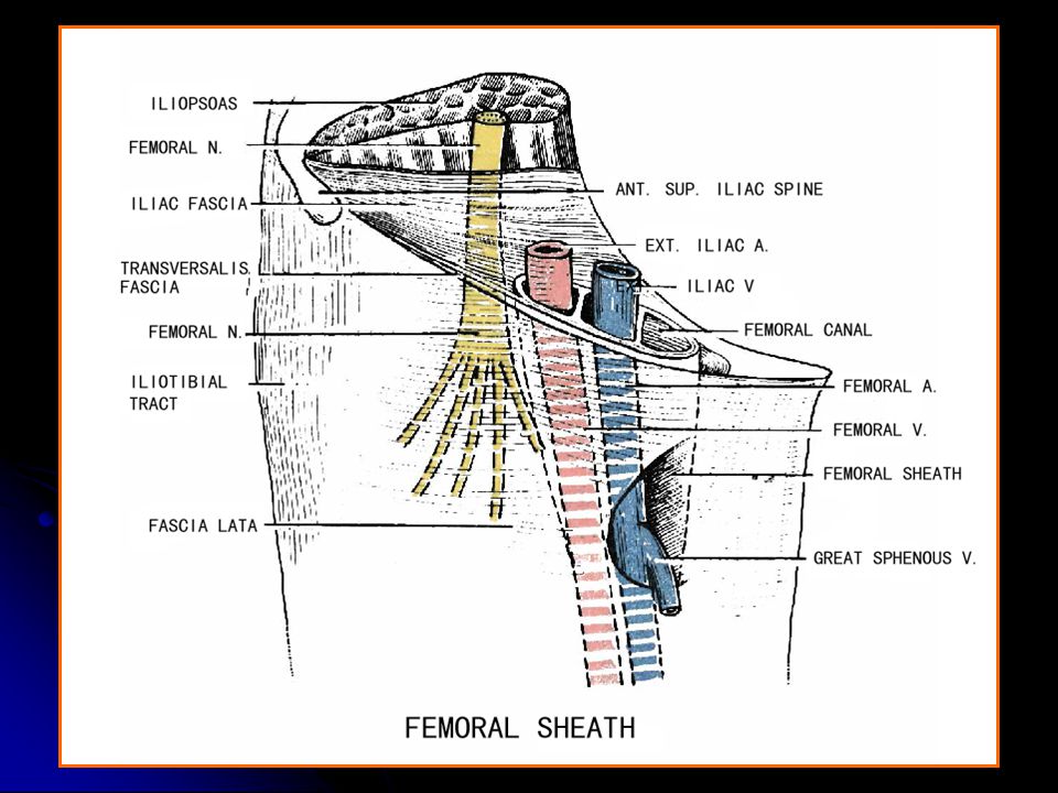
Femoral sheath

81
Femoral canal 1-1.5 cm Boundaries:
Ant. : inguinal lig. superior corner of falciform margin and femoral septum Post. : pectineal lig., pectineus Lat. : fibrous septum of femoral v.

82
femoral ring ant. : inguinal lig. med. : lacunar lig.,
post. : pectineal lig. lat. : fibrous septum of medial side of femoral v.

85
A 63-year-old female patient says that she has pain in her groin and upper thigh. Upon examination, you palpate a lump located below the inguinal ligament lateral to its attachment to the pubic tubercle. You suspect that this may be a hernia passing through the: femoral canal adductor hiatus obturator canal deep inguinal ring superficial inguinal ring A

86
The femoral canal contains the:
Deep inguinal lymph node(s) Femoral artery Femoral nerve Femoral vein Ilioinguinal nerve A
 Femoral artery. Femoral nerve. Femoral vein. Ilioinguinal nerve. A.")
87
The pulse of the femoral artery is best felt at which superficial reference point?
Anterior to the ankle joint Femoral triangle Mid-thigh Popliteal fossa Right lateral portion of the hypogastrium B

88
At which site could one expect to enter the femoral vein with a simple percutaneous (through the skin) introduction of an instrument? Above the middle of the inguinal ligament Lateral to the femoral arterial pulse Lateral to the pubic tubercle Medial to the femoral arterial pulse Medial to the pubic tubercle D

89
A serious complication of fractures of the femoral neck is avascular necrosis of the femoral head. This usually results from rupture of which artery? Acetabular branch of obturator Deep circumflex iliac Descending branch of lateral circumflex femoral Medial circumflex femoral Second perforating branch of lateral circumflex D

90
A ruptured aneurysm in the most proximal portion of the deep femoral artery would result in a hematoma located initially in the: Adductor canal. Femoral canal. Femoral triangle. Inguinal canal. Popliteal fossa.

91
Childhood immunizations are sometimes given via intramuscular injections into the quadriceps muscles of the anterior thigh. At the mid-thigh level, a needle passing into the space deep to the sartorius muscle might pierce the femoral vessels as they lie in the: Adductor canal Adductor hiatus Adductor triangle Femoral canal Femoral ring

92
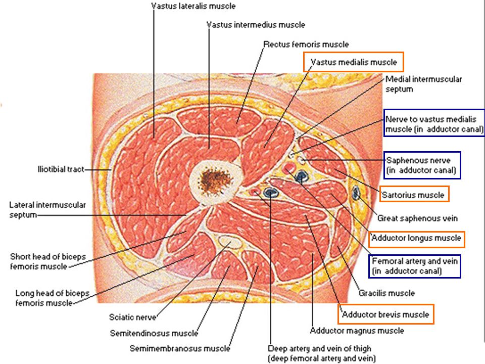
Which of the following is NOT located within the adductor canal?
Saphenous nerve Femoral artery Nerve to vastus medialis Femoral vein Deep femoral artery

93
THANKS!

Similar presentations


 Joint>")


