Download presentation
Presentation is loading. Please wait.

1
MCC NURSING DIANA BLUM MSN
Vascular Common disorders in America: hypertension atherosclerosis arterial occlusive disease abdominal aortic aneurysms (AAA) deep vein thrombosis (DVT) venous insufficiency MCC NURSING DIANA BLUM MSN
 deep vein thrombosis (DVT) venous insufficiency. MCC NURSING. DIANA BLUM MSN.")
2
hormones C reactive protein is a marker for cardiac inflammation
Increases mean: risk of damage Homocysteine: protein that promotes coagulation by increasing factor 5 and factor 11 while depressing activation of protein C and increasing thrombus formation risk Vitamin b6 and b12 and folate lowers homocysteine levels hormones

3
Arterial diseases: Arteriosclerosis (atherosclerosis)
Aneurysm formation Arteriosclerosis obliterans Raynaud’s phenomenon Arterial embolism Thromboangiitis obliterans Diabetic arteriosclerotic disease hypertension Arterial diseases:

4
Manifestations :ARTERIAL (50% occulsion before symptoms)
Prolonged capillary refill: - 3 seconds or more Ulcers: - open lesions on feet from diminished distal perfusion Ischemia (reduced oxygenation) - leads to pain Paresthesia (decreased sensation in extremities = tingling/numbing) Pain (in feet/leg muscles = burning, throbbing, cramping) -usually from exercise BUT also with elevation of lower extremities Hallmark sign: Intermittent claudication (pain in exercising muscles – usually in calf - directly related to decreased blood supply during activity & recedes with rest Temperature: (COLD) Skin color changes: skin pale on elevation but red dependent Reactive hyperemia: (reduced blood flow to extremity results in arteriolar dilation so when the blood supply is restored, the affected area becomes warm/red from congestion Pulse changes: Peripheral diminished or absent
 - leads to pain. Paresthesia (decreased sensation in. extremities = tingling/numbing) Pain (in feet/leg muscles = burning, throbbing, cramping) -usually from exercise BUT also. with elevation of lower extremities. Hallmark sign: Intermittent claudication (pain in. exercising muscles – usually in calf. - directly related to decreased. blood supply during activity & recedes with rest. Temperature: (COLD) Skin color changes: skin pale on. elevation but red dependent. Reactive hyperemia: (reduced blood flow. to extremity results in arteriolar dilation so when the blood supply is restored, the affected area becomes warm/red. from congestion. Pulse changes: Peripheral diminished or. absent.")
5
Arteriosclerosis -describes arterial disorders in which
degenerative changes result in decreased blood flow Atherosclerosis: - most common form of arteriosclerosis, excessive accumulation of lipids Arteriosclerosis Hypertension (MOST SIGNIFICANT) Cigarette smoking (nicotine has DIRECT vasoconstricting effect) Elevated serum cholesterol (fat causes obstructive plaques) Obesity (increased work to heart) Diabetes (hyperglycemia causes damage to vessel wall) Other: increase age, inactivity, family hx
 Cigarette smoking (nicotine has DIRECT. vasoconstricting effect) Elevated serum cholesterol (fat causes. obstructive plaques) Obesity (increased work to heart) Diabetes (hyperglycemia causes damage to vessel wall) Other: increase age, inactivity, family hx.")
6
Most common affected areas from arteriosclerosis:
Heart: coronary arteries (angina, MI, death) Brain (transient ischemic attacks =TIAs CVA, death) Kidneys (renal arterial stenosis lead to chronic renal failure) Extremities (gangrene of digits & intermittent claudication)
 Brain (transient ischemic attacks =TIAs. CVA, death) Kidneys (renal arterial stenosis lead to. chronic renal failure) Extremities (gangrene of digits & intermittent claudication)")
7
Pathophysiology of atherosclerosis
-inflammatory process, begins as fatty streaks that are deposited in the intima of the arterial wall Genetics and environment play a factor in the progression Elastic arteries: aorta, carotid, lg & med. sized muscular arteries (popliteals) most susceptible arteries. Endothelial injury: may be initiated by smoking, hypertension, diabetes, hyperlipidemia, Pathophysiology of atherosclerosis
 most. susceptible arteries. Endothelial injury: may be initiated by smoking, hypertension, diabetes, hyperlipidemia, Pathophysiology of atherosclerosis.")
8
Inflammatory cells(including macrophages) become attracted to the wall
Macrophages infiltrate wall and ingest lipid which turns them into foam cells They then release biochemical substances that cause further damage and attract platelets which then causes clots to form

9
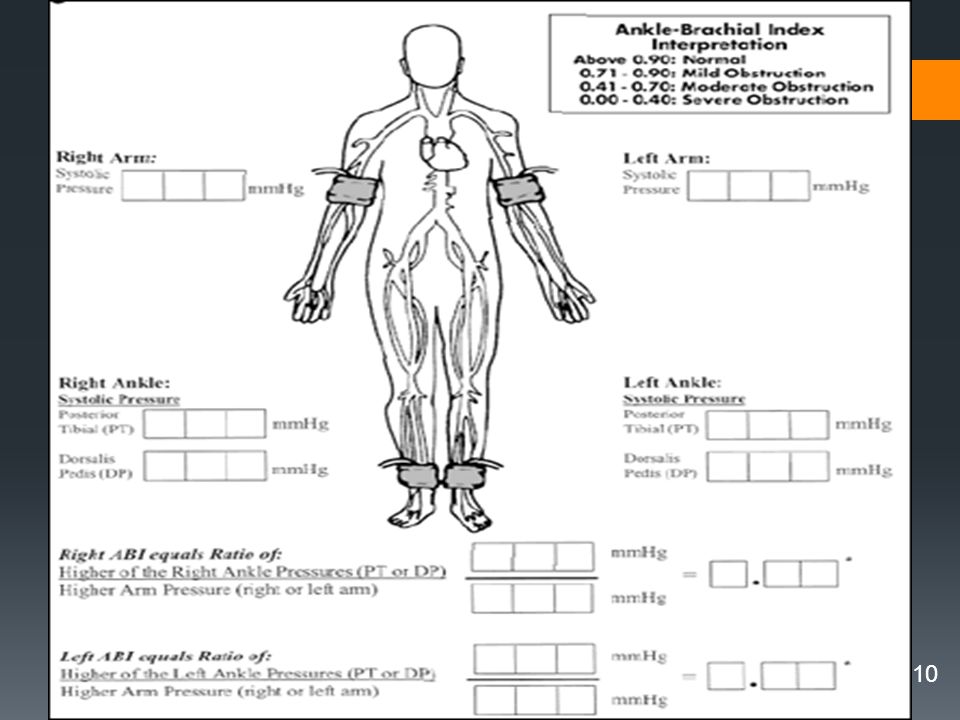
Ankle-brachial index of blood pressure: Used to diagnose peripheral vascular disease
-compares the blood pressure at ankle with that of the arm. -normally these should be the same (with a ratio of 1) -lesser number than 1 shows decreased blood pressure at the ankle compared to upper extremity = = which indicates peripheral vascular disease to lower extremities
 -lesser number than 1 shows decreased blood pressure at the ankle compared to upper extremity = = which indicates peripheral vascular disease to lower extremities.")
11
SURGERY Indications for fem-pop bypass: diabetes hypertension
vasculitis collagen disease Bueger’s disease Also, Embolectomy (surgical removal) SURGERY
 SURGERY.")
12
Fem-pop bypass

13
MEDICAL MANAGEMENT ANTIPLATELET THERAPY
Aspirin, ticlid, plavix, pletal, trental Beta blockers ARBs Statins Radiation therapy Angioplasty with stents

14
Nursing Interventions
Monitor BP for difference between arms Could be indicative of aortic coarctation Narrowing of aorta lumen Monitor for carotid bruits Assess cap refill, pulses,skin

15
Acute arterial stenosis
Monitor for the 5 P’s pain, sudden pallor pulselessness paresthesias paralysis

16
Acute peripheral arterial occlusion
may result from rupture and thrombosis of an atherosclerotic plaque, an embolus from the heart or thoracic or abdominal aorta, an aortic dissection, or acute compartment syndrome Symptoms and signs are sudden

18
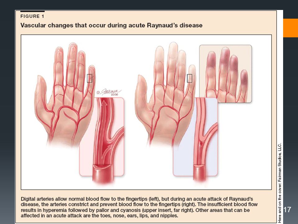
Buerger Disease Autoimmune disease
Recurrent inflammation of small arteries and veins of the extremities resulting in thrombus formation and occlusion. Unknown cause Men years old All races Link to heavy smoking/chewing tobacco s/s: rubor (reddish blue) color to foot, no Pedal pulse, discolored legs when dangled, eventually gangrene sets in
 color to foot, no Pedal pulse, discolored legs when dangled, eventually gangrene sets in.")
19
Aneurysms of Central Arteries
Enlargement of artery least 2X its normal Aortic dissection Medial & intimal layers separate Risk Factors: -hypertension -cocaine use - Marfan syndrome 85% are caused by atherosclerosis More frequent in men b/w years old Most common site for dissection 1/3 of pts with this die from rupture s/s: Asymptomatic Pain is primary symptom—constant Dyspnea Cough Hoarseness Stridor Aphonia (weakness or complete loss of voice) Unequal pupils Diagnostics: Chest x-ray TEE CT Aneurysms of Central Arteries
 Unequal pupils. Diagnostics: Chest x-ray. TEE. CT. Aneurysms of Central Arteries.")
20
Aortic Dissections: Type III most common type

21
Abdominal Aortic Aneurysm Size and Rupture Risk*
AAA Diameter (cm) Rupture Risk (%/yr) < 4 4–4.9 1% 5–5.9* 5–10% 6–6.9 10–20% 7–7.9 20–40% > 8 30–50% *Elective surgical repair should be considered for aneurysms > 5.0–5.5 cm.
 Rupture Risk (%/yr) < 4. 4–4.9. 1% 5–5.9* 5–10% 6– –20% 7– –40% > 8. 30–50% *Elective surgical repair should be considered for aneurysms > 5.0–5.5 cm.")
22
Aortic dissection

23
Signs/symptoms of aortic dissection:
n/v, diaphoresis with pain “tearing” pain Sudden onset not relieved with change of position Dissection of ascending aorta: anterior CP with radiation to neck, throat, jaw Dissection of descending: interscapular back pain radiation to lower back or abdomen Signs/symptoms of aortic dissection:

24
Treatment of hypertension for aortic dissection:
IV propranolol Nitropresside drip after beta blocker ( nitropresside by itself causes tachycardia AND left vent. contractility that is why a beta-blocker should be given first, then start nitropresside drip) Diagnosis: CXR (but 10% normal) see medialstinal widening Contrast CT MRI GOAL: to keep blood pressure to lowest possible but yet allows tissue perfusion Per physican recommendations
 Diagnosis: CXR (but 10% normal) see medialstinal. widening. Contrast CT. MRI. GOAL: to keep blood pressure to lowest. possible but yet allows tissue perfusion. Per physican recommendations.")
25
Surgery for distal dissections:
Mortality in 1st 48 hrs if unrepaired proximal aortic dissections is 40% Usually distal dissections treated medically unless: rapid expansion saccular formation persistent pain hemodynamic compromised blood leakage impending rupture

27
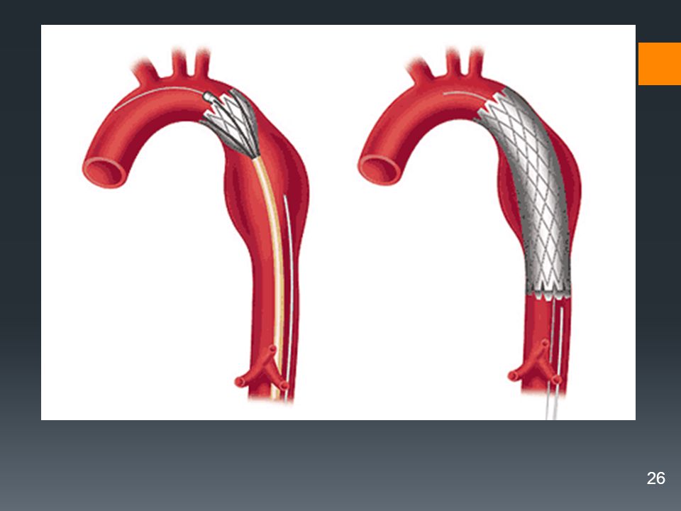
Dacron tube

28
Abdominal Aortic Aneurysm (AAA)
75% of all aneurysms Located between renal arteries & aortic bifurcation Symptoms from pressure exerted in surrounding structures. Many nonsymtomatic until ruptures Look for pulsating abdominal mass With rupture: hypovolemic shock & mortality around 90% Monitor growth: freq. CT scans Antihypertensives SURGICAL: graft

29
Post-op nursing interventions for graft:
Vitals Pulses distal to graft Report: changes in pulse cool extremities distal to graft white/blue to extremities distal to graft severe pain abd. distention decreased UO

30
Post-op nursing intervention (continued) Post graft
Elevation of head to 45° or less Renal function lab Respiratory status Paralytic ileus (NG tube) Assess for dysrhythmias post thoracic
 Assess for dysrhythmias post thoracic.")
31
Venous manifestations:
Skin color changes: reddened or cyanotic Edema: pooling of fluid results in edema Venous stasis ulcers: skin breakdown due to increased pressure from chronic pooling of blood Decreased mobility: may result from the edema Pain: - in feet/ leg muscles; aching/throbbing - results from venous stasis & increases as day progresses (esp with sitting or standing) Temperature changes: - warm to touch since blood can enter but cannot leave affected parts What dx are we talking about? Venous thrombosis (thrombophlebitis) known as DVT Varicose veins Venous stasis ulcers DVT risk for pulmonary embolism - legs - seen post hip surgery, knee replacement pregnancy, ulcerative colitis, hrt failure, immobility Venous manifestations:
 Temperature changes: - warm to touch since blood can enter. but cannot leave affected parts. What dx are we talking about Venous thrombosis (thrombophlebitis) known as DVT. Varicose veins. Venous stasis ulcers. DVT risk for pulmonary embolism. - legs. - seen post hip surgery, knee replacement. pregnancy, ulcerative colitis, hrt failure, immobility. Venous manifestations:")
32
DVT : Groin tenderness/pain Unilateral sudden onset edema leg
Homan’s sign (appears in only 10% of pt with DVT) Ultrasonography
 Ultrasonography")
33
DVT interventions: Rest (do NOT massage area)
Low-molecular weight heparin Coumadin TPA ****Contraindications to anticoagulant therapy Pt compliance, bleeding, aneurysms, trauma, alcohol, recent surgery, liver or kidney disease, hazard jobs, pregnancy DVT interventions:

34
Nursing cares Monitor for hemorrhage Monitor PT/PTT
Heparin is therapeutic b/w on ptt Coumadin is therapeutic b/w 2-3 on PT/INR Monitor for Thrombocytopenia Monitor Platelets s/s; purpura, bruising, hematomas Provide bedrest Ted Hose or ace wraps for prevention of DVT SCDs for prevention of DVT Pain meds Nursing cares

35
- excessive tension exerted on arterial walls which places pts at increased risk for target organ damage -asymptomatic until complications develop - elevation may be systolic or diastolic or both - normal <120 mmHg systolic <80 mmHg diastolic Often none Occipital headache more severe on rising Lightheadedness Epistaxis Known as the ‘Silent Killer’ What factors determine arterial pressure? Cardiac output which is the volume of blood pumped by the heart in 1 minute Peripheral vascular resistance which is the force in the peripheral blood vessels that the left ventricular must overcome to eject blood out of the heart Hypertension

36
Pathophysiologic processes for hypertension:
BP=CO X peripheral resistance Elevated BP is direct result of increased peripheral resistance, increased CO or both Renin-angiotensin-aldosterone system Aldosterone: increased water/Na+ retention thus increasing ECF volume which leads to increased CO with subsequent increase BP

37
Possible Causes of PVR Narrowing of blood vessels, PVD, CAD, kidney disease: > renin/angiotensin =vasoconstriction Release of catecholamine (epinephrine and adrenalin) = vasoconstriction > blood volume= more work to pump > Blood viscosity=harder to pump Ability of blood vessel to stretch 95% of cases of hypertension are 1st degree (essential) 2nd degree hypertension: CHAPS Cushing’s syndome Hyperaldosteronism Aortic coarctation Pheochromocytoma Stenosis of renal arteries Complications of Htn and pvr are Damage to blood vessels of the eyes, heart, kidney, brain resulting in: Stroke CHF AMI Renal failure Blindness
 = vasoconstriction. > blood volume= more work to pump. > Blood viscosity=harder to pump. Ability of blood vessel to stretch. 95% of cases of hypertension are 1st degree (essential) 2nd degree hypertension: CHAPS. Cushing’s syndome. Hyperaldosteronism. Aortic coarctation. Pheochromocytoma. Stenosis of renal arteries. Complications of Htn and pvr are. Damage to blood vessels of the eyes, heart, kidney, brain resulting in: Stroke. CHF. AMI. Renal failure. Blindness.")
38
Target Organ Disease from hypertension
Large vessels: aneurysmal dilation accelerated atherosclerosis aortic dissection Cardiac: acute= pulm edema, MI chronic= LVH Cerebrovascular: acute= Intracranial bleed, coma, seizure mental status changes, TIA, stroke chronic=TIA, stroke Target Organ Disease from hypertension

39
Target organ disease from hypertension:
Renal: acute=hematuria, azotemia chronic=elevated creatinine proteinuria Retinopathy: acute=papilledema, hemorrhages chronic=hemorrhages,exudates, Target organ disease from hypertension:

40
Treatment of hypertension:
Lifestyle modification ABCD: ACE inhibitors; ARB B-blockers Calcium channel blockers Diuretics

41
Hypertensive Crisis: Treatment
Parenteral agents for immediate redux of BP In ICU for monitoring Arterial line Drug of choice: sodium nitroprusside =direct acting arterial & venous vasodilator = reduces BP rapidly but lower mean arterial pressure no more than 25% over 1st 2 hours = easily titratable = monitor closely for hypotension = shield this drip from light Sometimes rare sometimes fatal Diastolic BP Causes vascular damage Can be caused by renal failure, HTN, Med withdrawal

42
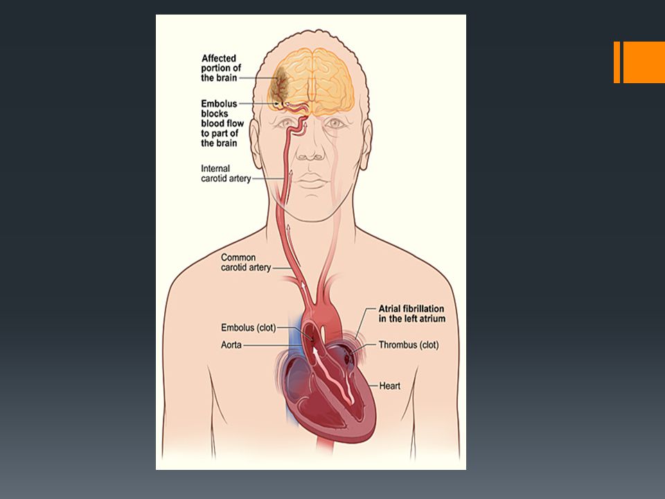
STROKE: occlusion of cerebral vasculature
DUE TO: 1. emboli that lodges in cerebral vasculature (from a-fib, vegetations on an infect valve) 2. atherosclerotic plaque (occludes carotid arteries) 3. venous occlusion (secondary to thrombosis) 4. arterial dissection (in carotid or vertebrobasilar system) 5. severe hypotension ( infarct in cerebral areas) 6. hemorrhage :occurs during activity Chapter 22
 2. atherosclerotic plaque (occludes carotid arteries) 3. venous occlusion (secondary to thrombosis) 4. arterial dissection (in carotid or vertebrobasilar system) 5. severe hypotension ( infarct in cerebral areas) 6. hemorrhage :occurs during activity. Chapter 22.")
43
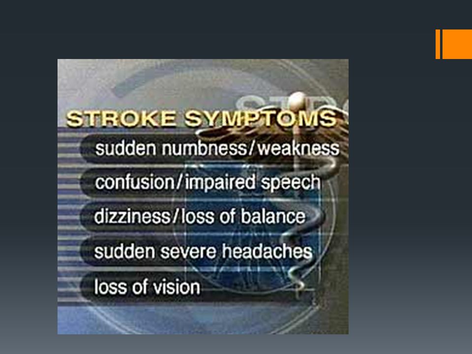
Sudden loss of function resulting from disrupted blood supply to area in brain
5 types: Large artery Caused by atherosclerosis Small penetrating artery Most common Also called lacunar strokes because it creates a cavity Cardiogenic emboli Usually from afib Cryptogenic No known cause Other Caused from Drug use, migraines,spontaneous TIA

45
Hemorrhagic stroke Bleeding into brain tissue or ventricles, subdural, or subarachnoid spaces due to ruptured aneurysm or from severe hypertension VASOSPASM (after a bleed) 4-14 days post hemorrhage Management is difficult Manifestations: Severe headache LOC Tinnitus Dizziness Hemiparesis Prognosis: variable Diagnostics: CT Lumbar puncture Angiography
 4-14 days post hemorrhage. Management is difficult Manifestations: Severe headache. LOC. Tinnitus. Dizziness. Hemiparesis. Prognosis: variable. Diagnostics: CT. Lumbar puncture. Angiography.")
46
Prevention Manage HTN Avoid alcohol Increase public awareness

48
Assessment Tools Neurological assessment upon admission or change in client status, including: Level of consciousness Orientation Motor ability Pupils Speech/language Vital signs Blood glucose Risk assessment for complications including fall, pressure ulcer, painful hemiparetic shoulder, spasticity/contractures, and deep vein thrombosis Pain assessment Administration and interpretation of dysphagia screen Nutrition and hydration screening Screening for alterations in cognition, perception, and language using validated tools Assessment of activities of daily living (ADL) using validated tools Assessment of bowel and bladder function Depression screening using a validated tool Assessment/screening of caregiver burden using a validated tool Screening of stroke clients and their partners for sexual concerns Assessment of stroke client and their caregivers' learning needs, abilities, learning preferences and readiness to learn Referral for further assessment and management, as indicated Documentation of all assessments and screenings
 using validated tools. Assessment of bowel and bladder function. Depression screening using a validated tool. Assessment/screening of caregiver burden using a validated tool. Screening of stroke clients and their partners for sexual concerns. Assessment of stroke client and their caregivers learning needs, abilities, learning preferences and readiness to learn. Referral for further assessment and management, as indicated. Documentation of all assessments and screenings.")
49
Treatment for stroke: (Note similar to measures for myocardial ischemia/MI)
Thrombolysis (who is not a candidate?) Lower BP Quit smoking Decrease cholesterol Antiplatelet (ASA)
 Lower BP. Quit smoking. Decrease cholesterol. Antiplatelet (ASA)")
50
Stroke treatment (continued)
ASA Heparin (SQ or IV contin infusion) Low-molecular wt heparin (lovenox) Warfarin (coumadin) Obtain PT, PTT prior to therapy PT: monitor oral anticoag : goal=1.5 to 2 times pt baseline PTT: monitor heparin: goal=1.5 to 2 times pt baseline INR: monitor Warfarin: goal=2 to 3
 Low-molecular wt heparin (lovenox) Warfarin (coumadin) Obtain PT, PTT prior to therapy. PT: monitor oral anticoag : goal=1.5 to 2 times pt baseline. PTT: monitor heparin: goal=1.5 to 2 times pt baseline. INR: monitor Warfarin: goal=2 to 3.")
51
More stroke treatment:
Carotid artery angioplasty Arteriovenous Malformation (gamma radiation through Gamma knife) Aneurysms (coils) Craniotomy for clot removal Assessments while on anticoags.: Observe for bleeding Also, antiplatelet meds (Plavix, Persantine) cause bruising, hemorrhage, liver disease (need liver function tests) GIVE clopidogrel (Plavix) with food
 Aneurysms (coils) Craniotomy for clot removal. Assessments while on anticoags.: Observe for bleeding. Also, antiplatelet meds (Plavix, Persantine) cause. bruising, hemorrhage, liver disease (need liver function tests) GIVE clopidogrel (Plavix) with food.")
52
Nursing Diagnosis Impaired physical mobility: -flaccid, spasticity
Disturbed sensory perception: -vision, proprioception, sensation Unilateral neglect: - use both sides of body (dress affected side first) Impaired verbal communication:: -expressive, receptive, both Impaired swallowing: must be evaluated, must prevent aspiration !!! But yet meet caloric needs Urinary and/or bowel incontinence Nursing Diagnosis
 Impaired verbal communication:: -expressive, receptive, both. Impaired swallowing: must be evaluated, must prevent aspiration !!! But yet meet caloric needs. Urinary and/or bowel incontinence. Nursing Diagnosis.")
53
Complications Rebleed Vasospasm Hydrocephalus Hypoxia of brain

54
Nursing interventions
Administer oxygen Provide adequate hydration Evaluate swallow function Frequent neuro checks Strict I/O Seizure precautions Monitor ICP Monitor BP closely Teach stress reduction techniques Manage agitation

55
Surgery and complications
Evacuation of blood via craniotomy Goal of surgery is to prevent further rupture/bleed Post op complications Disoriented Amnesia Korsaff’s syndrome (psychosis caused by lack of thiamine) Personality changes Intraop emboli Electrolyte disturbances GI bleed Surgery and complications
 Personality changes. Intraop emboli. Electrolyte disturbances. GI bleed. Surgery and complications.")
56
QUESTIONS???

Similar presentations


 CAD is most common form of heart disease and causes premature death. In UK, 1 in 3 men and.>")
 Embolism Trauma Crush injuries.>")


