Download presentation
Presentation is loading. Please wait.

1
Chapter 12: DNA and RNA

2
KWL Chart What I KNOW already about DNA What I WANT to know about DNA
What I have LEARNED about DNA

3
Learning Targets for Section 12-1
Summarize the relationship between genes and DNA? Describe the overall structure of DNA?

4
12–1 Research Behind DNA Griffith and Transformation
In 1928, British scientist Frederick Griffith tried to determine which bacteria produced pneumonia. Griffith isolated two different strains. 1. disease-causing = smooth colonies 2. harmless strain = rough colonies.

5
Griffith's Experiment Griffith injected mice
1. disease-causing bacteria- mice developed pneumonia and died. 2. harmless strain - didn’t get sick at all

6
Experiment cont. Griffith’s then mixed heat-killed, disease-causing bacteria with live, harmless ones and injected the mixture into mice Mice developed pneumonia and many died. Found their lungs filled with the disease-causing bacteria

7
Figure 12–2 Griffith’s Experiment
Section 12-1 Heat-killed, disease-causing bacteria (smooth colonies) Harmless bacteria (rough colonies) Control (no growth) Harmless bacteria (rough colonies) Heat-killed, disease-causing bacteria (smooth colonies) Disease-causing bacteria (smooth colonies) Dies of pneumonia Dies of pneumonia Lives Lives Live, disease-causing bacteria (smooth colonies) Go to Section:
 Harmless bacteria (rough colonies) Control (no growth) Harmless bacteria (rough colonies) Heat-killed, disease-causing bacteria (smooth colonies) Disease-causing bacteria (smooth colonies) Dies of pneumonia. Dies of pneumonia. Lives. Lives. Live, disease-causing bacteria (smooth colonies) Go to Section:")
8
Figure 12–2 Griffith’s Experiment
Section 12-1 Heat-killed, disease-causing bacteria (smooth colonies) Harmless bacteria (rough colonies) Control (no growth) Harmless bacteria (rough colonies) Heat-killed, disease-causing bacteria (smooth colonies) Disease-causing bacteria (smooth colonies) Dies of pneumonia Dies of pneumonia Lives Lives Live, disease-causing bacteria (smooth colonies) Go to Section:
 Harmless bacteria (rough colonies) Control (no growth) Harmless bacteria (rough colonies) Heat-killed, disease-causing bacteria (smooth colonies) Disease-causing bacteria (smooth colonies) Dies of pneumonia. Dies of pneumonia. Lives. Lives. Live, disease-causing bacteria (smooth colonies) Go to Section:")
9
Figure 12–2 Griffith’s Experiment
Section 12-1 Heat-killed, disease-causing bacteria (smooth colonies) Harmless bacteria (rough colonies) Control (no growth) Harmless bacteria (rough colonies) Heat-killed, disease-causing bacteria (smooth colonies) Disease-causing bacteria (smooth colonies) Dies of pneumonia Dies of pneumonia Lives Lives Live, disease-causing bacteria (smooth colonies) Go to Section:
 Harmless bacteria (rough colonies) Control (no growth) Harmless bacteria (rough colonies) Heat-killed, disease-causing bacteria (smooth colonies) Disease-causing bacteria (smooth colonies) Dies of pneumonia. Dies of pneumonia. Lives. Lives. Live, disease-causing bacteria (smooth colonies) Go to Section:")
10
Griffith’s Conclusion:
Griffith hypothesized some factor transformed harmless cells into the heat-killed harmful cells Griffith movie

11
Avery and DNA Avery and his colleagues repeated Griffith’s experiment then: Used enzymes that destroyed proteins, lipids, carbohydrates, and other molecules, including the nucleic acid RNA When they destroyed the nucleic acid (DNA), transformation did not occur
, transformation did not occur.")
12
Avery’s Conclusion: Avery and other scientists discovered that DNA is the nucleic acid that stores and transmits the genetic information from one generation of an organism to the next

13
The Hershey Chase Experiment
Martha Chase and Alfred Hershey

14
The Hershey Chase Experiment
Studied viruses, nonliving particles smaller than a cell that can infect living organisms Hershey and Chase reasoned that if they could determine which part of the virus—the protein coat or the DNA core—entered the infected cell, they would learn whether genes were made of protein or DNA They grew viruses in cultures of radioactive isotopes of phosphorus-32 (32P) and sulfur-35 (35S).
 and sulfur-35 (35S).")
15
The Hershey Chase Experiment
Proteins contain almost no phosphorus and DNA contains no sulfur If 35S was found in the bacteria, it would mean that the viruses’ protein had been injected, If 32P was found in the bacteria, then it was the DNA that had been injected

16
Figure 12–4 Hershey-Chase Experiment
Section 12-1 Bacteriophage with phosphorus-32 in DNA Phage infects bacterium Radioactivity inside bacterium Bacteriophage with sulfur-35 in protein coat Phage infects bacterium No radioactivity inside bacterium Go to Section:

17
Figure 12–4 Hershey-Chase Experiment
Section 12-1 Bacteriophage with phosphorus-32 in DNA Phage infects bacterium Radioactivity inside bacterium Bacteriophage with sulfur-35 in protein coat Phage infects bacterium No radioactivity inside bacterium Go to Section:

18
Figure 12–4 Hershey-Chase Experiment
Section 12-1 Bacteriophage with phosphorus-32 in DNA Phage infects bacterium Radioactivity inside bacterium Bacteriophage with sulfur-35 in protein coat Phage infects bacterium No radioactivity inside bacterium Go to Section:

19
Hershey and Chase’s Conclusion:
Hershey and Chase concluded that the genetic material of the bacteriophage was DNA, not protein.

21
REVIEW In your science journal:
List as many scientists you can remember And describe their contribution to our understanding of DNA

22
The Structure of DNA DNA is a long molecule made up of units called nucleotides Each nucleotide is made up of three parts: 1. a 5-carbon sugar called deoxyribose, 2. a phosphate group, 3. and a nitrogenous (nitrogen- containing) base
 base.")
23
There are four kinds of nitrogenous bases in DNA
Purines: Adenine: Expressed A Guanine: Expressed G Pyrimidines: Thymine: Expressed T Cytocine: Expressed C

24
Figure 12–5 DNA Nucleotides
Section 12-1 Purines Pyrimidines Adenine Guanine Cytosine Thymine Phosphate group Deoxyribose Go to Section:

25
Chargaff’s Rules Erwin Chargaff, an American biochemist, discovered that the percentages of guanine [G] and cytosine [C] are almost equal in any sample of DNA The same thing is true for adenine [A] and thymine [T] Despite the fact that DNA samples from organisms obeyed this rule, neither Chargaff nor anyone else had the faintest idea why

27
X-Ray Evidence In the early 1950s, a British scientist named Rosalind Franklin began to study DNA using a technique called X-ray diffraction The X-shaped pattern in the image shows that the strands in DNA are twisted around each other like the coils of a spring, a shape known as a helix

29
The Double Helix At the same time Francis Crick, and James Watson, were trying to understand the structure of DNA by building three-dimensional models of the molecule In 1953, Watson was shown a copy of Franklin’s X-ray pattern. In his book The Double Helix, Watson wrote: “The instant I saw the picture my mouth fell open and my pulse began to race.”

32
Watson and Crick’s Conclusion:
- DNA is a double helix in which two strands are wound around each other. - Each strand is made up of a chain of nucleotides. - The two strands are held together by hydrogen bonds between adenine and thymine and between guanine and cytosine.

33
Structure of DNA

34
Learning Targets for Section 12.2
Summarize the events that happen in DNA replication Relate the DNA molecule to chromosome structure.

35
12–2 Chromosomes and DNA Replication
DNA and Chromosomes Most prokaryotes have a single circular DNA molecule that contains nearly all of the cell’s genetic information

36
Prokaryotic Chromosome Structure
Section 12-2 Chromosome E. coli bacterium Bases on the chromosome Go to Section:

37
Differences between Prokaryotes and Eukaryotes
Eukaryotic DNA is a bit more complicated. Many eukaryotes have as much as 1000 times the amount of DNA as prokaryotes DNA Length DNA molecules are surprisingly long The chromosome of the prokaryote E. coli, contains 4,639,221 base pairs

39
Length of DNA This means that the nucleus of a human cell contains more than 1 meter of DNA How can so much DNA be packed into each and every cell in our body?

40
Chromosome Structure Eukaryotic chromosomes contain both DNA and protein, tightly packed together to form a substance called chromatin Chromatin consists of DNA that is tightly coiled around proteins called histones

41
Chromosome Structure Together, the DNA and histone molecules form a beadlike structure called a nucleosome This allows the chromosomes to be very tightly coiled up in the nucleus

42
Figure 12-10 Chromosome Structure of Eukaryotes
Section 12-2 Chromosome Nucleosome DNA double helix Coils Supercoils Histones Go to Section:

43
DNA Structure Activity

44
DNA Replication When Watson and Crick discovered the double helix structure of DNA, there was one more remarkable aspect that they recognized immediately. The structure explained how DNA could be copied, or replicated Each strand of the DNA double helix has all the information needed to reconstruct the other half by the mechanism of base pairing

46
DNA Replication During DNA replication, the DNA molecule separates into two strands Then produces two new complementary strands following the rules of base pairing. Each strand of the double helix of DNA serves as a template, or model, for the new strand

47
Figure 12–11 DNA Replication
Section 12-2 Original strand New strand DNA polymerase Growth DNA polymerase Growth Replication fork Replication fork Nitrogenous bases New strand Original strand Go to Section:

48
DNA Replication DNA replication animation DNA Replication animation

49
How DNA Replicates Start with a double strand of DNA
DNA replication is carried out by a series of enzymes. which “unzip” a molecule of DNA

50
How DNA Replicates A – T G – C C – G T – A
I’ve deleted the sugar-phosphate backbone for easier drawings Hydrogen bonds

51
DNA Unzips A – T A - T G – C G - C A – T A T C – G DNA C G
T – A unzips T A C – G C G

52
A – T G – C DNA PolymeraseIII A T C G T A C G
DNAP III moves along each strand adding a base pair at a time

53
A – T G – C A T T C G G T T A C CG DNAP III continues to moves along each strand adding a base pair at a time

54
"Replication Fork" A – T G – C A T A T C G C G T A TA C G CG

55
Finally get 2 new strands, exact copies
AT AT GC GC CG CG TA TA

56
STOP: Practice Replicate the following strand of DNA
ATG GGA CCG TAT ACG GAG TAC CCT GGC ATA TGC CTC

57
DNA and Enzymes DNA replication involves a host of enzymes and regulatory molecules The principal enzyme involved in DNA replication is called DNA polymerase In addition to replication DNA polymerase also “proofreads” each new DNA strand, helping to maximize the odds that each molecule is a perfect copy of the original DNA

58
Learning Targets for Section 12.3
What are the three main types of RNA? What is transcription? What is translation?

59
12–3 RNA and Protein Synthesis
The double helix structure explains how DNA can be replicated but it does not explain how a gene works Genes are coded DNA instructions that control the production of proteins

61
RNA The first step is to copy part of the nucleotide sequence from DNA into RNA a process called transcription RNA molecules then carry out the process of making proteins. RNA molecule is a working copy of a single gene. Using RNA makes it possible for a single gene to produce hundreds or even thousands of RNA molecules

62
The Structure of RNA There are three main differences between RNA and DNA: The sugar in RNA is ribose instead of deoxyribose RNA is generally single-stranded RNA contains uracil in place of thymine

63
Types of RNA There are three main types of RNA: messenger RNA, ribosomal RNA, and transfer RNA. messenger RNA (mRNA) serves as “messengers” from DNA to the rest of the cell. ribosomal RNA (rRNA) is the site where Proteins are assembled on ribosomes transfer RNA (tRNA) transfers each amino acid to the ribosome during the construction of a protein.
 serves as messengers from DNA to the rest of the cell. ribosomal RNA (rRNA) is the site where Proteins are assembled on ribosomes. transfer RNA (tRNA) transfers each amino acid to the ribosome during the construction of a protein.")
64
Transcription During transcription, RNA polymerase binds to DNA and separates the DNA strands. RNA polymerase then uses one strand of DNA as a template from which nucleotides are assembled into a strand of RNA

65
Figure 12–14 Transcription
Section 12-3 Adenine (DNA and RNA) Cystosine (DNA and RNA) Guanine(DNA and RNA) Thymine (DNA only) Uracil (RNA only) RNA polymerase DNA RNA Go to Section:
 Cystosine (DNA and RNA) Guanine(DNA and RNA) Thymine (DNA only) Uracil (RNA only) RNA polymerase. DNA. RNA. Go to Section:")
66
Transcription Transcription movie

67
Practice Transcribing
Remember A goes with T, C goes with G and Uracil takes the place of Thymine TAC GCA CCA TAT CCG ATT AUG CGU GGU AUA GGC UAA

68
Where to Begin? The enzyme will bind only to regions of DNA known as promoters, which have specific base sequences. Promoters are signals in DNA that indicate to the enzyme where to bind to make RNA Similar signals in DNA cause transcription to stop when the new RNA molecule is completed

69
Editing RNA Many RNA molecules have sections, called introns, edited out of them before they become functional. The remaining pieces, called exons, are spliced together. Then, a cap and tail are added to form the final RNA molecule.

71
The Genetic Code The “language” of mRNA instructions is called the genetic code. RNA contains four different bases: A, U, C, and G. In effect, the code is written in a language that has only four “letters.”

72
STOP THINK! What is the process of transcription making?
Where does transcription take place? What enzymes are used to complete transcription? Compare and Contrast DNA and RNA?

73
Genetic Code The genetic code is read three letters at a time
Each three-letter “word” in mRNA is known as a codon A codon consists of three consecutive nucleotides that specify a single amino acid that is to be added to the polypeptide

74
Genetic Code Because there are four different bases, there are 64 possible three-base codons (4 × 4 × 4 = 64). The codon, AUG, that can either specify methionine or serve as the initiation, or “start,” codon for protein synthesis There are three “stop” codons that do not code for any amino acid

76
Translation During translation, the cell uses information from messenger RNA to produce proteins This process is known as translation

77
Translation Before translation can occur, messenger RNA must first be transcribed from DNA in the nucleus and released into the cytoplasm Translation begins when an mRNA molecule in the cytoplasm attaches to a ribosome

78
Figure 12–18 Translation Section 12-3 mRNA Go to Section: Nucleus
Messenger RNA Messenger RNA is transcribed in the nucleus. mRNA Lysine Phenylalanine tRNA Transfer RNA The mRNA then enters the cytoplasm and attaches to a ribosome. Translation begins at AUG, the start codon. Each transfer RNA has an anticodon whose bases are complementary to a codon on the mRNA strand. The ribosome positions the start codon to attract its anticodon, which is part of the tRNA that binds methionine. The ribosome also binds the next codon and its anticodon. Methionine Ribosome mRNA Start codon Go to Section:

79
Translation As each codon of the mRNA molecule moves through the ribosome, the proper amino acid is brought into the ribosome and attached to the growing polypeptide chain That job is done by transfer RNA

80
Translation The codon matches up with complementary bases on the tRNA to tell it which amino acid to bring in The three bases on the tRNA molecule, called the anticodon, are complementary to one of the mRNA codons

81
Translation The polypeptide chain continues to grow until the ribosome reaches a stop codon on the mRNA molecule At that point the protein is released to be modified in the Golgi apparatus or to be shipped out to perform its function

82
Figure 12–18 Translation (continued)
Section 12-3 The Polypeptide “Assembly Line” The ribosome joins the two amino acids—methionine and phenylalanine—and breaks the bond between methionine and its tRNA. The tRNA floats away, allowing the ribosome to bind to another tRNA. The ribosome moves along the mRNA, binding new tRNA molecules and amino acids. Growing polypeptide chain Ribosome tRNA Lysine tRNA mRNA Completing the Polypeptide The process continues until the ribosome reaches one of the three stop codons. The result is a growing polypeptide chain. mRNA Translation direction Ribosome Go to Section:

83
Practice Translating original DNA ATG CCC AGT TCA TAA
Compl DNA = ___ ___ ____ ___ ____ mRNA = ____ ___ ____ ____ ____ Amino acid ______________, _______________, _____________, ______________, _____________.

84
Check these answers first
original DNA ATG CCC AGT TCA TAA Compl DNA = TAC GGG TCA AGT ATT mRNA = AUG CCC AGU UCA UAA

86
Amino Acid Answers Amino acid methionine, proline,
serine, serine, stop

88
BELLRINGER 11/11/09 COMPARE/CONTRAST RNA TO DNA
*** Include as much information about each that you can think of THEN DESCRIBE what happens during TRANSCRIPTION And DESCRIBE WHAT HAPPENS DURING TRANSLATION

89
12.4 Learning Targets Describe how gene mutations and chromosomal mutations occur Understand the difference between point mutations and frameshift mutations

90
Ways variations can arise
What is a mutation and where can it occur? Inheritable change in genetic code * 99.9 % are harmful. Only 0.1% are helpful!! What is a chromosomal mutation? Changes in number or structure of chromosome When do chromosomal mutations occur? During Meiosis – A cell division that produces gametes

91
TYPES OF MUTATIONS What is a frameshift mutation?
Change that shifts the genetic message through inserting or deleting a nucleotide What is a point mutation? Change at one point of a chromosome.

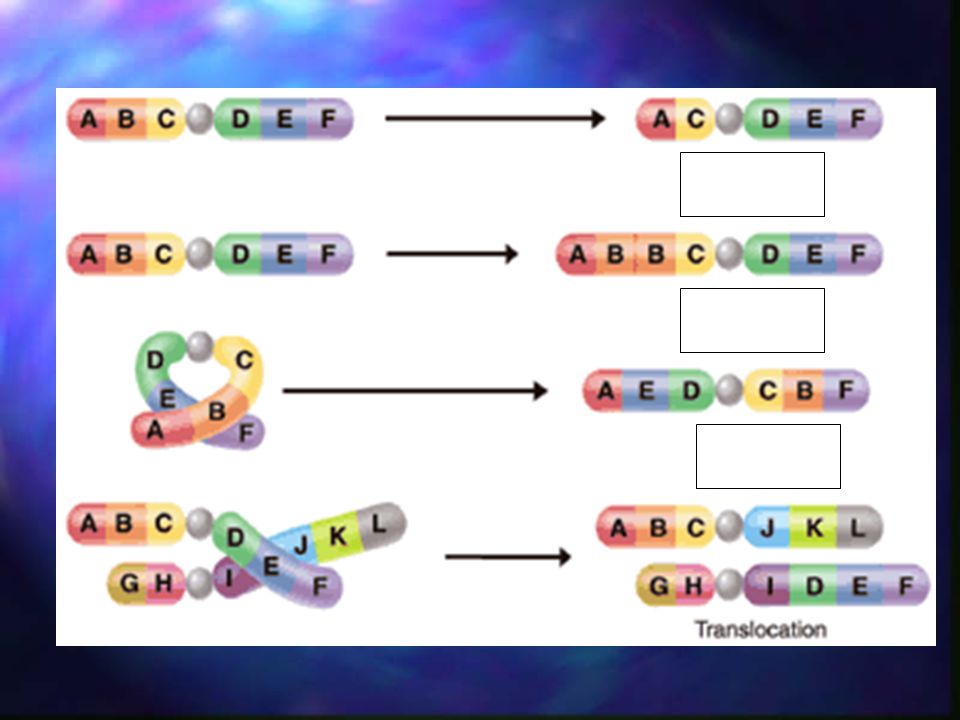
92
What is a deletion mutation?
Loss of one or more genes What is a duplication mutation? One or more genes are copied twice What is an inversion mutation? Part of a chromosome gets turned the wrong way

Similar presentations


 Harmless bacteria (rough colonies) Heat-killed, disease- causing bacteria (smooth colonies) Control (no growth)>")


