Download presentation
Presentation is loading. Please wait.

3
What Can a Skeleton Tell You
Aging Sexing Population Affinity Diet Pathology Trauma

4
Determining Age of Infant Prior to Birth
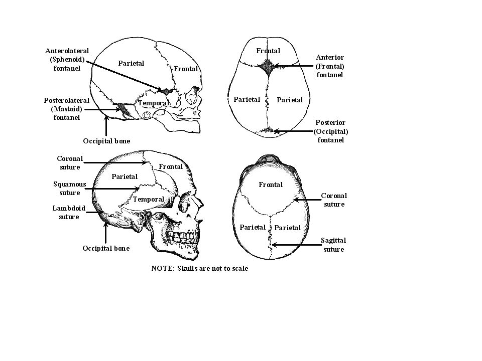
Cold Case Files are records kept for criminal cases that were never solved, often after several years. In some cases, however, a murder case may go years before it is even discovered. In these cases, most portions of the body will have long since either decayed or have been scavenged. The one portion of the body that takes the longest to decay is the skeleton. These cases would be, uh, cold, but in a different way . . . A person who studies skeletons in order to determine information about the person before she/he died is called a Forensic Anthropologist. Despite the neglect the dead body endures, there are still quite a number of things that one can learn. First of all, in the area of the skull, one can determine many things. In terms of infants, we can determine the age, in weeks, fairly accurately by examining the development of the skull: Notice, apart from the difference in size, how the fontanels (the soft spots that ultimately become the sutures, or fixed joints between the bones in the skull) change over time, gradually becoming smaller. Click HERE for the full size image Original image from Used with permission.
 change over time, gradually becoming smaller. Click HERE for the full size image Original image from Used with permission.")
5
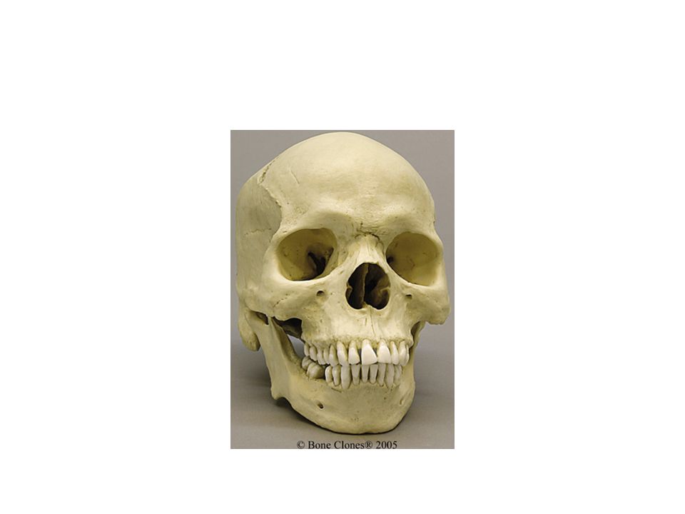
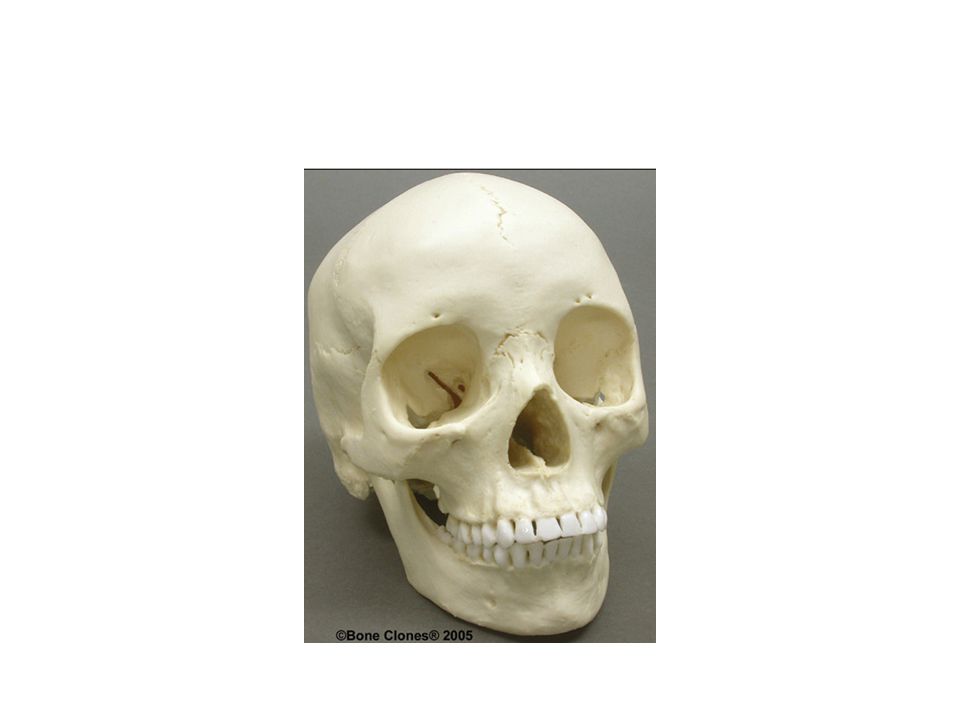
Try it Out Aged 31 weeks, 32 weeks, and 40 weeks (full term)
One must, however, be careful. Take the following two images, and try to figure out the approximate age of the second image, after examining the first image. Aged 31 weeks, 32 weeks, and 40 weeks (full term)
")
6
What about this one? If you had a hard time, perhaps it is due to the fact that the 2nd image was of a chimpanzee skull!

7
Bear Claw vs. Human Hand If you are curious, the human hand is on the left. In terms of forensic anthropologists, they are often called out to identify a bone, only to find that it is not even human, and thus not a crime. The most common non-human bone found in Connecticut? Ever heard of Spiral Ham?!

8
Like Fingerprints… just can’t be seen until antemortem
Examine the image below to see how the fontanels, described above, grow and fuse with the other bones to form the sutures. Although these sutures are as unique as fingerprints, as they cannot be seen antemortem (not without surgery, or a violent accident), they are not useful in terms of identification.
, they are not useful in terms of identification.")
9
When you don’t floss The sutures are, however, useful in terms of determining the approximate age of a skull. In terms of the sutures, as we grow older, the sutures ultimately completely fill in. The less filled in, the younger the person. This does mean that aging has a habit of making us all a bit more hard-headed; this explains a lot about your teachers . . . We also often tend to lose teeth as we age, and if we really do a bad job flossing, we lose BONE as well, as in the geriatric (elderly) skull below:
 skull below:")
10
Aging In cases where cultures have bound the skull, the sutures have had to move as the bones take on the new shape forced by the binding of the skull. Note the change in the coronal suture in the skull below: Peruvian Female, 100 BCE Original image from Used with permission

11
Arthritis Age can also be determined, in some cases, by the degree to which the bones show arthritis. Arthritis can be broken down to the prefix arthr- = joint, and -itis = inflammation. Arthritis is therefore the swelling of the joints. One of the effects of arthritis is to change the shape of the bones at the point where the joints are swollen, as you can see in the images below: Arthritic on the Left, and Normal on the Right Original image from Used with permission.

12
Arthritis Top Normal Bottom

13
Which One Has Arthritis

14
Osteoarthritis Top: a vertebra fused with the sacrum Bottom: the manubrium (top of the sternum) fused with the clavicle (shown cut here) Original image from Used with permission. Another aspect of aging involves the fusion of certain bones with one another, often as a result of a form of arthritis known as osteoarthritis, in which the cartilage in the joints ossifies, which means that it turns into bone. As there is cartilage between bones (Cartilage that connects bone to bone are called ligaments; cartilage connecting muscle to bone are called tendons.), this process, in essence, creates a larger bone made of the fusion between two bones. Some common examples in the elderly are shown below:
 fused with the clavicle (shown cut here) Original image from Used with permission. Another aspect of aging involves the fusion of certain bones with one another, often as a result of a form of arthritis known as osteoarthritis, in which the cartilage in the joints ossifies, which means that it turns into bone. As there is cartilage between bones (Cartilage that connects bone to bone are called ligaments; cartilage connecting muscle to bone are called tendons.), this process, in essence, creates a larger bone made of the fusion between two bones. Some common examples in the elderly are shown below:")
15
Vertebrate At the other end of the scale, there are changes in the body of the vertebrae (the largest portion of the bone) throughout the teenage years, which can be used to determine approximate age at time of death: Set of 16 vertebral centra unions for determining age for teenage and young adult skeletons by observations of epiphyseal union of the thoracic and first two lumbar vertebral centra.
 throughout the teenage years, which can be used to determine approximate age at time of death: Set of 16 vertebral centra unions for determining age for teenage and young adult skeletons by observations of epiphyseal union of the thoracic and first two lumbar vertebral centra.")
16
Youth Child's Wrist and Hand, showing the clear lines at the end (epiphysis) of the long bones. These areas, which are made of cartilage, are the epiphyseal plates, where growth occurs.
 of the long bones. These areas, which are made of cartilage, are the epiphyseal plates, where growth occurs.")
17
Adult In an adult hand (i.e., by the early to mid twenties), however, the growth plate has completely ossified (turned to bone). At that point, the bones stop growing. On the x-ray, these epiphyseal lines will appear as white lines in the same location as the plates were in the child's x-ray:Adult's Wrist and Hand, showing the white lines at the end (epiphysis) of the long bones. These areas, called the epiphyseal lines, form when the growth plates turn to bone.
, however, the growth plate has completely ossified (turned to bone). At that point, the bones stop growing. On the x-ray, these epiphyseal lines will appear as white lines in the same location as the plates were in the child s x-ray:Adult s Wrist and Hand, showing the white lines at the end (epiphysis) of the long bones. These areas, called the epiphyseal lines, form when the growth plates turn to bone.")
18
Healing After Brain Surgery?
This growth of bone is also evident in the repair of bone after injuries. Surgery to the skull is followed by the healing of the bone. In some rare instances, patients have even survived ancient brain surgery: Bolivian Female, Brain Surgery Survivor, 800 AD Peruvian Male, Bound Skull & Brain Surgery Survivor, 7000 BCE

19
Trephination Cont Unfortunately, not everyone survived the surgery. Survival, believe it or not, was even more unlikely during the 19th century, when surgeons would go straight from the morgue to the operating theater (and yes, it often was a theater, with seats rising on all sides), without washing their hands! The greatest proponent, although not the first, of hand washing to reduce infection? Dr. James Lister! View of wound in skull after trephination and removal of shattered bone, shown at bottom left. From Charles Bell, The Great Operations of Surgery, London, Etching by Thomas Landseer, after Bell. Inca skull after Trephination. The lack of bone growth after the surgery indicates that the treatment was likely worse than the disease
, without washing their hands! The greatest proponent, although not the first, of hand washing to reduce infection Dr. James Lister! View of wound in skull after trephination and removal of shattered bone, shown at bottom left. From Charles Bell, The Great Operations of Surgery, London, Etching by Thomas Landseer, after Bell. Inca skull after Trephination. The lack of bone growth after the surgery indicates that the treatment was likely worse than the disease.")
20
Defects as Indicators Sternal Defect Scoliosis
Defects in a person's skeleton can sometimes be used to identify an individual, but this often requires a previous x-ray to match the identity, rather like that required of using dental records to make a match. Some specific defects follow below: Sternal Defect Scoliosis

21
Cradleboarding A prematurely fused sagittal suture, forcing the skull to elongate to allow for the expanding brain during growth

22
Surgical Techniques In some cases the defects themselves are nothing more than the result of surgical techniques. In the case below, bone grafts from the pelvis were used to repair bones in the forearm (the radius and the ulna):
:")
23
Amputation Another example of a defect following surgery is that of the amputated limb. The repair of the bone at the distal end (the site of the amputation),. shows that the amputation happened antemortem:
,. shows that the amputation happened antemortem:")
24
Perimortem Note the sharp edges of the cuts above.
In a perimortem injury, however, the bones have no time to heal, so the breaks have clean, sharp edges, as in this case of an American Indian woman who was hit by a truck: Note the sharp edges of the cuts above.

25
Perimortem

26
Speaking of healing, broken bones will exhibit a thickened area at the site where the bone healed. This area is called the external callus. This can be useful in terms of determining whether a broken bone in a child is an example of an isolated incident, or a pattern of long term abuse

27
Machete Wounds, African Male

28
Broad Axe Trauma, Male Spanish Conquistador, 1680 AD Original image from Used with permission.

29
Male Roman Gladiator, with Blunt Force Trauma NOTE: Above eyes and on either side of the nose. Original image from

30
Hammer Wounds

31
Shotgun pellets

32
.410 Caliber

33
Large Caliber GSW

34
Rib started to grow around the .22 caliber bullet. That's antemoretem

35
Racial Characteristics – Sex Set 1

37
Set 2

39
Set 3

40
4

41
5

42
6

43
7

44
QUESTIONS: 1. What are two ways, other than size, to determine whether a skeleton is from a teenager or an adult? 2. How can you tell whether an injury occurred perimortem (around the time of death) or antemortem (well before the time of death)? 3. What are 5 things that you can determine about an individual adult from that person's intact skull?
 or antemortem (well before the time of death) 3. What are 5 things that you can determine about an individual adult from that person s intact skull")
45
RACE The arch of the maxilla can be found in three basic shapes: hyperbolic, parabolic, and rounded. Each of the the following three races have their own shape: (1) African = hyperbolic, (2) European = parabolic, and (3) Asian = rounded.
 African = hyperbolic, (2) European = parabolic, and (3) Asian = rounded.")
46
-These two categories are: (1) shovel-shaped, and
(2) spatulate, or spatula-shaped. -As there is more than one race with spatulate incisors, other indicators are necessary to positively identify race, although this single feature can be used to eliminate one of the possibilities. -Each of the the following three races have their own shape: African = spatulate European = spatulate Asian = shovel-shaped.
 spatulate, or spatula-shaped. -As there is more than one race with spatulate incisors, other indicators are necessary to positively identify race, although this single feature can be used to eliminate one of the possibilities. -Each of the the following three races have their own shape: African = spatulate. European = spatulate. Asian = shovel-shaped.")
47
Circle the Appropriate Answer
Arch Shape Hyperbola, Parabola, or Rounded Incisor Spatulate or Shovel-shaped RACE African Asian Caucasian

48
African ancestry, the nasal opening is more flared
African ancestry, the nasal opening is more flared. Another example is that of the zygomatic arch (or cheek bone), which is angled more forward in people of Asian ancestry, thus giving the person a slightly more flattened face..
, which is angled more forward in people of Asian ancestry, thus giving the person a slightly more flattened face..")
49
Gender- Pelvis

50
Try it out Angle > 90 degrees or < 90 degrees
Sacrum Forward or Backward Pelvic Outlet Small or Large Ilia Close or Spread Female or Male

51
Skull Landmarks Female Male Chin Rounded Square
Mastoid Process (Behind Ear) Small Large External Occipital Protuberance (Back of Skull) Small (Not Prominent) Large (Prominent) General Anatomy Gracile (i.e., Graceful) Robust Forehead Vertical Receding (Careful with the comments . . .) Brow Ridges (Location of Eyebrows) Slightly Developed Prominent Muscle Lines Orbital Margins (Edge of Eye Socket) Sharp Angle of Ascending Ramus (Back Corner of the Jaw) Obtuse Close to 90 degrees
 Small. Large. External Occipital Protuberance (Back of Skull) Small (Not Prominent) Large (Prominent) General Anatomy. Gracile (i.e., Graceful) Robust. Forehead. Vertical. Receding (Careful with the comments . . .) Brow Ridges (Location of Eyebrows) Slightly Developed. Prominent. Muscle Lines. Orbital Margins (Edge of Eye Socket) Sharp. Angle of Ascending Ramus (Back Corner of the Jaw) Obtuse. Close to 90 degrees.")
53
Circle the Appropriate Answer
Chin Rounded or Square Mastoid Process Small or Large Occipital Protuberance Small or Large General Anatomy Gracile or Robust Forehead Vertical or Receding Brow Ridges Slight or Prominent Muscle Lines Slight or Prominent Orbital Margins Sharp or Rounded Angle of Ramus 90 degrees or Obtuse Gender Female or Male Gender Female or Mal

54
Aging

55
Circle the Appropriate Answer
Adult skull has no remaining suture (called the frontal suture) in the middle of the Frontal bone. Remember, also, that all the sutures ultimately become more filled-in ("closed") as we age. Circle the Appropriate Answer Frontal Suture Present or Absent Other Sutures "Open" or "Closed" Adolescent or Adult
 in the middle of the Frontal bone. Remember, also, that all the sutures ultimately become more filled-in ( closed ) as we age. Circle the Appropriate Answer. Frontal Suture Present or Absent. Other Sutures Open or Closed Adolescent or Adult.")
56
Please also note that there is a great deal of cartilage at the end of each of the long bones, an area called the epiphysis (see the image below). (If each end is called the epiphysis, how do we show one end of the humerus from the other end in the name? Easy: Proximal epiphysis& Distal epiphysis!) The cartilage at all the epiphyses (pl.) indicates that a great deal of growth in long bones is actually happening at the ends (thus making the bones longer. Another way to determine age is to look at the epiphysis (end) of a long bone (the shape of which should be self-explanatory).
. (If each end is called the epiphysis, how do we show one end of the humerus from the other end in the name Easy: Proximal epiphysis& Distal epiphysis!) The cartilage at all the epiphyses (pl.) indicates that a great deal of growth in long bones is actually happening at the ends (thus making the bones longer. Another way to determine age is to look at the epiphysis (end) of a long bone (the shape of which should be self-explanatory).")
57
An x-ray image (radiograph) of a child will reveal a dark area where the growth plates are still made of cartilage (more x-rays can pass through cartilage, which is less dense, thus making a dark area); these areas are the epiphyseal plates. An x-ray radiograph of an adult will reveal a white area where the growth plates have been turned into bone (fewer x-rays can pass through bone, which is more dense, thus making a white line); these areas are the epiphyseal lines.
; these areas are the epiphyseal lines.")
58
Circle the Appropriate Answer
Epiphyseal Plate or Line Adult or Child

59
QUESTIONS: What is the easiest way to determine the gender (using the skeleton) of an individual, and why? What is the easiest way to tell (using the skeleton) whether a teenager is lying about her/his age, and why? Why can determining gender from a skull be difficult? Why should a forensic anthropologist use more than one bone (if possible) to determine the height of an individual? What other issue is important to question four, especially if there is only one bone from which to work?
 whether a teenager is lying about her/his age, and why Why can determining gender from a skull be difficult Why should a forensic anthropologist use more than one bone (if possible) to determine the height of an individual What other issue is important to question four, especially if there is only one bone from which to work")
Similar presentations


 Traumas (injuries)>")
 Anthropology.>")


