Download presentation
Presentation is loading. Please wait.

1
MITOSIS REVIEW Chapter 10 Test

2
ESSAY #1 How is cancer related to the cell cycle?
Do not have a normally functioning cell cycle

3
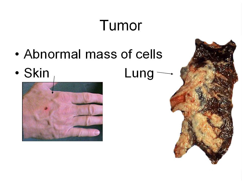
How are cancer cells different from most cells?
Divide excessively and can invade other tissue

9
ESSAY #2 STEM CELLS Cells that can turn into almost any kind of cell (not yet differentiated). Formed a few days after an egg and sperm join.

10
Stem Cell Animations "How Embryonic Stem Cell Lines are Made" Biology Animation Library :: Dolan DNA Learning Center Stem Cell Animation

11
Essay #2 (cont.) SOURCES Umbilical cord blood Fetal tissue
Adult bone marrow Embryonic stem cells

12
Essay #2 (cont.) PROS treat disorders like spinal damage, Parkinson’s disease, leukemia Make heart and nerve tissue in the lab

13
Essay #2 CONS Use embryos (kill them) Don’t have a lot of success yet
Who funds it (private v. government)
")
14
1. Sexual and Asexual ASEXUAL One parent Two Identical offspring
Two parents 4 different offspring

15
2. Chromatin, chromosomes, chromatids (all DNA + protein)
Interphase – loose chromatin Prophase –tightly coiled sister chromatids form through metaphase Anaphase + Telophase –sister chromatids separate to single chromosomes

16
3. Nucleosomes and Histones
8 histone proteins are wrapped with chromosomes to tightly coil into chromatids Histones + chromosomes = nucleosome

17
4. Asexual Reproduction Prokaryotes (no nucleus) Binary fission
Eukaryotes (nucleus) mitosis
 mitosis.")
18
5. Phases of Mitosis PMAT Prophase Metaphase Anaphase Telophase

19
6. Nuclear Envelope Changes
Prophase = nuclear envelope dissolves Telophase = nuclear envelope reforms

20
7. Diff Cytokinesis = Division of the cytoplasm Mitosis
= Division of nucleus

21
8. Cytokinesis Animal Cells Cleavage furrow Plant Cells Cell Plate

22
9. G1, S, G2 Interphase includes G1, S, and G2
G1 = organelle growth, and growth of cell S = DNA synthesis (replication) G2 = centriole and spindle growth, and growth of cell
 G2 = centriole and spindle growth, and growth of cell.")
23
10. Locate on a dividing cell:
Chromatids Centrioles Centromeres Spindle fibers Asters

24
10. Locate on a dividing cell:
Chromatids Centromeres Centrioles Spindle fibers Asters

25
centrioles chromatids Asters Spindle fibers centromeres

26
11. What makes chromatids move to poles?
Contraction of spindle fibers

27
12. What are cyclins (and Cdk’s)?
Protein regulators of the cell cycle

28
13. Cells Dividing A lot Not after formed Blood Skin Nerve
Digestive tract Not after formed Nerve muscle

29
13. B Cancer Cells Cancer cells due to an abnormal cell cycle
Cells grow abnormally and do not stop, even if there are too many Breast cancer cells

30
What phase? Chromatin thickens? Prophase Nuclear envelope disappears
Nuclear envelope reappears telophase

31
What phase? Centrioles move to opposite poles Prophase
Spindle fibers form Cell plate forms Cytokinesis

32
Which phase? Chromosomes line up at the equator metaphase
Cytoplasm divides Cytokinesis Nucleoli break down Prophase Nucleoli reform Telophase

33
15. As the cell increases in size
The surface area to volume ratio decreases

34
16. Why do cells divide? cell membrane could not keep up with bringing in enough oxygen/nutrients DNA can’t keep up

35
17. How many chromosomes Are in each human body cell? 46

36
18. How many times is the reduction
In length of the chromatid than it is in the chromosome form? 10,000 times

37
19. A cell spends what % of time in interphase?
90%

38
What is the purpose of p53? It is the tumor suppressor gene.
It checks that the DNA is OK. If not, it repairs it or kills the cell. IF it is faulty, it leads to a lot of cancer.

39
How does a cell respond to growth
When it comes in contact with other cells? Stops growing

Similar presentations


: The Process of Cell Division>")


