Download presentation
Presentation is loading. Please wait.

1
SOUTH AUSTRALIAN MATERNAL SERUM ANTENATAL SCREENING (SAMSAS© ) PROGRAM
Robert Cocciolone, Head, Antenatal Screening, WCH Genetics & Molecular Pathology Directorate, SA Pathology

2
“providing obstetric support”
Robert Cocciolone Khoa Lam Renata Bird Chris Rothe Lyn Raniolo “providing obstetric support” Eva Martin Gerry Slack

3
SOUTH AUSTRALIAN MATERNAL SERUM ANTENATAL SCREENING PROGRAM (SAMSAS© )
Longest running program in Australia, Neural Tube Defect (NTD) screening -1990/91 Down syndrome & other pregnancy pathologies(15-20wks) June 2001 First Trimester Down syndrome screening (10-14wks) April 2009 Integrated Testing in Second Trimester (9-20wks) Develop and manage own software and algorithms Service SA, TAS, NT with screening service support to PMH in WA Integrated with Neonatal Screening database Electronic access to Cytogenetic and Ultrasound reports Yearly audits published in the SA Birth Defects Register Report
 screening /91 Down syndrome & other pregnancy pathologies(15-20wks) June 2001 First Trimester Down syndrome screening (10-14wks) April 2009 Integrated Testing in Second Trimester (9-20wks) Develop and manage own software and algorithms. Service SA, TAS, NT with screening service support to PMH in WA. Integrated with Neonatal Screening database. Electronic access to Cytogenetic and Ultrasound reports. Yearly audits published in the SA Birth Defects Register Report.")
4
Google SAMSAS www.wch.sa.gov.au/samsas.html www.wch.sa.gov.au Services
A for Antenatal Screening or B for Birth Defects Register Google SAMSAS

7
about Maternal Serum Screening
MSS is offered as a program with access to pre & post test information, genetic counselling and diagnostic services – cvs, amnio & ultrasound. Information for Health Professionals about Maternal Serum Screening

8
SAMSAS©

9
Kit based fluoroimmunoassays ~ DELFIA system

11
Screening “the systematic application of a test procedure to identify individuals at sufficient risk to warrant diagnostic investigations” CVS 12 wks Amniocentesis 15+wks Morphology Ultrasound 18+wks Aim is to maximise detection of affected pregnancies and minimise false +ves

12
SAMSAS© Markers; Other Markers; Maternal age Gestational age
1st Trimester nd Trimester Integrated Test AFP AFP free hCG free hCG free hCG uE3 uE3 Papp-A Papp-A Nuchal Nuchal Translucency Translucency Other Markers; Maternal age Gestational age Other Variables

14
Fetus must occupy 3/4 of the image
Fetus must be in a Neutral position Hyperextension of fetal neck can increase the NT by 0.6 mm Flexion of the neck can decrease the NT by 0.4 mm Umbilical cord round the fetal neck ( 5 -10% of cases) can increase the NT by 0.8 mm Amniotic membrane and Nuchal membrane must be separate Measure the max thickness of the subcutaneous translucency
 can increase the NT by 0.8 mm. Amniotic membrane and Nuchal membrane must be separate. Measure the max thickness of the subcutaneous translucency.")
15
Increased nuchal translucency and exomphalos in a trisomy 18 fetus at 12 weeks of gestation

16
Turners syndrome - cystic hygroma

17
Severe asymmetrical growth restriction in a 13-week fetus with triploidy.

18
- NT is an ultrasonographic feature visible in 1st Tr of
NUCHAL TRANSLUCENCY - NT is an ultrasonographic feature visible in 1st Tr of pregnancy - NT results from an accumulation of fluid at the base of fetal neck NT thickness increases with GA (0.8 to 1.6 mm) Non specific marker, thickness >2.5(3.0) mm associated with an increased risk of aneuploidy, cardiovascular & pulmonary defects, skeletal dysplasia, renal, metabolic defects & congenital infections & fetal demise.
 Non specific marker, thickness >2.5(3.0) mm associated with an increased risk of aneuploidy, cardiovascular & pulmonary defects, skeletal dysplasia, renal, metabolic defects & congenital infections & fetal demise.")
19
Variables Population Medians / MoM Gestational Age Maternal Age
Recurrence Risk Singleton vs Twins Maternal Weight Ethnicity Diabetes Smoking Analytical Imprecision Risk Calculation ~ NT ~ Biochemistry ~ Combined ~ Integrated OUTCOME

21
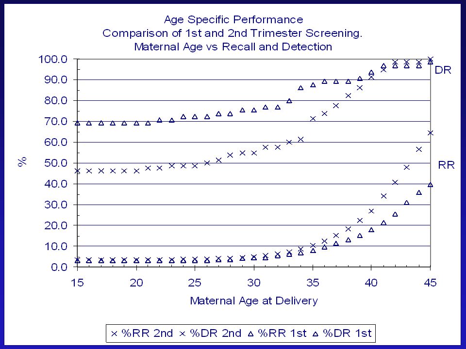
Effectiveness of different methods of screening
Screening for trisomy 21 Effectiveness of different methods of screening 100,000 pregnancies Method of screening Number detected Detection rate Screen positive * 5% N=5,000 Trisomy 21 N=200 * 2.5% Screen positive Integrated Test Maternal age 60 30% Serum biochemistry at 16 wks 130 65% Nuchal translucency (NT) at 12 wks 75% 150 Fetal NT & ß-hCG & PAPP- A at 12 wks 180 *90% Nicolaides KH. Fetal nuchal translucency. Am J Obstet Gynecol 2004
 at 12 wks. 75% 150. Fetal NT & ß-hCG & PAPP- A at 12 wks *90% Nicolaides KH. Fetal nuchal translucency. Am J Obstet Gynecol")
22
SAMSAS© Maternal Age Screening alone
Maternal Age Screening alone Second trimester biochemical screening First trimester combined screening Integrated Screening Amniocenteses performed per case detected 200 50 15 8 Fetal loss per case of Down syndrome detected 1 : 1 1 : 4 1 : 13 1 : 25 Procedure related fetal loss rates are difficult figures to obtain. Corrections need to be made for the background spontaneous fetal loss.

23
Background Risk Gestational age 20 40 60 80 100 10 15 25 30 35
65% Trisomy 21 20 40 60 80 100 10 15 25 30 35 15% Trisomy 13 12% Trisomy 18 <1% Triploidy 95% 47xxx/xxy/xyy 20% 45x % Nicolaides et al., The week scan, London 1999 Snijders et al 1995

24
Background Risk Maternal age 0.0001 0.001 0.01 0.1 1 10 20 25 30 35 40
44 Years Risk % Trisomy 21 Trisomy 18 Trisomy 13 xxx/xxy/xyy 45x Triploidy Nicolaides et al., The week scan, London 1999 Snijders et al 1995

25
Previous Chromosomal Abnormality
Assessment of Risk Previous Chromosomal Abnormality Trisomy 21 Trisomy 18 Trisomy 13 } + 0.75% Age Risk } 45XO 47XXY/XXX Triploidy

26
Marker Levels Change Significantly with Gestation
Measured levels are converted to Multiples Of the Population Median or MoM values. 1 MoM = Reference for all markers beta hCG at 10 wks GA Patient IU/L = 2 multiples Median IU/L MoM values are independent of gestational age and concentration Units LogMoM values are used in calculations as they exhibit a Gaussian distribution ( Mean +/- SD)
")
28
Adjustments AFP_MoM 1st AFP_MoM 2nd Beta_MoM 1st Beta_MoM 2nd PappA_MoM Ue3_MoM Risk Odds IVF /1.08 /1.09 /0.89 /0.92 Race Caucasian Aboriginal /0.9 /0.8 /1.06 Race South Asian /0.94 /0.925 /0.91 /1.082 /1.07 Indian, Pakistani, Thai, Malyasian Oriental /1.061 /1.093 Chineses, Korean, Japanese Afro-Caribbean /1.17 /1.107 /1.553 /0.99 Diabetes IDDM /0.96 /0.79 /0.93 Smoker /1.03 /0.81 Twins /2.13 /2.10 /1.9 /1.86 /1.67 Previous T21 +0.75% to T21 Risk odds Previous T18/T13 +0.75% to T18 Risk odds Previous T21 + T18/T13 +0.75% to both odds

29
Maternal Weight Noveux 1996, Graaf/Cuckle 2000, SAMSAS/Murdoch 2002

30
SAMSAS© Increasing Incidence of Twins (1990–2003) 1:70 to 1:55
* Assisted reproduction * Rate of twinning increases with age. >20% (2006) of pregnancies are now to women 35yrs or over, up from <9% in 1990.
 of pregnancies are now to women 35yrs or over, up from <9% in")
31
Twins Management Screening and Chorionicity
Nuchal discordance (mono and dichorionic) Diagnostic tests (CVS and Amniocentesis) Selective feticide Twin to twin transfusion Laser ablation of vessels Delivery
 Diagnostic tests (CVS and Amniocentesis) Selective feticide. Twin to twin transfusion. Laser ablation of vessels. Delivery.")
35
Diandric Digynic

36
(GA Overestimate) Diandric Digynic
 Diandric Digynic")
38
Other Not NTD & Not DS: AFP < 2 MoM & DS risk is < 1 in 300
RISKS Open Neural Tube Defects (NTD) Down syndrome / Aneuploidy Other NTD Down syndrome / Aneuploidy 2nd Trimester 1st & 2nd Trimester ↑ AFP ≥ 2 MoM ↑ Risk ≥ 1 in 300 Independent of Maternal Age Age Dependent Morphology scan CVS / Amnio ~ 1/30 will have a NTD ~ 1/20 or 1/40 will have DS Other Not NTD & Not DS: AFP < 2 MoM & DS risk is < 1 in 300 But Biochemical results fall outside the Normal expected.
 Down syndrome / Aneuploidy. Other. NTD Down syndrome / Aneuploidy. 2nd Trimester 1st & 2nd Trimester. ↑ AFP ≥ 2 MoM ↑ Risk ≥ 1 in 300. Independent of Maternal Age Age Dependent. Morphology scan CVS / Amnio. ~ 1/30 will have a NTD ~ 1/20 or 1/40 will have DS. Other Not NTD & Not DS: AFP < 2 MoM & DS risk is < 1 in 300. But. Biochemical results fall outside the Normal expected.")
39
Not NTD & Not Downs Profiles
SAMSAS screened pregnancies N=62,563 1st Trimester N= 26,914 2nd Trimester N= 35,649 Profile Non Downs N= 206 (0.77%) Profile Not NTD Not Downs N= 123 (0.35%) Total N = 329 (0.53%) OUTCOMES NOT KNOWN N = 25 (7.6%) NORMAL N = 103 (31.3%) FETAL DEATH N = 171 (52%) ABNORMAL N = 30 ( 9.1%) 9 x Triploidy, 9 x T18 3 x T15, 3 x Anenceph.1st Tr 2 x Turners, 2 x Mult. Abn. 2 x Metabolic
 Profile. Not NTD Not Downs. N= 123 (0.35%) Total N = 329 (0.53%) OUTCOMES. NOT KNOWN. N = 25 (7.6%) NORMAL. N = 103 (31.3%) FETAL DEATH. N = 171 (52%) ABNORMAL. N = 30 ( 9.1%) 9 x Triploidy, 9 x T18. 3 x T15, 3 x Anenceph.1st Tr. 2 x Turners, 2 x Mult. Abn. 2 x Metabolic.")
40
Not viable at time of screen. No NT measured
Not Known= 4 Normal= 2 (Obese~no NT) Fetal Death= 137 Abnormal= 6 (1xTriploidy,2xXO,2xT15,1xAnencephaly) Anomalies Found 143/145 Odds 1/1 ( 98.6%) N= 24 Not Known= 2 Normal= 0 Fetal Death= 18 Abnormal= 4 (2xT18,1xAnencephaly,1xMultiple Con. Abn.) Anomalies Found 22/22 Odds 1/1 (100%)
 Fetal Death= 137. Abnormal= 6 (1xTriploidy,2xXO,2xT15,1xAnencephaly) Anomalies Found 143/145. Odds 1/1 ( 98.6%) N= 24. Not Known= 2. Normal= 0. Fetal Death= 18. Abnormal= 4 (2xT18,1xAnencephaly,1xMultiple Con. Abn.) Anomalies Found 22/22. Odds 1/1 (100%)")
41
Viable at time of screen. Viable at time of screen.
Not Known= 12 Normal= 62 Fetal Death= 13 Abnormal= 12 (4xTrip.,6xT18,1xMultiple Con. Abn.,1xMetabolic) Anomalies Found 25/87 Odds 1/3.5 ( 28.6%) N= 57 Not Known= 7 Normal= 39 Fetal Death= 3 Abnormal= 8 (4xTriploidy,1xT18,1xOther,1xAnencephaly,1xRenal) Anomalies Found 11/50 Odds 1/4.5 ( 22%)
 Anomalies Found 25/87. Odds 1/3.5 ( 28.6%) N= 57. Not Known= 7. Normal= 39. Fetal Death= 3. Abnormal= 8 (4xTriploidy,1xT18,1xOther,1xAnencephaly,1xRenal) Anomalies Found 11/50. Odds 1/4.5 ( 22%)")
42
Detected Detected

43
The best way to detect these pregnancies is through discriminatory algorithms and distributions , utilizing multiple markers. Y= 0.425*ln_beta_MoM – 0.631*ln_Papp-a_MoM *ln_NT_MoM False -ves

44
What does a risk report mean?
Reassurance of 99.8 % & Anomaly Risk of 2.0 – 25% Summary data ~ SAMSAS yearly audits SA Birth defects Register Reports

45
DS Risk = Mat. Age Risk x LR

46
LRaff = h2/h1 Unaffected Affected h2 Detection h1 Rate False -ve
Y= 0.425*ln_beta_MoM – 0.631*ln_Papp-a_MoM *ln_NT_MoM Unaffected Affected h2 Detection Rate h1 False -ve False +ve’s

47
DS Risk =Mat. Age Risk × LR
SAMSAS© DS Risk =Mat. Age Risk × LR 20 yrs = 1 in × 2 = 1 in 800 30 yrs = 1 in × 2 = 1 in 550 35 yrs = 1 in 500 × 2 = 1 in 250 40 yrs = 1 in 156 × 2 = 1 in 78 45 yrs = 1 in 40 × 2 = 1 in 20

49
BENEFITS OF AN EARLY SCAN
SAMSAS© BENEFITS OF AN EARLY SCAN confirms the fetus is alive permits accurate dating of pregnancy allows early diagnosis of multiple pregnancy & chorionicity detects major structural abnormalities and missed abortion

50
Why did we introduce 1st trimester combined screening into SA ?
SAMSAS© Why did we introduce 1st trimester combined screening into SA ?

51
Aust N Z J Obstet Gynaecol 2008; 48: 492-500
Down Syndrome Detect Recall %35 %PG Age 2nd Tr % 4.9% 9% % % 6.6% 15% % % 7.4% % % 30 SAMSAS© Primigravida 1st Tr % % % IntG % % % Combining first and second trimester markers for Down syndrome screening: Think twice Robert Cocciolone, Kate Brameld, Peter O'Leary, Eric Haan, Peter Muller, Karen Shand Aust N Z J Obstet Gynaecol 2008; 48:

52
Advantages of 1st trimester screening
benefits of an early scan less false +ves & -ves less normal fetuses lost higher detection personal benefits to patients from earlier diagnosis Disadvantages cost of the nuchal translucency scan logistically more difficult to manage

53
Logistical considerations?
1TR & 2TR screening will coexist. Management of reports & requests. How many risks, by whom and when? Audits and ongoing programme evaluation.

54
How can these be addressed?
Centralised service & database for, - all patient demographics - biochemical results - NT measurements & providers - reporting - recalculation of risks - retrievable data for analysis Logical software - risks linked to gestation (1TR & 2TR algorithms)
")
55
Trends in state/based Down syndrome screening and invasive prenatal testing with the introduction of first trimester combined Down syndrome, South Australia Muller PR, Cocciolone R, Haan EA, et al Am J Obstet Gynecol 2007;196:315.e1-315.e7.

56
Utilization of maternal serum Down syndrome screening % of all confinements
69-79% NS

57
Utilization of second trimester maternal serum (∆) and first trimester combined Down syndrome screening (□), % of all confinements * P < 0.001 75% 49% * * 25% 0.8%

58
% confinements ≥ 35 years (•) of age vs % of confinements undergoing invasive prenatal tests (∆)
9.3% * 7.6%

59
Confinements ≥ 35 years of age undergoing invasive prenatal tests
43% * 24.8% * P < 0.001

60
Number of invasive prenatal tests to detect one DS fetus
Year Down syndrome cases Rate of DS per invasive procedure* 1995 32 1/86 1996 1/94 1997 41 1/83 1998 46 1/61 1999 42 1/71 2000 37 1/79 2001 36 1/90 2002 44 1/66 2003 1/46 2004 34 1/49 2005 47 1/40 * * p < 0.001

61
Number of invasive prenatal tests to detect one aneuploid fetus
Year Aneuploidy cases Rate of aneuploidy per invasive procedure* 1995 51 1/35 1996 46 1/47 1997 50 1/38 1998 65 1/31 1999 60 1/33 2000 56 1/32 2001 55 1/30 2002 2003 64 1/24 2004 1/21 2005 91 1/15 * * p < 0.001

62
Overall Prenatal Detected Down Syndrome cases (%)
Year Down syndrome cases Prenatal detected DS cases (%)** 1995 32 71.9 1996 81.3 1997 41 70.7 1998 46 73.9 1999 42 71.4 2000 37 70.3 2001 36 58.3 2002 44 65.9 2003 81.0 2004 34 88.2 2005 47 83.0 ** p = 0.21
** ** p =")
63
Demonstrated an improved efficiency in the utilization of invasive prenatal tests
Despite the increase in gravid women ≥ 35 years of age the number of invasive prenatal tests in this age group has significantly declined Despite the significant decrease in invasive prenatal tests the overall antenatal detection of Down syndrome did not decrease, and appears to increase once the utilization of first trimester combined Down syndrome screening reaches > 30% of confinements

64
To review changes in the utilization and effectiveness of state/population-based antenatal screening for NTDs in South Australia from 1986 to 2004

66
Overall antenatal detection of NTD(%)
Year Average births/Yr (#) Average NTDs/Yr Overall antenatal detection of NTD(%) 19,714 41 86 19,437 32* 88.8 17,867 24 94.5 ** *State-wide educational drive on the benefits of Folic Acid supplementation for the reduction of NTDs 1994 ** p<0.001
 Average NTDs/Yr. Overall antenatal detection of NTD(%) , , * , ** *State-wide educational drive on the benefits of Folic Acid. supplementation for the reduction of NTDs ** p<")
67
Despite a significant decrease in the utilization of MSAFP screening, the population-based detection of NTDs has increased significantly in South Australia. The decreased utilization of second trimester MSAFP represents improve clinician confidence in second trimester ultrasound for the detection of NTDs

68
….future directions SAMSAS© new biochemical markers
fetal cells in maternal circulation fetal DNA in maternal circulation complex protein profiles - mass spectrometry

69
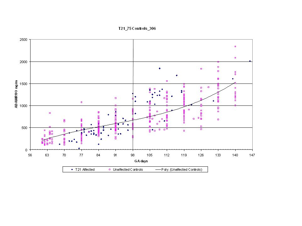
ADAM12, a disintegrin and metalloprotease
The superfamily of zinc peptidases – Metzincins Reprolysins/Adamalysins SVMPs (snake venom proteases) ADAMs (a disintegrin and metalloprotease) ADAMTS (with thrombospondin motifs) Matrix metalloproteases (MMPs) Astacins Serralysins ADAM12 is a multidomain protein with protease, cell adhesion, fusion and signaling activities. ADAM12 is a member of the large family of ADAM12 proteins, of which more than 30 have been identified. 19 ADAM genes are currently known in humans. Human ADAM12 is encoded by a single gene located on chomosome 10q26.3, where ADAM8 also resides. ADAM12 gene has 2 distinct splice variants, producing the long ADAM12-L form and the shorter ADAM12-S form: ADAM12-S consists of 718 amino acids (secreted form), ADAM12-L consists of 881 amino acids (prototype transmembrane form) ADAM12 is one of only a few ADAMs that exist in two forms Note that ADAM12 cannot be measured from EDTA plasma!
 ADAMs (a disintegrin and metalloprotease) ADAMTS (with thrombospondin motifs) Matrix metalloproteases (MMPs) Astacins. Serralysins. ADAM12 is a multidomain protein with protease, cell adhesion, fusion and signaling activities. ADAM12 is a member of the large family of ADAM12 proteins, of which more than 30 have been identified. 19 ADAM genes are currently known in humans. Human ADAM12 is encoded by a single gene located on chomosome 10q26.3, where ADAM8 also resides. ADAM12 gene has 2 distinct splice variants, producing the long ADAM12-L form and the shorter ADAM12-S form: ADAM12-S consists of 718 amino acids (secreted form), ADAM12-L consists of 881 amino acids (prototype transmembrane form) ADAM12 is one of only a few ADAMs that exist in two forms. Note that ADAM12 cannot be measured. from EDTA plasma!")
70
ADAMs are focus in asthma, alzheimer and cancer research
In humans ADAM mRNA is present at low levels in most adult tissues human placenta expresses very high levels of ADAM12 A large proportion of human carcinomas express ADAM12 breast carcinoma tissue and urine of breast cancer patients, liver metastases of colon carcinoma Overall, it appears that ADAMs are mainly expressed during growth and development Note that our license covers Maternal Health applications

72
ADAMs are of high research focus
pro- metalloprotease disintegrin transmembrane cytoplasmic cysteine-rich EGF-like Several studies has shown tha ADAMS are mainly located inside cells Translocation from intracellular storage to the cell surface might be regulated ADAM12 metalloprotease is activated inside the cell unlike matrix metalloproteases which are secreted as proforms and become activated outside the cell ADAM12-S cleaves IGFBP-3 and IGFBP-5 via its cysteine-rich domain, which may regulate bioavailability of IGF (PAPP-A is a protease for IGFBP-4) ADAM12 releases soluble HB-EGF acivating epidermal growth factor and thus promotes cardiac hypertrophy ADAM12 interacts with integrin and syndecan adhesion receptors ADAM12 has one of the longest cytoplasmic domains compared to other ADAMs; the tail is at least involved in the regulation of ADAM12 localization (inside the cell or at the cell surface) 19 genes in the human
 ADAM12 releases soluble HB-EGF acivating epidermal growth factor and thus. promotes cardiac hypertrophy. ADAM12 interacts with integrin and syndecan adhesion receptors. ADAM12 has one of the longest cytoplasmic domains compared to other ADAMs; the tail is at least involved in the regulation of ADAM12 localization (inside the cell or. at the cell surface) 19 genes in the human.")
Similar presentations


>")


