Download presentation
Presentation is loading. Please wait.

1
X-rays and image production

2
What are x-rays? They are high energy photons. Typically 0.01nm.Way beyond ultraviolet. 700nm=infra red 400nm=blue. Gamma rays are naturally occurring radiation in the same range.

3
How do we produce x-rays?
Accelerate electrons at high energies (typically at over 70 kilo electron volts>70KeV ) into a tungsten target. 99% of the energy is released as heat and light.1% of energy is released as electromagnetic radiation in the desired range (useful x-rays). This energy is produced by the braking electron as they interact with other electrons convert their energy into electromagnetic radiation. Mean energy of the x-ray photons is 1/2-1/3 of KeV. Output is proportional Zx(KV)2
 into a tungsten target. 99% of the energy is released as heat and light.1% of energy is released as electromagnetic radiation in the desired range (useful x-rays). This energy is produced by the braking electron as they interact with other electrons convert their energy into electromagnetic radiation. Mean energy of the x-ray photons is 1/2-1/3 of KeV. Output is proportional Zx(KV)2.")
4
How are x-rays attenuated?
This occurs by absorption and scattering of the x-rays. Compton Scatter.-interaction with free electrons. Most of the electrons in Carbon, Nitrogen and Oxygen are free. The photons are essentially scattered. The higher the energy of the photons, the more the scattering is in a forward direction. Dependant on Z. Photoelectric effect.-the energy of the photons displaces electrons in the K shell and is absorbed. In soft tissue PE=Compton in image contribution at about 30KeV Photoelectric absorption increases tissue contrast but at a higher dose to the patient. Apart from mammography most diagnostic tests involve x-ray generated at 70 KeV or more. The amount of signal generated at the receptor (or film blackening) is proportional to (KV)2.mA Therefore if you have a poor image and there is a marked difference between radio density of the tissue being differentiated (e.g. insertion of pacing wires), then turn up the KV not the mA (less dose to the patient).
 is proportional to (KV)2.mA. Therefore if you have a poor image and there is a marked difference between radio density of the tissue being differentiated (e.g. insertion of pacing wires), then turn up the KV not the mA (less dose to the patient).")
5
Forming the image Traditionally, the x-rays fix silver bromide crystal on the film to produce a radiograph (not x-ray). In fact the x-rays activate fluorescent screens, which give off light, which then interact with the silver bromide crystals. In digital films the x-rays activate multiple small detectors to produce an image. You should end up with an image which allows you to differentiate between bone, fat, soft tissue and gas

7
Anyone can tell you that the bowel is dilated.
But your knowledge of normal anatomy tells you that the haustral pattern is abnormal. If you look at the soft tissue around the bowel you can tell that bowel wall is thickened.

9
Toxic effects on patients
Non stochastic (dose related). These should all be avoided in diagnostic radiology. Damage to skin, loss of hair, cataracts, radiation sickness. Stochastic (probability related). Carcinogenesis Increased probability with increased dose. Latent period of at least 5-10years.
. These should all be avoided in diagnostic radiology. Damage to skin, loss of hair, cataracts, radiation sickness. Stochastic (probability related). Carcinogenesis Increased probability with increased dose. Latent period of at least 5-10years.")
10
Tissues in order of sensitivity
Gonads Breast Bone marrow Lung Thyroid bone

11
We measure the dose in Sieverts
1Sv given to a thousand patients will produce 125 deaths. Chest radiograph mSv (flying to the US is worth about 7CXRs) Lumbar spine mSv IVU mSv Most tests are mSv Barium Enema/CT Abdo-pelvis 8-10mSv (this increases your cancer risk by 1 in 2000 or perhaps slightly higher)
 Lumbar spine 2.15mSv. IVU 5mSv. Most tests are 0.5-5mSv. Barium Enema/CT Abdo-pelvis 8-10mSv (this increases your cancer risk by 1 in 2000 or perhaps slightly higher)")
12
Computerised Axial Tomography

15
King Tut having a CT scan

16
A fan of x-ray beams of high KV is produced
A fan of x-ray beams of high KV is produced. Essentially solve lots of simultaneous equations along the line of x-ray beam. In fact use a process of back projection and Fourier analysis.

17
CT IMAGES Typically at >150KeV. A lot of Compton scatter.
Essentially we divide the field being imaged into lots of voxels (little cubes) and each tiny cube is allocated an attenuation value. We solve loads of equations to work out each attenuation value. Typically 512 x512, occasionally 1024 by 1024. The spatial resolution is less than what is seen on plane film.
 and each tiny cube is allocated an attenuation value. We solve loads of equations to work out each attenuation value. Typically 512 x512, occasionally 1024 by The spatial resolution is less than what is seen on plane film.")
18
Windowing and Houndsfield units
Each voxel is given a value. Densest bone (petrous temporal)-3000Hu. Soft tissue typically 30-50Hu. Water 0 Hu. Fat -50 to-100Hu. Air-1000Hu. The problem is displaying all these values in an image that we can interpret. The human eye can only use black through gray to white to show what we are interested in. We pick an appropriate level and window width. We deal with this problem by using different settings to interrogate different areas of interest.
-3000Hu. Soft tissue typically 30-50Hu. Water 0 Hu. Fat -50 to-100Hu. Air-1000Hu. The problem is displaying all these values in an image that we can interpret. The human eye can only use black through gray to white to show what we are interested in. We pick an appropriate level and window width. We deal with this problem by using different settings to interrogate different areas of interest.")
19
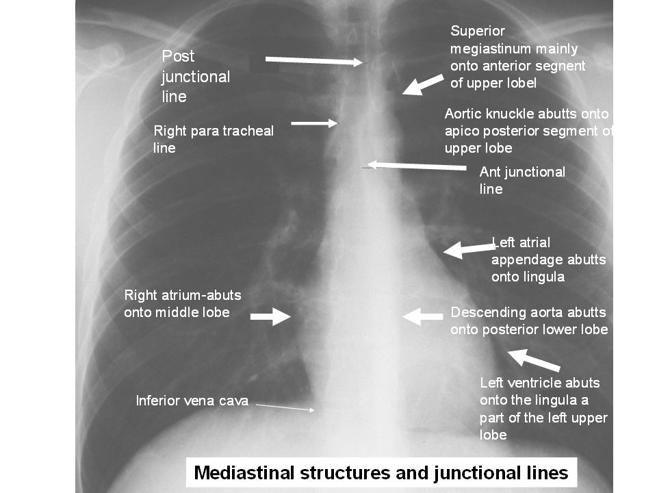
Looking at the mediastinum and soft tissue of the chest.
What is a typical value of soft tissue? Answer 30-40Hu,set this as our level. What is the range of values we are interested in? 150 would be enough. 40+150/2=115 which is bony. 40-150/2=-35 which is fat. This would be useless for the lungs as they look uniformly black. This would be useless for bone as it would al uniformly appear white.

20
A B C T3

22
Left major fissure more posterior

23
Left pulm artery arching over the Left main bronchus
Right main pulm artery travelling in front of Right main bronchus

Similar presentations


 and her.>")


