Download presentation
Presentation is loading. Please wait.

1
Cathe management of Acute Coronary Syndrome

2
Outline : -Objective - Statistics -Atherosclerosis -Pathophysiology -Risk factor -Acute coronary syndrome -Angina pectoris -Pathophysiology principle -Classification of angina -cardiac catheterization -Use of cardiac catheterization -Technique of cardiac catheterization - Type of cardiac catheterization -Nursing intervention of cardiac catheterization -Self management after cardiac catheterization

3
Objective : 1-Describe the pathophysiology of atherosclerosis
Objective : 1-Describe the pathophysiology of atherosclerosis Describe the risk factors Determine the acute coronary syndrome 4- Determine the angina and the classification of angina 5- determine the cardiac catheterization 6- describe the technique and the type oa cardiac catheterization

4
Statistics Cardiovascular diseases: number one cause of death globally In 2005: an estimated 17.5 million people died from cardiovascular disease, 30 % of all global deaths. Of these deaths, 7.6 million were due to heart attacks and 5.7 million due to stroke. About 80% of these deaths occurred in low- and middle- income countries. By 2015 an estimated 20 million people will die from cardiovascular disease (mainly from heart attacks and strokes).
.")
5
Atherosclerosis A major cause of cardiovascular disease.
A complex, insidious process, beginning long before symptoms occur.

6
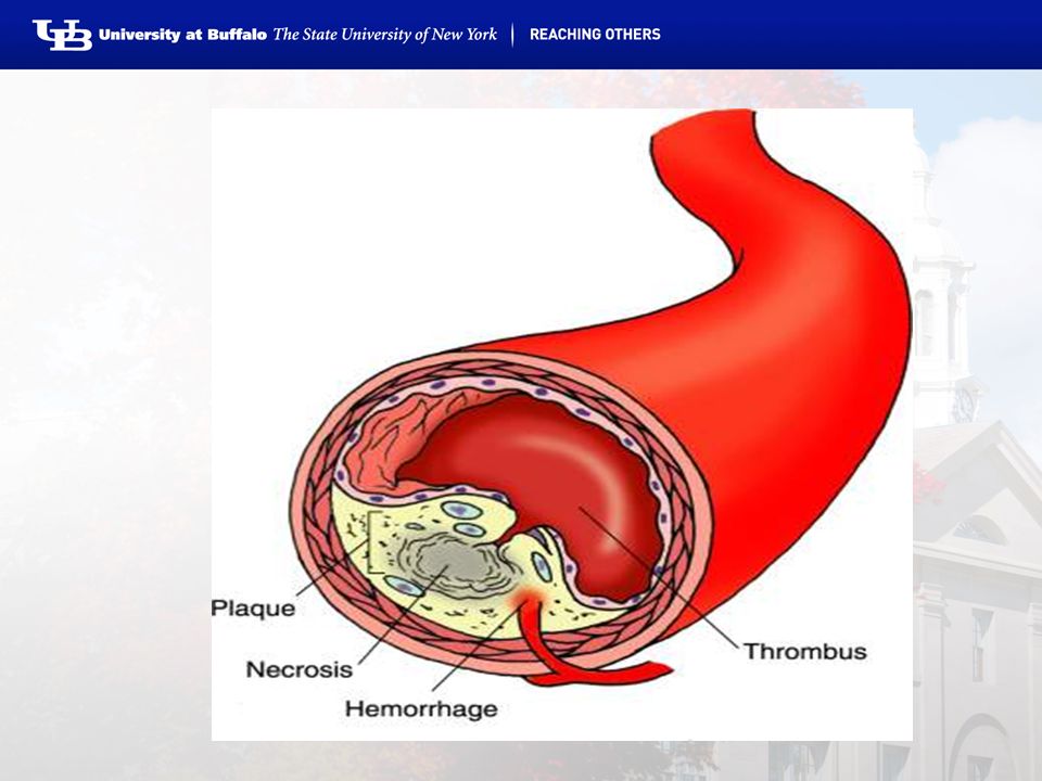
Pathophysiology Injury to endothelium
Increased levels of cholesterol/triglycerides Hypertension Cigarette smoking Deposits in the lining of the artery Cholesterol, cellular waste, calcium, and fibrin Atheroma (lipid plaque with a fibrous covering) Keeps building to partial or complete blockage
 Keeps building to partial or complete blockage.")
8
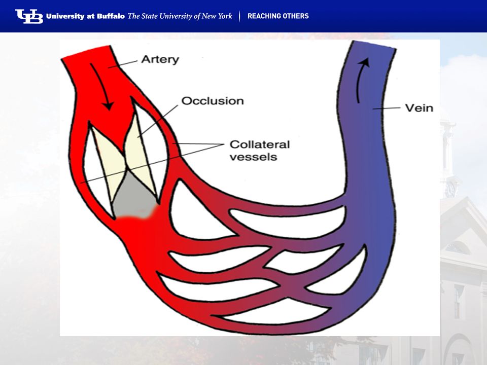
Fibrous plaques are most often found in the coronary, popliteal, and internal carotid arteries and in the abdominal aorta. Because of the fibrous plaque, the amount of blood flow through the artery is reduced, resulting in decreased supply of oxygen to tissues. Symptoms often do not occur, however, until 75% or more of the blood supply to the area is occluded. The occurrence of symptoms may depend to an extent on the development of collateral circulation.

10
Risk Factors The cause of atherosclerosis is not clearly known
Major Risk Factors Non-modifiable Age Heredity Combination of environmental and genetic influences Race Rates of hypertension, obesity, and diabetes Sex Modifiable Cigarette smoking even second hand High cholesterol-LDL vs. HDL Hypertension Physical inactivity Obesity Diabetes mellitus

11
Contributing Risk Factors
Stress (smoking, overeating) Sex hormones (Female hormones and HDL) Birth Control Pills (old vs new) (smoking & hypertension)
 Sex hormones. (Female hormones and HDL) Birth Control Pills (old vs new) (smoking & hypertension)")
12
Contributing Risk Factors
Excessive Alcohol Intake (hypertension, heart failure, stroke, high triglycerides, and dysrhythmias) Homocysteine Levels (increased platelet adhesiveness, enhances LDL deposition in the arterial wall, and activates the coagulation cascade)
 Homocysteine Levels. (increased platelet adhesiveness, enhances LDL deposition in the arterial wall, and activates the coagulation cascade)")
13
Acute coronary syndrome
A relatively new term used to describe patients who have clinical symptoms compatible with acute myocardial ischemia. Acute coronary syndrome includes unstable angina and acute MI.

14
Angina Pectoris Is the term used to describe chest pain or discomfort that results from coronary artery disease. The patient may describe the sensation as: pain, pressure, fullness, squeezing, or heaviness.

15
Pathophysiological Principles
Caused by transient, reversible myocardial ischemia. Myocardial oxygen demand ≠ myocardial oxygen supply Most commonly, decreased oxygen supply Atherosclerotic narrowing Dynamic obstruction: intense focal spasm of a coronary artery Arterial inflammation (arterial narrowing, plaque destabilization, rupture, and thrombogenesis) A marked increase in oxygen demand (fever, tachycardia, and thyrotoxicosis)
 A marked increase in oxygen demand (fever, tachycardia, and thyrotoxicosis)")
16
Classification of Angina
Stable – chronic stable angina, classic angina Paroxysmal, occurs with physical exertion (predictable) Relieved by rest or nitroglycerin
 Relieved by rest or nitroglycerin.")
17
Classification of Angina
Unstable – preinfarction angina or crescendo angina Unexpected chest pain or discomfort that usually occurs while at rest. More prolonged and severe Need to be treated immediately: risk for acute MI, cardiac dysrhythmias, or cardiac sudden death Variant – Prinzmetal’s angina, vasospastic angina Result of coronary artery spasm Occurs at rest (a form of unstable angina) Severe coronary atherosclerosis!!
 Severe coronary atherosclerosis!!")
18
Cardiac catheterization
Introduction : is an invasive diagnostic procedure in which radiopaque arterial and venous catheter are introduced into selected blood vessels of the right and left sides of the heart . -Catheter advancement is guided by fluoroscopy . -Most commonly , the catheters are inserted percutaneously through the blood vessels ,or via cutdown procedure if the patient has poor vascular access -pressure and oxygen saturation level in the four heart chambers are measured .

19
Use of cardiac catheterization : is most frequently used to diagnose CAD , assess coronary artery patency , and determine the extent of atherosclerosis and determine whether revascularization procedures , including PCI or coronary artery bypass surgery , may be of benefit to the patient . -Is also used to diagnose pulmonary arterial hypertension or to or to treat stenotic heart valves via percutaneous balloon valvuloplasty

20
The technique of cardiac catheterization :
-during cardiac catheterization , the patient has one or more IV lines in place for the administration of sedatives , fluid , heparin , and other medications . -The site of the catheter insertion in the femoral or radial artery , the patients anticoagulation status and other variable (eg, advanced age , obesity , bleeding disorder ) . -The use of smaller (4 or 6 fr ) catheter is associated with shorter recovery times . Several options to achieve arterial hemostasis after catheter removal , including manual pressure , mechanical compression devices such as the femostop (placed over puncture site for 30 minutes ) and percutaneously deployed devices , are used .
 . -The use of smaller (4 or 6 fr ) catheter is associated with shorter recovery times . Several options to achieve arterial hemostasis after catheter removal , including manual pressure , mechanical compression devices such as the femostop (placed over puncture site for 30 minutes ) and percutaneously deployed devices , are used .")
21
Cont: the product are placed over the puncture site as the catheter is removed and manual pressure is applied for 4 to 10 minutes . - patient hospitalized for angina or acute MI who require cardiac catheterization usually return to their hospital rooms recovery .

22
Type of cardiac catheterization : 1-Angiography 2-Aortography 3- Coronary arteriography 4- Right heart catheterization 5- left heart catheterization

23
Angiography Cardiac catheterization is usually performed with angiography, technique in which a contrast agent is injected into the vascular system to outline the heart and blood vessel .when a specific heart chamber or blood vessel is singled out for study , the procedure is know as selective angiography . Angiography makes use of cineangiograms , a series of rapidly changing films on an intensified fluoroscopic screen that record the passage of the contrast agent through the vascular site or sites . Recording allows for comparison of data over time . Common site for selective angiography are the aorta , the coronary arteries , and the right and left sides of the heart . Are used to evaluate the degree of atherosclerosis and to determine treatment . They are also used to study suspected congenital anomalies of coronary arteries .

24
Aortography Is a form of angiography that outlines the lumen of the aorta and the major arteries arising from it . In thoracic aortography , a contrast agent is used to study the aortic arch and its major branches . The catheter may be introduced into the aorta using the translumbar or retrograde brachial or femoral artery approach .

25
Coronary arteriography : coronary arteriography involves the introduction of a catheter into the right or left brachial or femoral artery , which is then passed into ascending aorta and manipulated into the right and left coronary arteries .

26
Right heart catheterization : right heart catheterization usually precedes left heart catheterization . It involves passage of a catheter from an antecubital or femoral vein into the right atrium ,right ventricle , pulmonary artery , and pulmonary arterioles . Pressures and oxugen saturation levels from each of these areas are obtained and recorded . Although right heart catheterization is considered relatively safe , potential complication include cardiac dysrhythmias , venous spasm , infection of the insertion site , cardiac perforation , and rarely cardiac arrest .

27
Left heart catheterization : is performed to evaluate the patency of the coronary arteries and the function of the left ventricle and the mitral and aortic valves . Potential complication include dysrhythmias , MI , perforation of the heart or great vessels , and systemic embolization . In this approach , the physician insert the catheter into the right brachial artery or a femoral artery and advances it into the aorta and left ventricle . After the procedure , the catheter is carefully withdrawn and hemostasis is achieved using manual pressure or other techniques previously described . If the physician performed an arterial or venous cutdown , the sutured and a sterile dressing is applied .

28
Nursing intervention : nursing responsibilities before cardiac catheterization include the following : 1- the patient is instructed to fast , usually for 8 to 12 hours ,before the procedure . If catheterization is to be performed as an outpatient procedure ,a friend , family member , or other responsible person must transport the patient home . 2- the patient is informed of the expected duration of the procedure and advised that it will involve lying on a hard table for less than 2 hours . 3- the patient is reassured that IV medication are given to maintain comfort . 4- the patient is informed about sensation that will be experienced during the catheterization . 5- the patient is encouraged to express fears and anxieties . The nurse provides teaching and reassurance to reduce apprehension .

29
Nursing intervention (cont ) : nursing responsibilities after cardiac catheterization may include the following : 1- the catheter access site is observed for bleeding or hematoma formation .peripheral pulses are assessed in the affected extremity (dorsalis pedis and posterior tibial pulses in the lower extremity , radial pulse in the upper extremity ) every 15 minutes for 1 hour , and then every 1 to 2 hours until the pulses are stable . 2- temperature , color ,and capillary refill of the effective extremity are frequently evaluated , per local nursing standards .the patient is assessed for effected extremity pain , numbness , or tingling sensation that may indicate arterial insufficiency . 3- dysrhythmias are carefully screened by observing the cardiac monitor or by assessing the apical and peripheral pulses for change in rate and rhythm . 4- bed rest is maintained for 2 to 6 hours after the procedure . If manual or mechanical pressure is used the patient must remain on bed rest for up to 6 hours with the affective leg straight and the head of the bed elevated no grater than 30 degrees .

30
Cont : 5- the patient is instructed to report chest pain and bleeding or sudden discomfort from the catheter insertion sites promptly the patient is monitored for contrast agent induced nephropathy by observing for elevations in serum creatinine levels patient safety is ensured by instructing the patient to ask for help when getting out of bed the first time after the procedure . the patient is monitored for bleeding from the catheter access site and for orthostatic hypotension , indicated by complaints of dizziness or lightheadedness .

31
Self management after cardiac catheterization : after discharge from the hospital for cardiac catheterization , guidelines for self-care include the following : 1- for the next 24 hours , do not bend at the waist (to lift any thing ) strain , or lift heavy objects . 2- avoid tub baths , but shower as desired . 3- talk with your physician about when you may return to work , drive , or resume strenuous activities . 4- call your physician if any of the following occur : bleeding , swelling , new bruising or pain from your procedure puncture site ,temperature of 38.6 or more . 5- if test result show that you have coronary artery disease , talk with your physician about options for treatment , including cardiac rehabilitation programs in your community . 6- talk with your physician and nurse about lifestyle changes to reduce your risk for further or future heart problem , such as quitting smoking , lowering your cholesterol level , initiating dietary changes , beginning an exercise program , or losing weight .

32
Cont : 7- your physician may prescribe one or more new medication depending on your risk factors ( medications to lower your blood pressure or cholesterol ; aspirin or clopidogrel to prevent blood clots ) , take all of your medications as instructed , if you feel that any of them are causing side effects , call your physician immediately ,do not stop taking any medications before talking to your doctor .
 , take all of your medications as instructed , if you feel that any of them are causing side effects , call your physician immediately ,do not stop taking any medications before talking to your doctor .")
Similar presentations


 CAD is most common form of heart disease and causes premature death. In UK, 1 in 3 men and.>")
Hypertension 2)Coronary Artery Disease - arteriosclerosis - atherosclerosis - angina - myocardial infarction.>")


 Bell Ringer: On a piece of paper, write your name and today’s date Do not use your notes!!! Write the process of how.>")