Download presentation
Presentation is loading. Please wait.

1
In-Text Art, Ch. 9, p. 166

2
In-Text Art, Ch. 3, p. 37

3
Figure 3.1 Nucleotides Have Three Components

4
Figure 3.2 Linking Nucleotides Together

5
In-Text Art, Ch. 3, p. 36

6
Figure 3.3 RNA

7
Figure 3.4 DNA

8
Figure 3.5 DNA Replication and Transcription

9
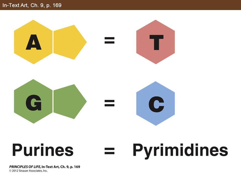
In-Text Art, Ch. 9, p. 169

11
Figure 9.5 DNA Is a Double Helix

12
Figure 9.6 Base Pairs in DNA Can Interact with Other Molecules

13
In-Text Art, Ch. 9, p. 172

14
Figure 9.7 Each New DNA Strand Grows by the Addition of Nucleotides to Its 3′ End

15
Figure 9.8 The Origin of DNA Replication

16
Figure 9.9 DNA Forms with a Primer

17
Figure 9.10 DNA Polymerase Binds to the Template Strand

18
Figure 9.11 The Two New Strands Form in Different Ways

19
Figure 9.12 The Lagging Strand Story

20
Figure 9.12 The Lagging Strand Story (Part 1)
")
21
Figure 9.12 The Lagging Strand Story (Part 2)
")
22
Figure 9.12 The Lagging Strand Story (Part 3)
")
23
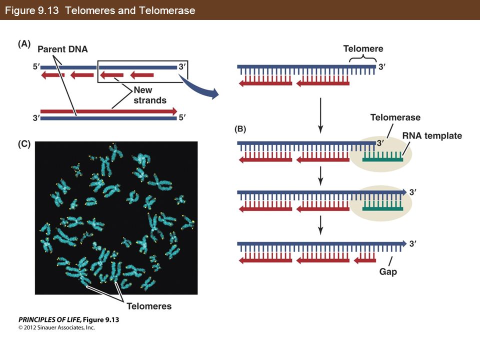
Figure 9.13 Telomeres and Telomerase

25
Figure 9.14 DNA Repair Mechanisms

26
Figure 9.14 DNA Repair Mechanisms (Part 2)
")
27
Figure 9.16 Mutation and Phenotype

28
Figure 9.18 Spontaneous and Induced Mutations

29
Figure 9.18 Spontaneous and Induced Mutations (Part 1)
")
30
Figure 9.18 Spontaneous and Induced Mutations (Part 2)
")
31
Figure 9.18 Spontaneous and Induced Mutations (Part 3)
")
32
Figure 9.19 5-Methylcytosine in DNA Is a “Hotspot” for Mutations

33
Figure 10.1 Metabolic Diseases and Enzymes

34
Figure 10.2 Gene Mutations and Amino Acid Changes

35
Figure 10.3 From Gene to Protein

36
Figure 10.5 DNA Is Transcribed to Form RNA

37
Figure 10.5 DNA Is Transcribed to Form RNA (Part 1)
")
38
Figure 10.5 DNA Is Transcribed to Form RNA (Part 2)
")
39
Figure 10.5 DNA Is Transcribed to Form RNA (Part 3)
")
40
Figure 10.5 DNA Is Transcribed to Form RNA (Part 4)
")
41
Figure 10.6 Transcription of a Eukaryotic Gene

42
Figure 10.6 Transcription of a Eukaryotic Gene (Part 1)
")
43
Figure 10.6 Transcription of a Eukaryotic Gene (Part 2)
")
44
Table 10.1 Differences between Prokaryotic and Eukaryotic Gene Expression

45
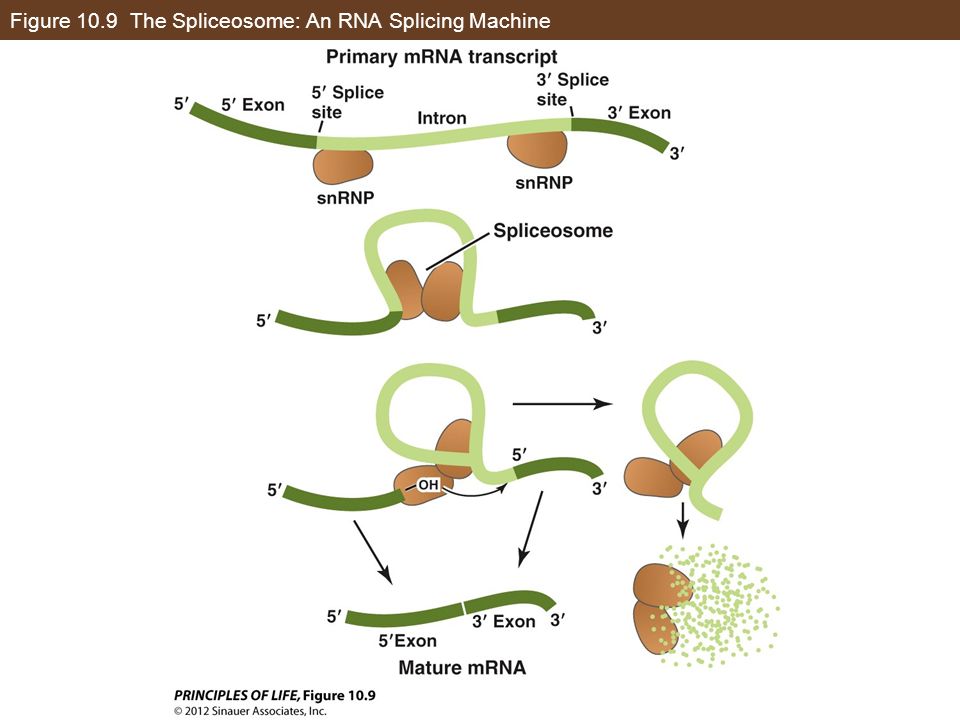
Figure 10.9 The Spliceosome: An RNA Splicing Machine

47
In-Text Art, Ch. 10, p. 195

48
Figure 10.11 The Genetic Code

49
Figure 10.12 Mutations

50
Figure 10.12 Mutations (Part 1)
")
51
Figure 10.12 Mutations (Part 2)
")
52
Figure 10.12 Mutations (Part 3)
")
53
Figure 10.12 Mutations (Part 4)
")
54
Figure 10.13 Transfer RNA

55
Figure 10.14 Ribosome Structure

56
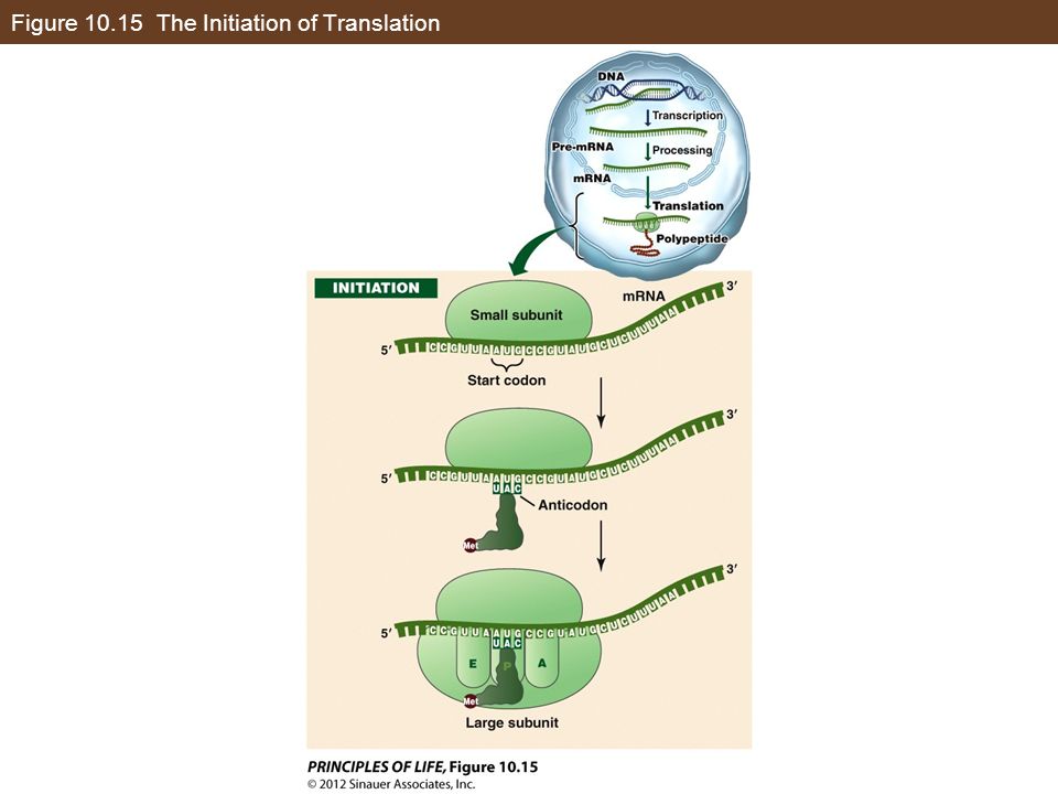
Figure 10.15 The Initiation of Translation

58
Figure 10.15 The Initiation of Translation (Part 1)
")
59
Figure 10.15 The Initiation of Translation (Part 2)
")
60
Figure 10.16 The Elongation of Translation

61
Figure 10.16 The Elongation of Translation (Part 1)
")
62
Figure 10.16 The Elongation of Translation (Part 2)
")
63
Figure 10.17 The Termination of Translation

64
Figure 10.17 The Termination of Translation (Part 1)
")
65
Figure 10.17 The Termination of Translation (Part 2)
")
66
Table 10.2 Signals that Start and Stop Transcription and Translation

67
Figure 10.18 A Polysome

68
Figure 10.18 A Polysome (Part 1)
")
69
Figure 10.18 A Polysome (Part 2)
")
70
Figure 10.19 Destinations for Newly Translated Polypeptides in a Eukaryotic Cell

71
Figure 10.19 Destinations for Newly Translated Polypeptides in a Eukaryotic Cell (Part 2)
")
72
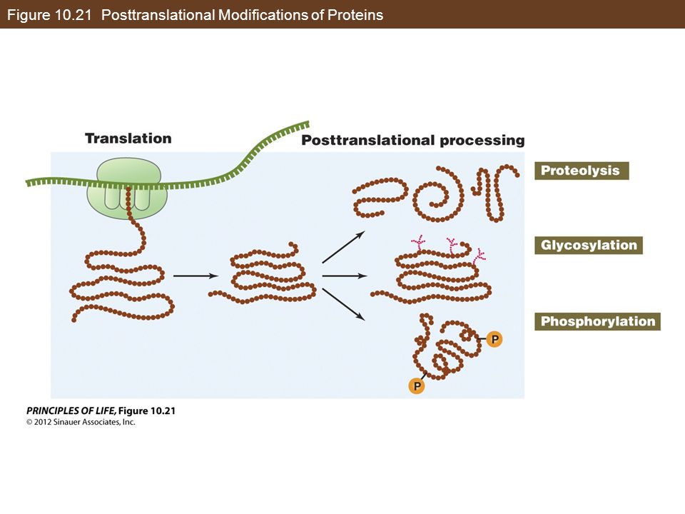
Figure 10.21 Posttranslational Modifications of Proteins

74
Figure 10.22 An Antibiotic at the Ribosome

Similar presentations