Download presentation
Presentation is loading. Please wait.

2
Digestive System

3
Basic Divisions – Digestive tract – Accessory organs: various exocrine glands

4
Digestive Processes – Ingestion – Mechanical Processing – Motility PeristalsisPeristalsis

5
Digestive Processes – Ingestion – Mechanical Processing – Motility Peristalsis

6
Digestive Processes – Ingestion – Mechanical Processing – Motility Peristalsis Segmentation movements

7
Digestive Processes, continued – Chemical digestion – Secretion – Absorption – Excretion and defecation

8
Non-Digestive Functions of Digestive Tract – Immunity – Storage of iron

9
Layers of the digestive tract – Mucosa Epithelium Lamina propria (areolar CT) Muscularis mucosae
 Muscularis mucosae")
10
Layers of the digestive tract, continued – Submucosa includes the submucosal plexus

12
Layers of the digestive tract, continued – Muscularis externa: responsible for peristalsis and segmentation movements longitudinal layer circular layer myenteric plexus

13
Layers of the digestive tract, continued – Serosa (the visceral peritoneum is an example) Simple squamous epithelium Areolar CT Within peritoneal cavity only
 Simple squamous epithelium Areolar CT Within peritoneal cavity only")
14
Layers of the digestive tract, continued – Adventitia Dense irregular CT Oral cavity, pharynx, esophagus, rectum

15
The Peritoneum – Parietal p. – Visceral p.

16
The mesenteries – Mesentery proper – Mesocolon – Greater omentum – Lesser omentum – Falciform ligament

21
Accessory structures of the oral (buccal) cavity – Teeth: will cover in lab – Tongue: read textbook – Salivary glands buccal glands lingual glands
 cavity – Teeth: will cover in lab – Tongue: read textbook – Salivary glands buccal glands lingual glands")
23
Oral (buccal) cavity – Salivary Glands buccal glands lingual glands major salivary glands – parotid
 cavity – Salivary Glands buccal glands lingual glands major salivary glands – parotid")
25
Oral (buccal) cavity – Salivary Glands buccal glands lingual glands major salivary glands – parotid – sublingual – submandibular
 cavity – Salivary Glands buccal glands lingual glands major salivary glands – parotid – sublingual – submandibular")
29
– Structure of salivary glands glandular epithelium merocrine cells

30
– Structure of salivary glands glandular epithelium merocrine cells compound tubulo-acinar

33
– Functions of saliva lubrication for swallowing, speaking re-mineralizes tooth enamel buffer antibodies (IgA) dissolves food molecules some chemical digestion
 dissolves food molecules some chemical digestion")
34
Pharynx – To be discussed with the respiratory system

35
Esophagus – Structural features muscular tube about 25 cm long posterior to larynx, trachea pierces diaphragm through esophageal hiatus

39
2 Esophogeal sphincters: – upper esophageal sphincter – lower esophageal sphincter

41
Histology highlights – mucosa – submucosa: lots of mucous glands – muscularis externa – adventitia (no serosa)
")
44
Gastroesophageal Reflux

45
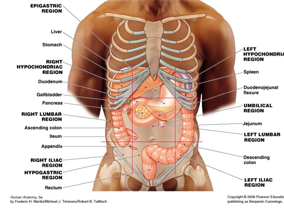
Stomach – Location

46
Stomach – Location: from epigastric and umbilical region

47
Stomach – Location: from epigastric and umbilical region to left hypochondriac regions

48
Gross Structural Features – cardiac region – fundus – body – pylorus – pyloric sphincter

49
Stomach motility videoStomach motility video

50
Stomach Histology – mucosa

51
Stomach Histology – mucosa: location of gastric glands

52
Stomach Histology – mucosa: location of gastric glands

53
Stomach Histology – mucosa: location of gastric glands gastric gland cells – mucous cells – parietal cells – chief cells – endocrine cells

54
Stomach Histology, continued – muscularis: three layers

55
Stomach Functions – food reservoir – formation of chyme – some chemical digestion – regulation of chyme entry into S.I. – intrinsic factor production – some absorption

56
Digestive System, review Basic Divisions – Digestive tract – Accessory organs: various exocrine glands

57
Pancreas – Location Umbilical region

59
Pancreas – Location Umbilical region Retroperitoneal

61
Pancreas Gross Structure – Head – Body – Tail

62
Pancreas Gross Structure – Head – Body – Tail – Ducts pancreatic accessory

63
– pancreas histology mostly glandular epithelium – exocrine pancreas – endocrine pancreas

64
– exocrine pancreas functions – sodium bicarbonate – digestive enzymes

66
– endocrine pancreas structure: thousands of islets of Langerhans

68
– endocrine pancreas function: hormone secretion – glucagon – insulin – somatostatin

69
Liver –Location: epigastric and right hypochondriac regions

70
Liver –Location: epigastric and right hypochondriac regions

72
Liver –Gross structure: 2 major lobes separated by the falciform ligament

74
Liver Blood Supply – Hepatic portal vein – Hepatic arteries

77
Liver Histology – Functional unit:

78
Liver Histology – Functional unit: liver lobule

81
Liver Histology – Functional unit: liver lobule

82
Liver Histology – Functional unit: liver lobule – Liver cells

83
Liver Histology – Functional unit: liver lobule – Liver cells hepatocytes Kupffer cells

85
– Each liver lobule supplied by branches of: hepatic arteries hepatic portal veins

87
– Liver Functions Maintains blood glucose levels Cholesterol synthesis HDL and LDL synthesis Plasma protein synthesis

88
– Liver Functions, continued Hormone and drug removal Phagocytosis Vitamin storage Iron storage Bilirubin excretion

91
– Liver Functions, continued Hormone and drug removal Phagocytosis Vitamin storage Iron storage Bilirubin excretion

92
– Liver Functions, continued Hormone and drug removal Phagocytosis Vitamin storage Iron storage Bilirubin excretion Bile salt secretion

94
Gall Bladder – Location: right lumbar region

96
Liver Blood Supply – Hepatic portal vein – Hepatic arteries

97
Gross Structural Features of Gall Bladder – Muscular sac – Mucosa folded into rugae – Bile enters and leaves through cystic duct

98
Gall Bladder Function – Stores and concentrates bile – Contracts during meals to force bile into SI

101
Biliary Pathway

102
Biliary Pathway: “plumbing” which drains bile

107
Small Intestine – 1 inch diameter – 10-20 ft. in length duodenum (10 in) jejunum (3-6 ft.) ileum (6-12 ft)
 jejunum (3-6 ft.) ileum (6-12 ft).")
108
Features of SI mucosa – Plica circularis

109
Features of SI mucosa – Plica circularis

110
Features of SI mucosa –Plica circularis –Villi

111
Features of SI mucosa –Plica circularis –Villi

112
Features of SI mucosa –Plica circularis –Villi

113
Features of SI mucosa –Plica circularis –Villi –Microvilli

114
Features of SI mucosa –Plica circularis –Villi –Microvilli

115
Features of SI mucosa, cont’d –Epithelial cell types: absorptive cells Goblet cells Endocrine cells Paneth cells

117
Features of SI mucosa, cont’d –MALT in lamina propria

118
Features of SI mucosa, cont’d –MALT in lamina propria

119
Features of SI mucosa, cont’d –MALT in lamina propria –Intestinal glands (“crypts”)
")
121
Features of SI submucosa –Submucosal glands in duodenum

122
Motility of SI –Segmentation movements –Peristalsis

123
Functions of small intestine – Completion of chemical digestion “brush-border” enzymes required – Absorption – Endocrine control of some digestive processes

124
Summary movie

125
Large Intestine

127
Large Intestine (large bowel)
")
128
–2.5 inches in diameter

129
5-6 feet long –Cecum –Colon Ascending Transverse Descending Sigmoid –Rectum –Anal canal

136
Features of LI mucosa – no villi – numerous intestinal glands – goblet and absorptive cells in epithelium – MALT in lamina propria

137
Structural Features of cecum and colon – Taeniae coli – Haustra – Epiploic appendages

139
Other Structural Features – Vermiform appendix

142
Other Structural Features – Vermiform appendix

143
Other Structural Features – Vermiform appendix – Ileocecal valve

145
Other Structural Features – Vermiform appendix – Ileocecal valve

146
Other Structural Features – Vermiform appendix – Ileocecal valve – Stretch receptors in rectum initiate defecation reflex

147
Other Structural Features – Vermiform appendix – Ileocecal valve – Stretch receptors in rectum initiate defecation reflex – Anal sphincters of anal canal

149
Motility of Large Intestine – From cecum to transverse colon: peristalsis – From transverse colon to rectum: mass movements

150
Functions of Large Intestine – Water and electrolyte absorption – Feces formation – Defecation

151
Large Intestinal Bacteria – Coat surface of mucosa – Examples: E. coli – Keep out pathogenic bacteria

152
48

Similar presentations