Download presentation
Presentation is loading. Please wait.

1
Functional Anatomy of Prokaryotic and Eukaryotic Cells

2
Size Comparisons of Selected Microorganisms/Parasites

3
Prions and Viroids Prions are proteins that are believed to cause diseases like BSE. Viroids are small RNA’s with no associated protein that causes diseases in plants. Neither fit the criteria for living organisms, but they are microbes and are therefore studied by microbiologists.

4
Viruses Consist of nucleic acid and protein.
Viruses are most simply a strand of DNA or RNA which may be single or stranded surrounded by a protein coat. These pictures are of the bacteriophage (bacteria virus) T4.
 T4.")
5
Viruses 2 Obligate intracellular parasites.
Some viruses have additional structure like a lipopolysacchride envelope that helps them to penetrate the host cell (as seen in this slide) or tail fibers that help them to attach to the host cell and inject their DNA (as seen on the previous slide).
 or tail fibers that help them to attach to the host cell and inject their DNA (as seen on the previous slide).")
6
Viruses 3 Are they really alive?
In general, are smaller than most prokaryotes.

7
Viruses 4 Reproduce using host cell mechanisms.
This slide illustrates the basic steps in a viruses life cycle. Beginning at the top right and going clockwise: 1. The virus will attach to the host cell. 2. Inject its DNA (or enter the host cell and release its DNA). 3. The DNA will circularize or if the virus contained RNA the viral protein reverse transcriptase will make a complementary double stranded DNA copy. 4. If the virus is one that has a lysogenic life cycle, the viral DNA will be incorporated into the host chromosome until environmental signals cause it to be excised. 5. The viral DNA is replicated and its genes are transcribed and translated to make the viral structural proteins. 6 & 7. The new viral particles are assembles into progeny. 8. The new viruses are released by the lysing of the host cell or by budding.
. 3. The DNA will circularize or if the virus contained RNA the viral protein reverse transcriptase will make a complementary double stranded DNA copy. 4. If the virus is one that has a lysogenic life cycle, the viral DNA will be incorporated into the host chromosome until environmental signals cause it to be excised. 5. The viral DNA is replicated and its genes are transcribed and translated to make the viral structural proteins. 6 & 7. The new viral particles are assembles into progeny. 8. The new viruses are released by the lysing of the host cell or by budding.")
8
Viruses 4 Adenovirus Rhinovirus
Copyright notice: This notice must accompany any copy of the images. The images must not be used for commercial purposes without the consent of the copyright owners. The images are not in the public domain. The images can be freely used for educational purposes. Copyright 1994 Veterinary Sciences Division. Rhinovirus Adenovirus and rhinovirus are both human cold viruses.

9
Prokaryotic Cells Comparing Prokaryotic and Eukaryotic Cells
Prokaryote comes from the Greek words for prenucleus. Eukaryote comes from the Greek words for true nucleus.

10
Prokaryote Eukaryote One circular chromosome, not in a membrane
No histones No organelles Peptidoglycan cell walls Binary fission Paired chromosomes, in nuclear membrane Histones Organelles Polysaccharide cell walls Mitotic spindle

11
Average size: µm µm Basic shapes:

12
Most bacteria are monomorphic A few are pleomorphic
Unusual shapes Star-shaped Stella Square Haloarcula Most bacteria are monomorphic A few are pleomorphic Figure 4.5

13
Arrangements Pairs: diplococci, diplobacilli Clusters: staphylococci
Chains: streptococci, streptobacilli

17
Glycocalyx Outside cell wall Usually sticky
A capsule is neatly organized A slime layer is unorganized & loose Extracellular polysaccharide allows cell to attach Capsules prevent phagocytosis Figure 4.6a, b

18
Flagella Outside cell wall Made of chains of flagellin
Attached to a protein hook Anchored to the wall and membrane by the basal body Figure 4.8

19
Flagella Arrangement Figure 4.7

20
Figure 4.8

22
Motile Cells Rotate flagella to run or tumble
Move toward or away from stimuli (taxis) Flagella proteins are H antigens (e.g., E. coli O157:H7)
 Flagella proteins are H antigens (e.g., E. coli O157:H7)")
23
Motile Cells Figure 4.9

24
Axial Filaments Endoflagella In spirochetes
Anchored at one end of a cell Rotation causes cell to move Figure 4.10a

25
Fimbriae allow attachment
Pili are used to transfer DNA from one cell to another Figure 4.11

26
Pili Short protein appendages smaller than flagella
Adhere bacteria to surfaces E. coli has numerous types K88, K99, F41, etc. Antibodies to will block adherance F-pilus; used in conjugation Exchange of genetic information Flotation; increase boyancy Pellicle (scum on water) More oxygen on surface
 More oxygen on surface.")
27
F-Pilus for Conjugation

28
Cell Wall Prevents osmotic lysis Made of peptidoglycan (in bacteria)
Figure 4.6a, b

29
Peptidoglycan Polymer of disaccharide N-acetylglucosamine (NAG) & N-acetylmuramic acid (NAM) Linked by polypeptides Figure 4.13a

34
Gram-Positive cell walls
Teichoic acids: Lipoteichoic acid links to plasma membrane Wall teichoic acid links to peptidoglycan May regulate movement of cations Polysaccharides provide antigenic variation Figure 4.13b

35
Teichoic Acids Gram + only Glycerol, Phosphates, & Ribitol
Attachment for Phages

37
Figure 4.13b, c

38
Gram-positive cell walls Gram-negative cell walls
Thick peptidoglycan Teichoic acids In acid-fast cells, contains mycolic acid Thin peptidoglycan No teichoic acids Outer membrane

39
Gram-Negative Outer Membrane
Lipopolysaccharides, lipoproteins, phospholipids. Forms the periplasm between the outer membrane and the plasma membrane. Protection from phagocytes, complement, antibiotics. O polysaccharide antigen, e.g., E. coli O157:H7. Lipid A is an endotoxin. Porins (proteins) form channels through membrane
 form channels through membrane.")
40
Gram-Negative Outer Membrane
Figure 4.13c

42
Lipopolysaccharide (LPS)
Endotoxin or Pyrogen Fever causing Toxin nomenclature Endo- part of bacteria Exo- excreted into environment Structure Lipid A Polysaccharide O Antigen of E. coli, Salmonella G- bacteria only Alcohol/Acetone removes

44
LPS (cont’d) Functions Toxic; kills mice, pigs, humans
G- septicemia; death due to LPS Pyrogen; causes fever DPT vaccination always causes fevers Adjuvant; stimulates immunity Heat Resistant; hard to remove Detection (all topical & IV products) Rabbits (measure fever) Amoebocytes Lyse in presence of LPS
 Rabbits (measure fever) Amoebocytes Lyse in presence of LPS.")
45
LPS (cont’d.) Appearance of Colonies
Mucoid = Smooth (lots of LPS or capsule) Dry = Rough (little LPS or capsule) O Antigen of Salmonella and E. coli 2,000 different O Ags of Salmonella 100’s different O Ags of E. coli E. coli O157 O Ags differ in Sugars, not Lipid A
 Dry = Rough (little LPS or capsule) O Antigen of Salmonella and E. coli. 2,000 different O Ags of Salmonella. 100’s different O Ags of E. coli. E. coli O157. O Ags differ in Sugars, not Lipid A.")
46
Damage to Cell Walls Lysozyme digests disaccharide in peptidoglycan.
Penicillin inhibits peptide bridges in peptidoglycan. Protoplast is a wall-less cell. Spheroplast is a wall-less Gram-positive cell. L forms are wall-less cells that swell into irregular shapes. Protoplasts and spheroplasts are susceptible to osmotic lysis.

47
Gram Stain Mechanism Crystal violet-iodine crystals form in cell
Gram-positive Alcohol dehydrates peptidoglycan CV-I crystals do not leave Gram-negative Alcohol dissolves outer membrane and leaves holes in peptidoglycan CV-I washes out

48
Bacterial Stains Bacteria are not always easy to see under a microscope. In order to see them better, stains are used. A negative stain is one that will stain the bacteria and make them stand out from the background. A Gram stain will stain bacteria based on their cell wall characteristics. The purple Gram stain will bind to certain bacteria which are designated Gram positive and the others are Gram negative. The Gram negative cells are generally stained with a pink negative stain so that they can be seen.

49
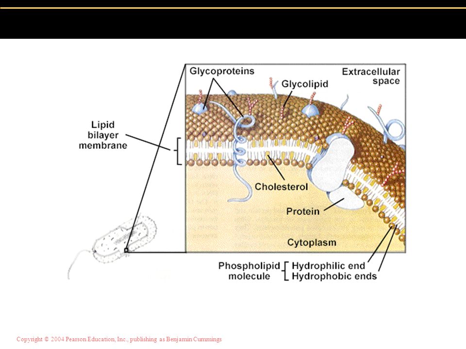
Plasma Membrane Figure 4.14a

50
Plasma Membrane Phospholipid bilayer Peripheral proteins
Integral proteins Transmembrane proteins Figure 4.14b

51
Fluid Mosaic Model Membrane is as viscous as olive oil.
Proteins move to function Phospholipids rotate and move laterally Figure 4.14b

55
Cytoplasm 80% Water {20% Salts-Proteins) Osmotic Shock important
DNA is circular, Haploid Advantages of 1N DNA over 2N DNA More efficient; grows quicker Mutations allow adaptation to environment quicker Plasmids; extra circular DNA Antibiotic Resistance No organelles (Mitochondria, Golgi, etc.)
")
56
Spores SPORE

58
Cross-section of Bacillus spore
The central protoplast, or germ cell, carries the constituents of the future vegetative cell, accompanied by dipicolinic acid --heat resistance of the spore. Surrounding the protoplast is a cortex consisting largely of peptidoglycan (murein), -- heat and radiation resistance of the spore. The inner layer, the cortical membrane or protoplast wall, becomes the cell wall of the new vegetative cell when the spore germinates. The spore coats, which constitute up to 50 percent of the volume of the spore, protect it from chemicals, enzymes, etc.
, -- heat and radiation resistance of the spore. The inner layer, the cortical membrane or protoplast wall, becomes the cell wall of the new vegetative cell when the spore germinates. The spore coats, which constitute up to 50 percent of the volume of the spore, protect it from chemicals, enzymes, etc.")
59
The developmental cycle of the Endospore.

61
Bacterial endospores. Phase microscopy of sporulating bacteria demonstrates the refractility of endospores, as well as characteristic spore shapes and locations within the mother cell.

62
Atypical Cell Walls Mycoplasmas Lack cell walls
Sterols in plasma membrane Archaea Wall-less, or Walls of pseudomurein (lack NAM and D amino acids)
")
63
Archaebacteria Newly added taxonomic kingdom.
The archaebacteria are prokaryotes, but they have been shown to be more closely related the the eukaryotes than are true bacteria through the comparison of the protein sequences of 16s ribosomal subunits and the DNA sequences.

64
Archeabacteria 2 Single celled prokaryotes. Extremophiles.
4 broad groups. Methanogens + extreme halophiles Methanogens only Sulfur dependent Thermophiles

65
Archeabacteria 3 The preceding Methanobacterim image is from the Department of Microbiology and Evolutionary Biology at the University of Nijmegen, the Netherlands. The picture on the left shows a methanogenic archaen, and those on the right show a thermophile.

66
Eukaryote Cell Structure

67
Algae Eukaryotes that may be unicellular, colonial or filamentous.
These images are from the Molecular Evolution and Organelle Genomics program at the University of Montreal. Copyright by Charles J. O’Kelly and Tim Littlejohn. Distribution for noncommercial purposes permitted so long as this copyright notice is included and acknowledgement is made. The algae shown are green algae which are thought to be the ancestors of green plants. The photo on the left shows the unicellular life stage of Halosphera minor. The one on the right shows the colonial stage of the same organism.

68
Algae 2 Aquatic or terrestrial. Mostly plant-like characteristics.
Photosynthetic. Great variety of types, but all contain chlorophyll. Some animal-like characteristics like phagocitosis of other organisms. In an aquatic environment, algae are a very important carbon dioxide fixer, oxygen supplier and food source. Phagocitosis means to ingest by surrounding. A commonly seen example of this is seen in the amoebae.

69
Algae 3 Important marine food source.
This picture is a diatom. These are a type of marine algae that are a very important food source.

70
Protozoa Single celled eukaryotes, but may form colonial aggregates.
Aquatic with animal-like characteristics. This image is from the Molecular Evolution and Organelle Genomics program at the University of Montreal. Copyright by Charles J. O’Kelly and Tim Littlejohn. Distribution for noncommercial purposes permitted so long as this copyright notice is included and acknowledgement is made.

71
Protozoa 2 Ingest organic matter for nutrients.
Vary greatly in size from 0.003mm to 5mm. Many are human parasites. Most are motile. Examples of human diseases caused by protozoa are malaria and dysentery. Motility in protozoa is accomplished in one of three ways. The use of flagella - as seen on the protozoa on the left in the illustration. The use of cilia - as seen on the protozoa in the middle. The use of cytoplasmic streaming (a flowing type motion of the whole organism) - as seen with the ameoba on the right in the illustration.
 - as seen with the ameoba on the right in the illustration.")
72
Fungi Very diverse group of eukaryotes. Not all are microbes.
Yeasts are unicellular and spherical. Molds are filamentous with branching. The preceding Pyromyces sp. image is from the Department of Microbiology and Evolutionary Biology at the University of Nijmegen, the Netherlands The micrograph on the left is of yeast and the one on the right is a microscopic mold.

73
Fungi 2 Non-photosynthetic.
Require the uptake of organic matter for nutrients. Saprophytic or parasitic. Propagate by spores. Saprophytes and parasites both use the complex molecules of another organism as nutrients. However, saprophytes live off of organisms that are already dead and decaying, whereas parasites live off of living organisms and are harmful to them.

Similar presentations