Download presentation
Presentation is loading. Please wait.

1
Copyright © 2010 Pearson Education, Inc. Figure 4.6 The Structure of a Prokaryotic Cell

2
Copyright © 2010 Pearson Education, Inc. Figure 24.12 Glycocalyx Outside cell wall Usually sticky (biofilms) Capsule: neatly organized Slime layer: unorganized and loose Extracellular polysaccharide allows cell to attach Capsules prevent phagocytosis Capsules were instrumental in determining that DNA was the genetic material (Fig. 8.24 p. 235)
 Capsule: neatly organized Slime layer: unorganized and loose Extracellular polysaccharide allows cell to attach Capsules prevent phagocytosis Capsules were instrumental in determining that DNA was the genetic material (Fig p. 235).")
3
Copyright © 2010 Pearson Education, Inc. Griffith’s experiments leading the way to discovering- what is the genetic (hereditary) material? The key was the GLYCOCALYX!
 material. The key was the GLYCOCALYX!.")
4
Copyright © 2010 Pearson Education, Inc. Griffith’s experiments leading the way to discovering- what is the genetic (hereditary) material?
 material .")
5
Copyright © 2010 Pearson Education, Inc. Griffith’s experiments leading the way to discovering- what is the genetic (hereditary) material?
 material .")
6
Copyright © 2010 Pearson Education, Inc. Griffith’s experiments leading the way to discovering- what is the genetic (hereditary) material?
 material .")
7
Copyright © 2010 Pearson Education, Inc. Figure 4.7 Arrangements of Bacterial Flagella

8
Copyright © 2010 Pearson Education, Inc. Figure 4.10a Axial Filaments Also called endoflagella or periplasmic flagella In spirochetes only The spirochetes are a … unique group of bacteria. This phylum contains not only many medically important species such as Treponema pallidum and Borrelia burgdorferi but others live inside arthropods such as termites and some that are free-living and reside in soil and water. http://jb.asm.org/cgi/content/full/182/23/6698 http://jb.asm.org/cgi/content/full/182/23/6698 Individual AF can be anchored at one end or the other of a cell. Rotation causes the cell to move.

9
Copyright © 2010 Pearson Education, Inc. Figure 4.11 Fimbriae and Pili: Made of a different protein (pilin) than flagella and are shorter, thinner, straighter. Fimbriae allow attachment: Important for some diseases (gonorrhea and E.coli 0157:H7) and biofilms.
 than flagella and are shorter, thinner, straighter. Fimbriae allow attachment: Important for some diseases (gonorrhea and E.coli 0157:H7) and biofilms..")
10
Copyright © 2010 Pearson Education, Inc. Pili: Some are used for movement and others for transfer of DNA Yikes! Resistance!

11
Copyright © 2010 Pearson Education, Inc. Figure 4.13a Peptidoglycan Linked by polypeptides

12
Copyright © 2010 Pearson Education, Inc. Gram Positive Cell Wall Many layers of peptidoglycan

13
Copyright © 2010 Pearson Education, Inc. Impetigo and Necrotizing fasciitis flesh- eating disease)
")
14
Copyright © 2010 Pearson Education, Inc. Figure 4.13c Gram-Negative Cell Wall

15
Copyright © 2010 Pearson Education, Inc. Gram - Sepsis

16
Copyright © 2010 Pearson Education, Inc. The Gram Stain Table 4.1 (a) Gram-Positive(b) Gram-Negative
 Gram-Positive(b) Gram-Negative.")
17
Copyright © 2010 Pearson Education, Inc. Figure 4.14b The Plasma Membrane Fig. 4.14 p. 90 -as viscous as olive oil Phospholipid bilayer Peripheral proteins Integral proteins Transmembrane proteins

18
Copyright © 2010 Pearson Education, Inc. Figure 4.17a Movement of Materials across Membranes Simple diffusion: Movement of a solute from an area of high concentration to an area of low concentration

19
Copyright © 2010 Pearson Education, Inc. Figure 4.17b-c Movement of Materials across Membranes Facilitated diffusion: Solute combines with a transporter protein in the membrane

20
Copyright © 2010 Pearson Education, Inc. Figure 4.17d Movement of water across Membranes Through lipid layer Aquaporins (water channels)
.")
21
Copyright © 2010 Pearson Education, Inc. Figure 4.18c–e The Principle of Osmosis Fig. 4.18 p. 93 The movement of water across a selectively permeable membrane from an area of high water concentration to an area of lower water concentration

22
Copyright © 2010 Pearson Education, Inc. Figure 4.6 The Nucleoid, Plasmids, & Ribosomes

23
Copyright © 2010 Pearson Education, Inc. Sporulation and Germination Figure 4.21a

24
Copyright © 2010 Pearson Education, Inc. Figure 4.22a The Eukaryotic Cell

25
Copyright © 2010 Pearson Education, Inc. Table 4.2 Cytoplasm

26
Copyright © 2010 Pearson Education, Inc. Figure 4.24a–b The Eukaryotic Nucleus

27
Copyright © 2010 Pearson Education, Inc. Organelles only found in Eukaryotic Cells Nucleus: Contains chromosomes ER: Transport network Golgi complex: Membrane formation and secretion Lysosome: Digestive enzymes Vesicles: Membrane sacs-storage, protection, transport Vacuole: Brings food/liquid into cells and provides support

28
Copyright © 2010 Pearson Education, Inc. Organelles only found in Eukaryotic Cells Mitochondrion: Cellular respiration Chloroplast: Photosynthesis Peroxisome: Oxidation of fatty acids; destroys H 2 O 2 Centrosome: Consists of protein fibers and centrioles (Utilized for cell division)
.")
29
Copyright © 2010 Pearson Education, Inc. Figure 10.2 Endosymbiotic Theory

30
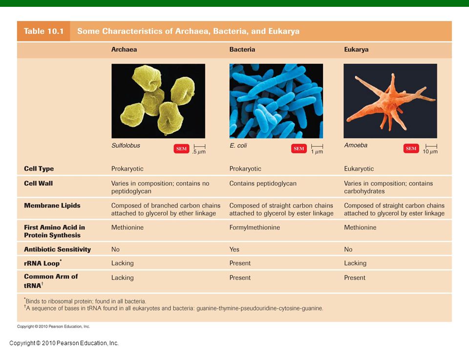
Copyright © 2010 Pearson Education, Inc.

Similar presentations