Download presentation
Presentation is loading. Please wait.

1
Western Blotting Lab. 7

2
Introduction Blotting is a technique by which a macromolecule such as DNA, RNA, or protein is resolved in a gel matrix, transferred to a solid support, and detected with a specific probe These powerful techniques allow us to identify and characterize specific molecules in a complex mixture of related molecules Some of the more common techniques include: Southern blotting (DNA) Northern blotting (RNA) and Western blotting (for protein)
 Northern blotting (RNA) and Western blotting (for protein)")
3
Introduction Western blotting, also known as immunoblotting or protein blotting, is a technique used to detect the presence of a specific protein in a complex protein mixture It is a core technique in cell biology, molecular biology, virology and others Western blots have become one of the most common analytical tools for the Detection of viral proteins characterization of monoclonal and Polyclonal antibody preparations and in determining the specificity of the immune response to viral antigens

4
Western Blot Applications for Medical Diagnosis
For HIV confirmatory HIV-test to detect anti-HIV antibody in a human serum sample For HBV confirmatory test for Hepatitis B infection For Herps detection of HSV infections

5
The Western blotting procedure relies upon three key elements to accomplish this task:
The separation of protein mixtures by size using gel electrophoresis The efficient transfer of separated proteins to a solid support; and the specific detection of a target protein by appropriately matched antibodies Once detected, the target protein will be visualized as a band on a blotting membrane, X-ray film, or an imaging system

6
Steps involved in western blotting
Sample preparation Gel Electrophoresis Blotting (or transfer) Blocking Antibody Probing Detection
 Blocking. Antibody Probing. Detection.")
7
1- Sample Preparation All sources of protein, from single cells to whole tissues, biological fluids and proteins secreted in vitro, are open to analysis by Western blotting In most cases, the cells are harvested, washed, and lysed to release the target protein For best results, all these steps should be carried out on ice This will minimize proteolysis, dephosphorylation, and denaturation, since all begin to occur once the cells are disrupted

8
1- Sample Preparation The choice of extraction method depends primarily on the sample and whether the analysis is targeting all the proteins in a cell or only a component from a particular subcellular fraction The endogenous proteases may be liberated upon cell disruption and may degrade the target molecule, the sample should be protected during cell disruption and subsequent purification by the use of a cocktail of protease inhibitors to avoid uncontrolled protein losses

9
1- Sample Preparation Numerous methods are available for disrupting cells and preparing their contents for analysis by Western blotting Detergent lysis The membranes are solubilized, lysing cells and liberating their contents Ultrasonication The sound waves generate a region of low pressure, causing disruption of the membranes of cells Freeze/thaw lysis Cells are disrupted by the repeated formation of ice crystals and the method is usually combined with enzymatic lysis Enzymatic digestion The enzymes dissolve cell walls, coats, capsules, capsids, or other structures To ensure that samples are in the proper range of detection for the assay, and so they can be compared on an equivalent basis, it is important to know the concentration of total protein in each sample

10
2- Gel Electrophoresis- Gel Preparation
Reagent 8% (Running Gel) 5% (Stacking Gel) Acrylamide/ Bisacrylamide (40%) * 4.0 mls 2.5 mls 1 M Tris-HCl 7.5 mls Distilled water 8.2 mls 9.7 mls 10% SDS 200 µl 10% Ammonium Persulfate 100 µl TEMED (added last) 10 µl * = 19:1 w:w ratio of acrylamide to N,N'-methylene bis-acrylamide The purpose of the stacking gel is to condense the protein into a tight band before running it through the separating gel. This allows better resolution in the separating gel. The stacking effect works because the stacking gel is made up of a lower concentration of polyacrylamide than the separating gel. The protein stacks because the proteins move very quickly through the stacking gel, only to run into the separating gel. The proteins at the top of the stacking gel then have time to catch up with the proteins at the bottom. [The other relevant difference between the stacking gel and the separating gel is pH, which affects the charge and thus the migration of glycine, a component of the electrophoresis running buffer. In the stacking gel (at pH 6.8), glycine "trails" the proteins and facilitates their stacking. At the separating gel pH of 8.8, glycine is negatively charged and thus runs at the dye front, leaving the proteins to separate out behind it.]
 5% (Stacking Gel) Acrylamide/ Bisacrylamide (40%) * 4.0 mls. 2.5 mls. 1 M Tris-HCl. 7.5 mls. Distilled water. 8.2 mls. 9.7 mls. 10% SDS. 200 µl. 10% Ammonium Persulfate. 100 µl. TEMED (added last) 10 µl. * = 19:1 w:w ratio of acrylamide to N,N -methylene bis-acrylamide. The purpose of the stacking gel is to condense the protein into a tight band before running it through the separating gel. This allows better resolution in the separating gel. The stacking effect works because the stacking gel is made up of a lower concentration of polyacrylamide than the separating gel. The protein stacks because the proteins move very quickly through the stacking gel, only to run into the separating gel. The proteins at the top of the stacking gel then have time to catch up with the proteins at the bottom. [The other relevant difference between the stacking gel and the separating gel is pH, which affects the charge and thus the migration of glycine, a component of the electrophoresis running buffer. In the stacking gel (at pH 6.8), glycine trails the proteins and facilitates their stacking. At the separating gel pH of 8.8, glycine is negatively charged and thus runs at the dye front, leaving the proteins to separate out behind it.]")
11
2- Gel Electrophoresis- Gel Preparation
Mix ingredients in the order shown above, ensuring no air bubbles form Pour the separating gel into glass plate assembly Overlay gel with water to ensure a flat surface and to exclude air Leave to polymerize for ~ 20 minutes Then prepare the stacking gel and pour on the running gel, insert comb and leave for 20 min

12
2- Gel Electrophoresis- Sample Buffer
A sample of protein, is boiled in sample buffer (at 95oC for 5 minutes) which contains: The β-mercaptoethanol reduces disulfide bonds SDS disrupts protein secondary and tertiary structure Glycerol to make samples sink into wells Bromophenol Blue dye to visualize samples The detergent binds to hydrophobic regions in a constant ratio of about 1.4 g of SDS per gram of protein, (every 3 aa)
 which contains: The β-mercaptoethanol reduces disulfide bonds. SDS disrupts protein secondary and tertiary structure. Glycerol to make samples sink into wells. Bromophenol Blue dye to visualize samples. The detergent binds to hydrophobic regions in a constant ratio of about 1.4 g of SDS per gram of protein, (every 3 aa)")
13
2- Gel Electrophoresis- Sample Buffer
The end result has two important features: All proteins contain only primary structure and All proteins have a large negative charge which means they will all migrate towards the positive pole when placed in an electric field. They migrate through a gel towards the positive pole at a rate proportional to their linear size

14
2- Gel Electrophoresis Loading Samples & Running the gel
Samples are loaded into separate wells A protein marker is also loaded Run at 200 volts for minutes Running Buffer (pH 8.3) contains Tris Base Glycine SDS
 contains. Tris Base. Glycine. SDS")
15
3- Blotting Following gel electrophoresis, the separated protein mixtures are transferred to a solid support for further analysis Transfer can be done in wet or semi-dry conditions Semi-dry transfer is generally faster Wet transfer is recommended for large proteins, >100 kD For both kinds of transfer, the membrane is placed next to the gel The two are sandwiched between absorbent materials, and the sandwich is clamped between solid supports to maintain tight contact between the gel and membrane

16
3- Blotting The system is connected to a power supply and blotting is run for 1 hour at 80 mA The buffer for wet transfer contains: Tris methanol (20%) Glycine 0.1% SDS
 Glycine. 0.1% SDS.")
17
3- Blotting- Blotting Membranes
The solid support onto which the separated proteins are transferred is usually of two types, both of which bind proteins with high affinity: Nitrocellulose membrane has excellent protein binding and retention capabilities is brittle and thus it is usually less effective when blots need to be reused Polyvinylidene fluoride (PVDF) membrane PVDF demonstrates superior mechanical strength making it suitable for stripping/reprobing
 membrane. PVDF demonstrates superior mechanical strength making it suitable for stripping/reprobing.")
18
3- Blotting- Visualization of proteins in membranes: Ponceau Red stain
Ponceau Red is a reversible stain with poor sensitivity Ponceau S is compatible with both nitrocellulose and PVDF membranes This is a quick and easy way to visualize proteins transferred to membranes Ponceau S is easily removed with water and is regarded as a “gentle” treatment that does not interfere with subsequent immunological detection steps

19
4- Blocking Blocking is a very important step in the immunodetection phase of Western blotting because it prevents non-specific binding of antibody to the blotting membrane The most commonly used blocking solutions contain 3-5% BSA or non-fat dried milk in a solution of PBS (phosphate buffered saline) or TBS (tris buffered saline) Often, a small amount of Tween 20 detergent is added to blocking and washing solutions to reduce background staining, and the buffer is known as PBST or TBST
 or TBS (tris buffered saline) Often, a small amount of Tween 20 detergent is added to blocking and washing solutions to reduce background staining, and the buffer is known as PBST or TBST.")
20
5- Antibody Probing Once the protein samples are separated and transferred onto a membrane, the protein of interest is detected and localized using a specific antibody The blot will be incubated in a dilute solution of antibody, usually for a few hours at room temperature or overnight at 4°C The antibody is diluted in wash buffer (PBST or TBST) or in the blocking solution, the choice depends upon the antibody Since antibody preparations vary in their levels of purity and specific binding properties, there will be differences in the level of dilution required The manufacturer’s datasheet should provide dilution recommendations for a particular preparation
 or in the blocking solution, the choice depends upon the antibody. Since antibody preparations vary in their levels of purity and specific binding properties, there will be differences in the level of dilution required. The manufacturer’s datasheet should provide dilution recommendations for a particular preparation.")
21
5- Antibody Probing Usually, Western blotting protocols utilize a non-labeled primary antibody directed against the target protein Wash the membrane several times in TBST while agitating, 5 minutes or more per wash, to remove residual primary antibody A species-specific, labeled secondary antibody directed against the constant region of the primary antibody is then used The secondary antibody serves not only as a carrier of the label but is also a mechanism to amplify the emitted signals, as many secondary antibodies can theoretically bind simultaneously to the primary antibody Secondary Ab is also diluted according to the manufacturer’s recommendations and incubated for 1 hour at RT

22
6- Detection with Substrate
The most common antibody label used in Western blots is HRP, a small, stable enzyme with high specificity and rapid turnover The signal is detected when HRP is exposed to a substrate solution in the final step of the immunodetection procedure Substrate solutions for Western blotting are chemical reagents that are acted upon by the enzyme to yield a signal that can be easily measured HRP label is typically detected with either colorimetric or chemiluminescent substrates

23
6- Detection with Substrate
Colorimetric substrates for HRP {eg. Tetramethylbenzidine(TMB)} produce purple/black bands directly on the surface of the blot These substrates are very easy to use and take from a few minutes to a few hours to produce visible bands Detection limits for colorimetric substrates are in the low nanogram range
} produce purple/black bands directly on the surface of the blot. These substrates are very easy to use and take from a few minutes to a few hours to produce visible bands. Detection limits for colorimetric substrates are in the low nanogram range.")
24
6- Detection with Substrate
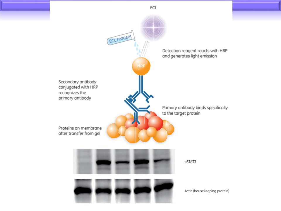
More routinely, HRP is used with ECL (enhanced chemiluminescence) detection For ECL detection, the substrate is luminol which is oxidized by HRP in the presence of H2O2 to produce light The emitted light is detected by exposing the Western blot to X-ray film, or by using a CCD camera for light capture The emitted light forms a band on the film, or on the screen of the imaging system, indicating where the HRP-labeled antibody has bound to the target protein ECL detection of HRP is extraordinarily sensitive, allowing for the visualization of picogram to femtogram amounts of target protein
 detection. For ECL detection, the substrate is luminol which is oxidized by HRP in the presence of H2O2 to produce light. The emitted light is detected by exposing the Western blot to X-ray film, or by using a CCD camera for light capture. The emitted light forms a band on the film, or on the screen of the imaging system, indicating where the HRP-labeled antibody has bound to the target protein. ECL detection of HRP is extraordinarily sensitive, allowing for the visualization of picogram to femtogram amounts of target protein.")
26
M

27
HIV BLOT 2.2 WESTERN BLOT ASSAY

28
Introduction Screening tests are widely available for detecting antibodies to both HIV-1 and HIV-2 Such tests can be extremely sensitive but have a potential for being less specific, leading to false positive interpretations Independent supplemental tests of high specificity are therefore necessary to further confirm the presence of antibodies to HIV-1 and/or HIV-2

29
Introduction The test is intended for use as a more specific supplemental test on human serum or plasma specimens found repeatedly reactive using ELISA The separated specific HIV-1 viral antigens incorporated onto the strips via electrophoretic and blotting procedures

30
Principles of the Procedure
The nitrocellulose strips are incorporated with separated, bound antigenic proteins from: partially purified inactivated HIV-1 plus a specific HIV-2 synthetic peptide on the same strips Individual nitrocellulose strips are incubated with diluted serum or plasma and controls Specific antibodies to HIV-1 and HIV-2 if present in the specimens will bind to the HIV-1 and HIV-2 proteins on the strips

31
Principles of the Procedure
The strips are washed to remove unbound materials Goat anti-human IgG conjugated with alkaline phosphatase is used as a secondary Ab and then the substrate BCIP/NBT is added for detection This method has the sensitivity to detect marginal amounts of HIV specific antibodies in serum or plasma

32
Controls Non-reactive Control Strong Reactive Control
Inactivated human serum with high titered antibodies to HIV-1 and HIV-2 Weak Reactive Control Inactivated human serum with low titered antibodies to HIV-1

33
Procedure

Similar presentations


 SDS PAGE Isoelectric Point Isoelectric focusing.>")


>")

![Lab#6 Western Blotting BCH 462[practical].](/18/6070629/big_thumb.jpg "Lab#6 Western Blotting BCH 462[practical].>")