Download presentation
Presentation is loading. Please wait.

1
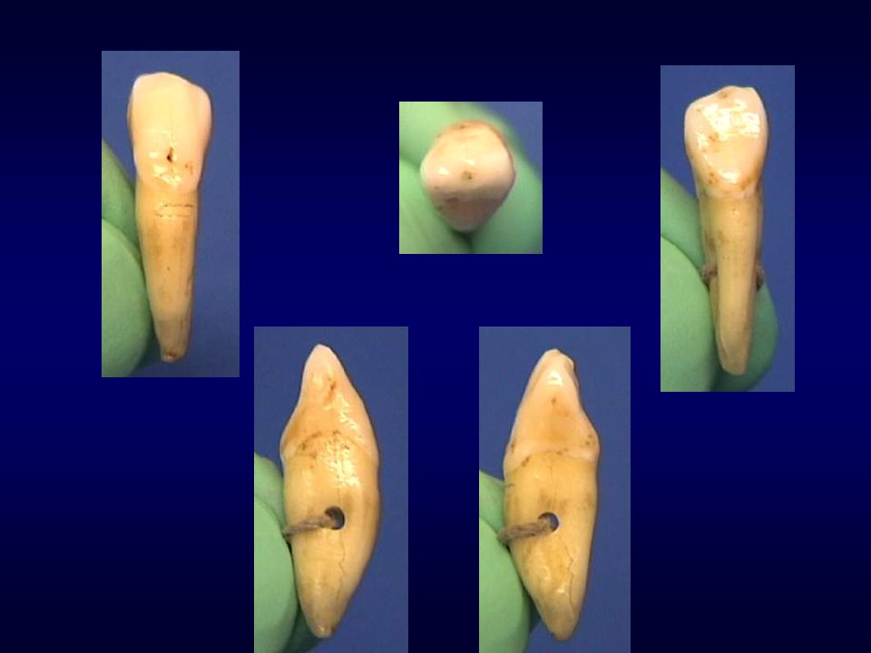
Permanent Canines

2
Introduction “Cornerstones of the mouth” - 3rd from midline (“eye-teeth”) Situated between incisors and premolars Form: single cusp (middle labial lobe) Function: Occlusion - piercing, tearing, lateral guidance Esthetics - facial support Phonetics - speech Greatest combined crown-root length (in either arch)
 Function: Occlusion - piercing, tearing, lateral guidance. Esthetics - facial support. Phonetics - speech. Greatest combined crown-root length (in either arch)")
3
Permanent Maxillary Canine

4
General Characteristics:
Arch position 3rd from midline Between anteriors and posteriors Universal #6 and #11 Single facial cusp Function: tearing, piercing, esthetics, and occlusion

5
Canine vs Central Crown length almost the same
M-D, canine narrower (by 1 mm) F-L, canine wider (by 1 mm)
 F-L, canine wider (by 1 mm)")
6
Canine vs Central: Root of canine longer
Cingulum of canine more prominent Middle lobe of canine more developed cusp form Canine has two lingual fossae (divided by lingual ridge)
")
7
Development Timeline:
Initial calcification: months Enamel completed: years Eruption: years Root completion: years

8
Labial View Pentagonal (5-sided) (trapezoidal geometric form)
Strong labial ridge with MF and DF developmental depressions Height of contour (HOC) at cervical third
 at cervical third.")
9
Labial view - mesial outline:
Generally convex from mesial HOC to cervical Contact area (HOC) at junction of incisal-middle third
 at junction of incisal-middle third.")
10
Labial view - distal outline:
Distal HOC at middle third Shorter than mesial outline from HOC to cervical Distal bulge with slight cervical concavity

11
Labial view - incisal outline:
2 cusp ridges Mesial cusp ridge (shorter) Distal cusp ridge (longer) With wear, mesial cusp ridge becomes longer* Cusp tip in line with midline of root
 Distal cusp ridge (longer) With wear, mesial cusp ridge becomes longer* Cusp tip in line with midline of root.")
12
Lingual View M, D, and I outlines similar to labial view
Bulky cingulum (bulkiest of anteriors) Ridges: M and D marginal ridges Lingual ridge Fossae: ML and DL fossa
 Ridges: M and D marginal ridges. Lingual ridge. Fossae: ML and DL fossa.")
13
Lingual view: Uncommonly, linguogingival groove and lingual pit present HOC at cervical third Cervical line slight distal offset

14
Mesial View Triangular crown
Wider F-L than incisors* (widest F-L of all anteriors) Cusp tip located just facial to long axis Labial outline convex, HOC at cervical third
 Cusp tip located just facial to long axis. Labial outline convex, HOC at cervical third.")
15
Mesial view: Lingual outline convex at gingival 1/2 and concave at incisal 1/2 Lingual HOC at cervical third Mesial HOC at junction of incisal-middle thirds

16
Distal View Cervical 1/2 usually concave
CE line curvature less than mesial HOC at middle third (most cervical of anteriors)
")
17
Incisal View Asymmetrical diamond shape outline Mesial half bulkier
Distal half appears “drawn” out Cingulum offset to distal Four cusp ridges: Mesial cusp ridge Distal cusp ridge Facial cusp ridge Lingual cusp ridge

18
Root Single, longest of all teeth* Wider F-L than M-D
Cervical cross-section is ovoid Normally straight, but may deflect distally

19
Permanent Mandibular Canine
Universal #22 and #27 General form and function similar to maxillary canine

20
Comparisons with maxillary canine:
Crown as long or longer (by 1 mm) M-D and F-L dimensions smaller (by .5 mm)
 M-D and F-L dimensions smaller (by .5 mm)")
21
Mandibular vs Maxillary Canine:
Total length about same* Root length slightly shorter* Lingual anatomy less developed

22
Development Timeline:
Initial calcification: months Enamel completed: years Eruption: years Root completed: years

23
Labial View Pentagonal: 5-sided Narrower M-D than maxillary canine
Crown is longer I-C (longest I-C of all teeth*)
")
24
Labial view - mesial outline:
Almost straight line from HOC to cervical and mesial outline of root HOC at incisal third, near mesioincisal angle (more incisal than max canine)
")
25
Labial view - distal outline:
More convex than mesial Rounded distoincisal corner HOC at junction of incisal-middle thirds

26
Labial view - incisal outline:
D cusp ridge longer than M With wear, D cusp ridge becomes longer* Cusp tip in line with root midline

27
Lingual View M, D, I aspects mimic labial view
CE line exhibits distal offset Anatomy not as pronounced as maxillary

28
Mesial View Labial outline convex, HOC at cervical third
Cusp tip is lingual to root midline Labial outline “moonshape”

29
Mesial view: Lingual outline less cingulum curvature, HOC cervical third Contact area at incisal third

30
Distal View Similar to mesial HOC at junction of incisal-middle thirds

31
Incisal View Similar to maxillary canine Bulky mesial half
Distal appears “pulled” or “pinched” Cingulum offset to distal

32
Root Longest root in mandibular arch*
Single, straight - bifurcation possible Wider F-L than M-D Cervical cross-section is “flattened” ovoid

33
How To Tell Maxillary Canine from Mandibular:
M-D dimension of maxillary wider Mesial outline of mandibular straight Facial outline of mandibular appears “moonshape” - location of cusp tip

34
Maxillary vs Mandibular:
M and D contact areas are more incisal with mandibular Lingual anatomy of maxillary more prominent Location of wear facets

35
How to tell right from left:
Incisal view, distal appears “pinched” Mesial cusp ridge shorter than distal CE line curvature more on mesial

Similar presentations


 Name the two dental arches.>")


