Download presentation
Presentation is loading. Please wait.

1
Human Anatomy, First Edition McKinley & O'Loughlin
Chapter 11 Lecture Outline: Axial Muscles

2
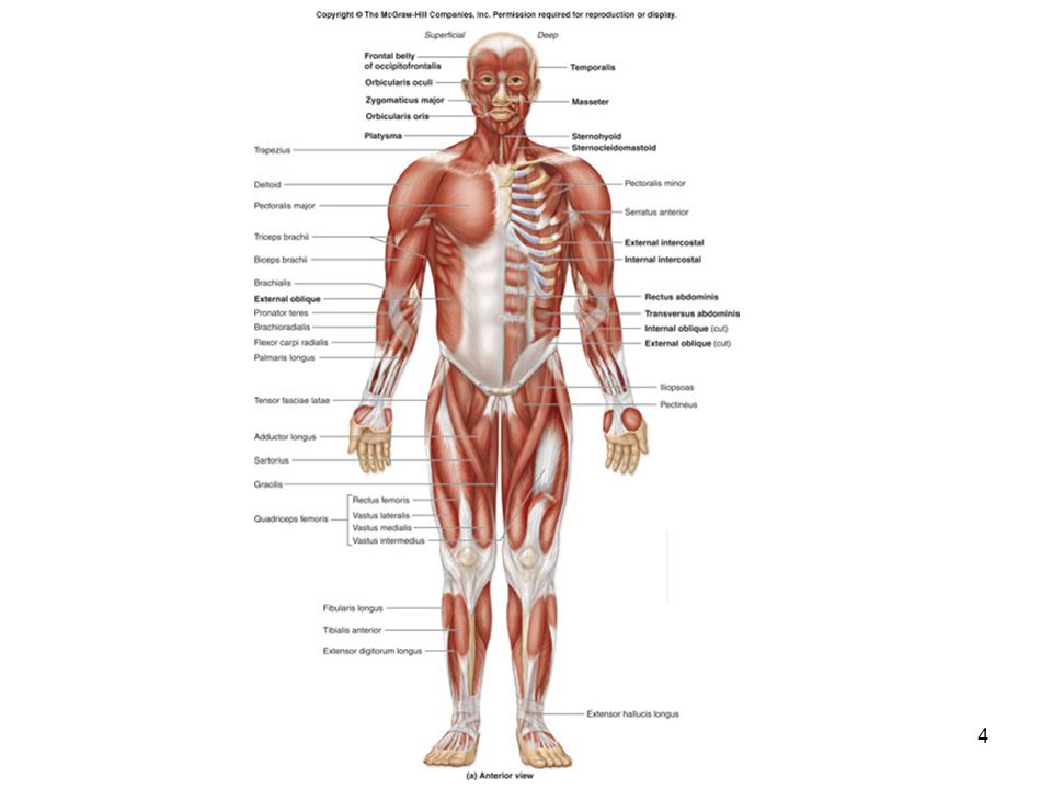
Axial Muscles Have both their origins and insertions on parts of the axial skeleton. Support and move the head and spinal column. Function in nonverbal communication by affecting facial features. Move the lower jaw during chewing. Assist in food processing and swallowing. Aid breathing. Support and protect the abdominal and pelvic organs. Are not responsible for stabilizing or moving the pectoral or pelvic girdles or their attached limbs.

6
Muscles of the Head and Neck
Separated into several specific groups. Almost all originate on either the skull or the hyoid bone.

7
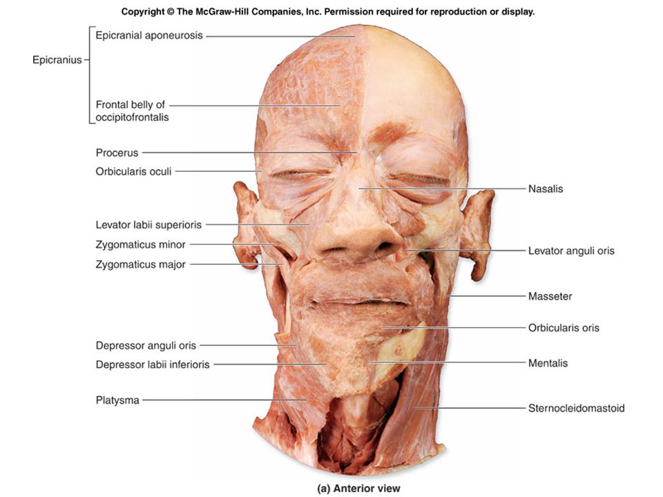
Muscles of Facial Expression
Originate in the superficial fascia or on the skull bones. Insert into the superficial fascia of the skin. Contort the skin causing it to move.

12
Muscles of Facial Expression
Several are associated with the nose. The mouth is the most expressive part of the face muscles in that area are very diverse Orbicularis oris consists of muscle fibers that encircle the opening of the mouth. when it contracts the mouth closes

14
Extrinsic Eye Muscles Often called extraocular muscles. Move the eyes.
Are termed extrinsic because they originate within the orbit and insert onto the sclera. Six extrinsic eye muscles. the rectus muscles (medial, lateral, inferior, and superior) the oblique muscles (inferior and superior)
 the oblique muscles (inferior and superior)")
19
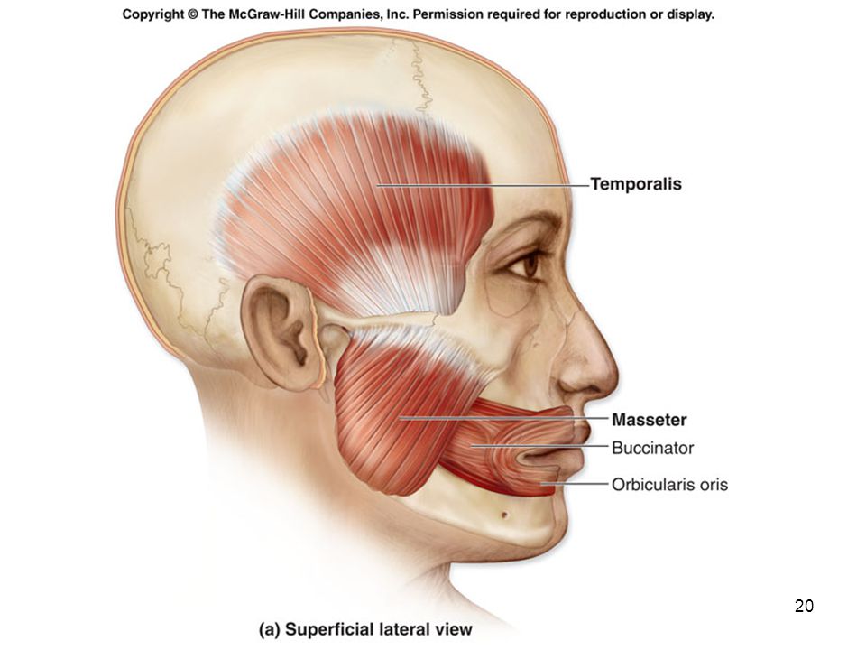
Muscles of Mastication
Refers to the process of chewing. Move the mandible at the temporomandibular joint. Four paired muscles of mastication temporalis masseter lateral pterygoids medial pterygoids

22
Muscles That Move the Tongue
The left and right genioglossus muscles originate on the mandible and protract the tongue. The left and right styloglossus muscles originate on the styloid processes of the temporal bone. elevate and retract the tongue (pull the tongue back into the mouth) The left and right hyoglossus muscles originate at the hyoid bone and insert on the sides of the tongue. Depress and retract the tongue The left and right palatoglossus muscles originate on the soft palate. elevate the posterior portion of the tongue
 The left and right hyoglossus muscles originate at the hyoid bone and insert on the sides of the tongue. Depress and retract the tongue. The left and right palatoglossus muscles originate on the soft palate. elevate the posterior portion of the tongue.")
24
Muscles That Move the Tongue
The tongue is an agile, highly mobile organ. It consists of intrinsic muscles that curl, squeeze, and fold the tongue during chewing and speaking. the tongue itself is a big muscle Extrinsic muscles of the tongue, originate on other head and neck structures and insert on the tongue. glossus = “tongue” Used in various combinations to accomplish the precise, complex, and delicate tongue movements required for proper speech. Manipulate food within the mouth in preparation for swallowing.

26
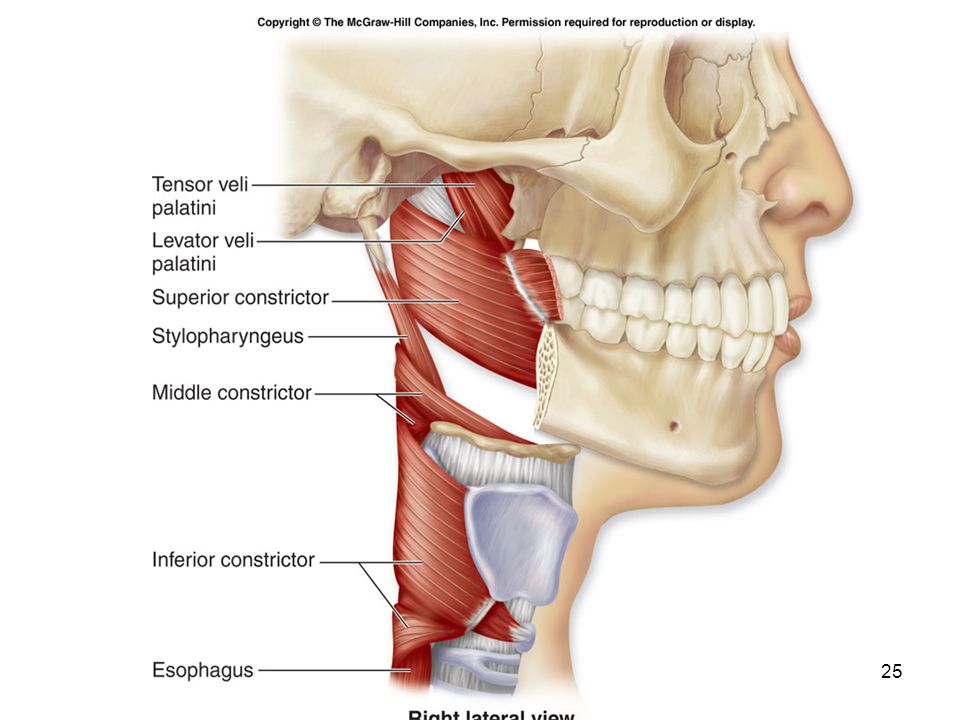
Muscles of the Pharynx Commonly known as the “throat.”
Is a funnel-shaped tube that lies posterior to both the oral and nasal cavities. Muscles help form or attach to this tube and aid in swallowing. Primary pharynx muscles are the pharyngeal constrictors (superior, middle, and inferior). Initiate swallowing and force the bolus inferiorly into the esophagus. Help elevate or tense the palate when swallowing.
. Initiate swallowing and force the bolus inferiorly into the esophagus. Help elevate or tense the palate when swallowing.")
27
Muscles of the Anterior Neck
The suprahyoid muscles are superior to the hyoid bone. The infrahyoid muscles are inferior to the hyoid bone.

30
Anterior and Lateral Neck Muscles
Flex the head and neck downward. “neck flexion” and “head flexion” refer to the same movement The main muscles are the sternocleidomastoid and the three scalenes.

32
Posterior Neck Muscles
Extend the head/neck. The trapezius attaches to the skull and helps extend the head/neck. Primary function is to help move the pectoral girdle.

33
Insert Fig

35
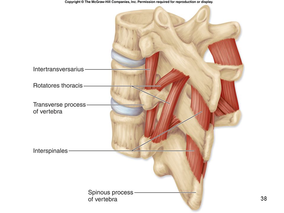
Muscles of the Vertebral Column
Very complex. Have multiple origins and insertions. Exhibit quite a bit of overlap. Are covered by the most superficial back muscles. trapezius and latissimus dorsi The “neck” is the cervical portion of the vertebral column. The muscles extend the cervical portion of the vertebral column.

39
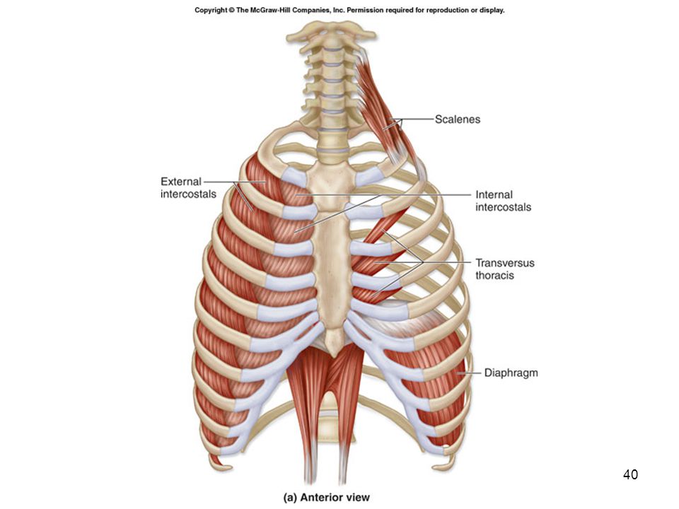
Muscles of Respiration
Respiration involves inhalation and exhalation. During inhalation, several muscles contract to increase the dimensions of the thoracic cavity as the lungs fill with air. The thoracic cavity expands both to cause the lungs to fill with air and to accommodate the expanding lungs. During exhalation, some respiratory muscles contract and others relax, collectively decreasing the dimensions of the thoracic cavity and forcing air out of the lungs. Are on the anterior and posterior surfaces of the thorax. Are covered by more superficial muscles that move the upper limb.

44
The Diaphragm Is an internally placed, dome-shaped muscle.
Forms a partition between the thoracic and abdominal cavities. The most important muscle associated with breathing. The muscle fibers converge from its margins toward a fibrous central tendon. A strong aponeurosis is the insertion tendon for all peripheral muscle fibers.

45
The Diaphragm When the diaphragm contracts, the central tendon is pulled inferiorly toward the abdominal cavity, thereby increasing the vertical dimensions of the thoracic cavity. As it compresses the abdominal cavity, it also increases intra-abdominal pressure. Also important in helping return venous blood to the heart from the lower half of the body.

46
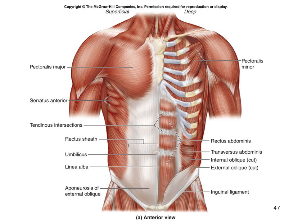
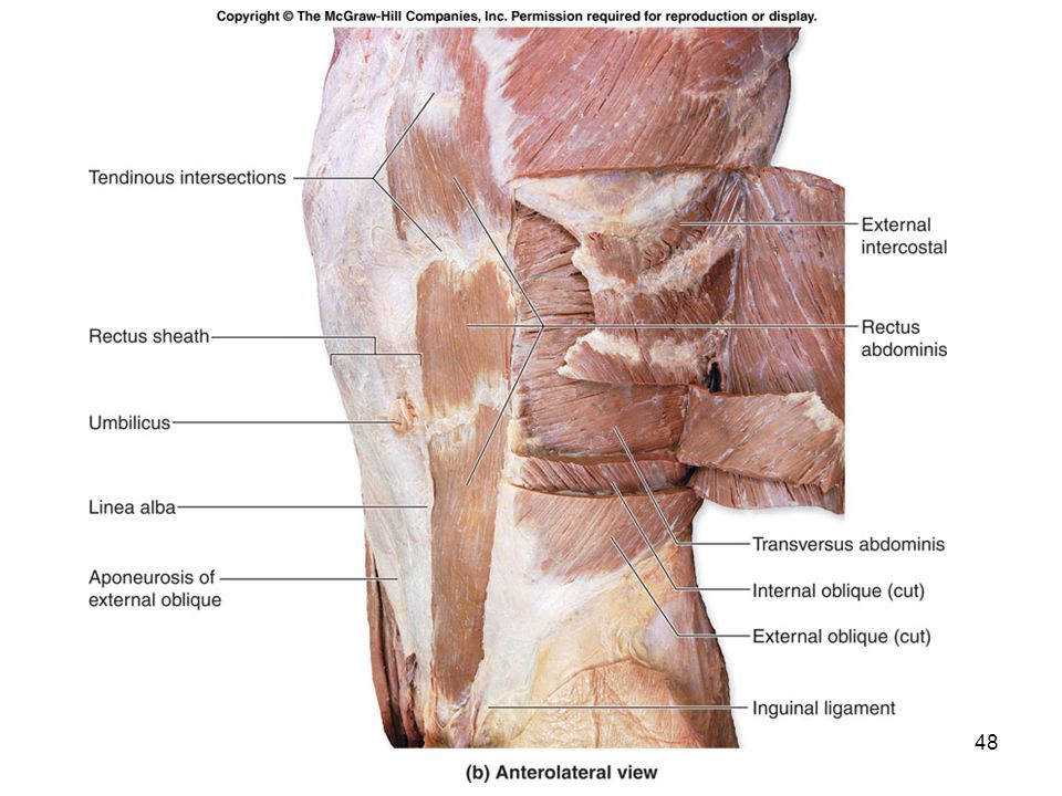
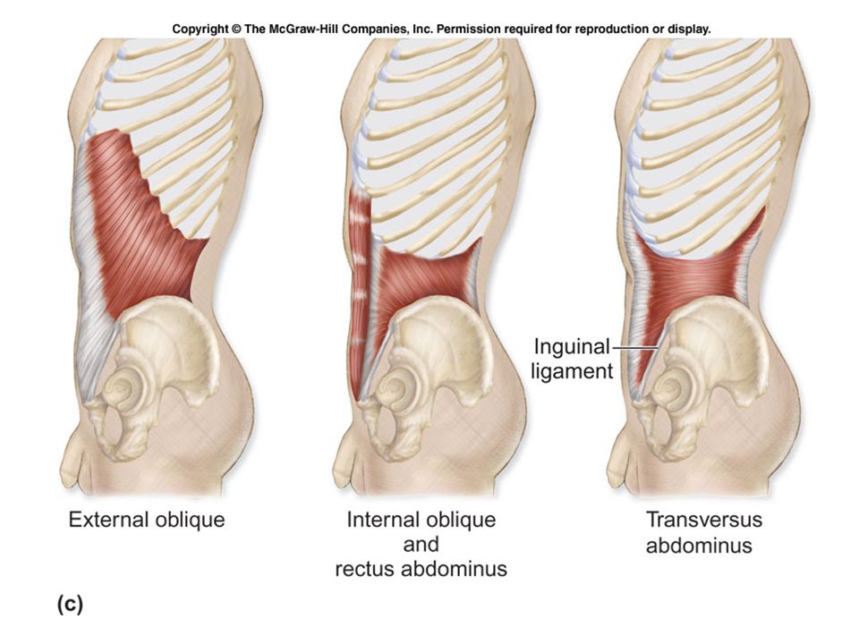
Muscles of the Abdominal Wall
Four pairs of muscles collectively compress and hold the abdominal organs in place. the external oblique internal oblique transversus abdominis rectus abdominis Work together to flex and stabilize the vertebral column. When they unilaterally contract they laterally flex the vertebral column.

50
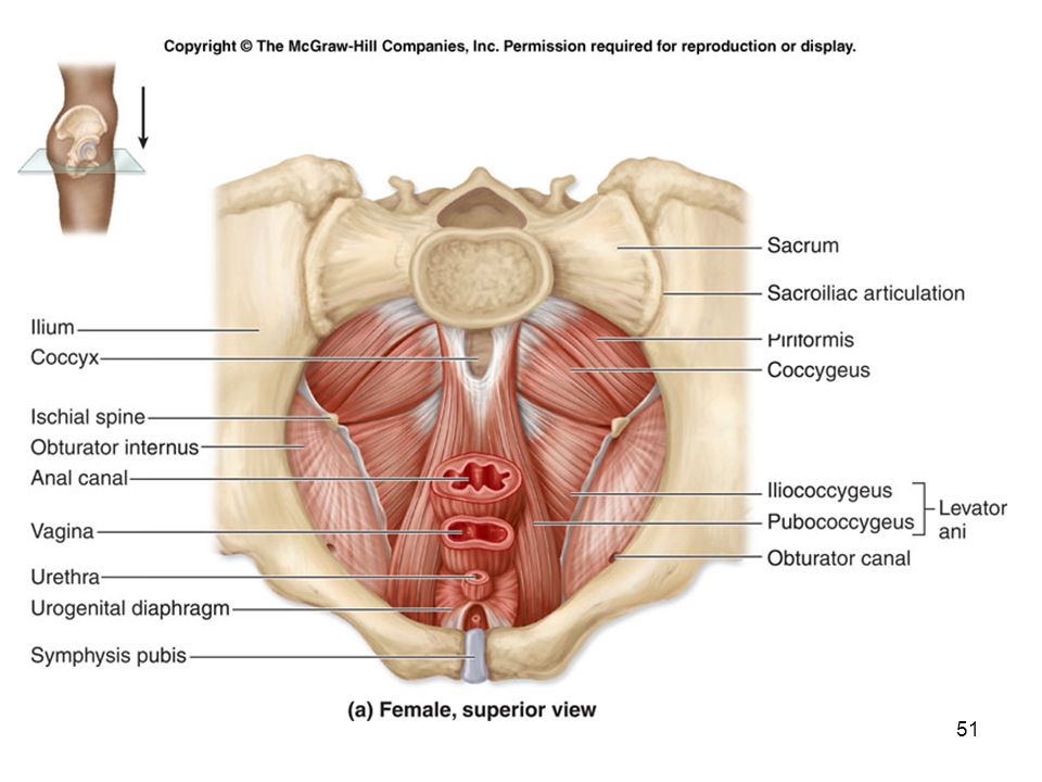
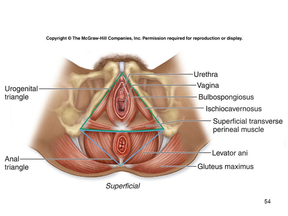
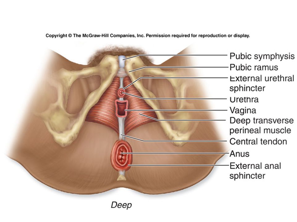
Muscles of the Pelvic Floor
Formed by three layers of muscles and associated fasciae, collectively known as the pelvic diaphragm. extends from the ischium and pubis of the ossa coxae across the pelvic outlet to the sacrum and coccyx Collectively form the pelvic floor and support the pelvic viscera the pelvic cavity floor is composed of muscle layers that form the urogenital and anal triangles, extend across the pelvic outlet, and support the organs in the pelvic cavity

56
Hernias A portion of the viscera protrudes through a weakened point of the muscular wall of the abdominopelvic cavity. Significant medical problem develops if the herniated portion of the intestine swells, becoming trapped. Blood flow to the trapped segment may diminish, causing that portion of the intestine to die. Called a strangulated intestinal hernia. is very painful and can be life-threatening

57
Two Types of Hernias There are two types of hernias.
inguinal hernias and femoral hernias An inguinal hernia is the most common type of hernia to require treatment. The inguinal region is one of the weakest areas of the abdominal wall.

58
Inguinal Hernia Males are more likely to develop inguinal hernias than females. Rising pressure in the abdominal cavity provides the force to push a segment of the small intestine into the canal. There are two types of inguinal hernia. direct inguinal hernia - the loop of small intestine protrudes directly through the superficial inguinal ring, but not down the entire length of the inguinal canal, and creates a bulge in the lower anterior abdominal wall indirect inguinal hernia - herniation travels down the entire inguinal canal and may even extend all the way into the scrotum

59
Femoral Hernia Occurs in the upper thigh, just inferior to the inguinal ligament, originating in the femoral triangle. Medial part of the femoral triangle is relatively weak and prone to stress injury, allowing a loop of small intestine to protrude. Women more commonly develop femoral hernias because of the greater width of their femoral triangle.

Similar presentations


>")

